Abstract
AbstractCortical neural activities lead to changes in the cerebral blood flow (CBF), which involves astrocytic control of cerebrovascular tone. However, the manner in which astrocytic activity specifically leads to vasodilation or vasoconstriction is difficult to determine. Here, cortical astrocytes genetically expressing a light-sensitive cation channel, channelrhodopsin-2 (ChR2), were transcranially activated with a blue laser while the spatiotemporal changes in CBF were noninvasively monitored with laser speckle flowgraphy in the anesthetised mouse cortex. A brief photostimulation induced a fast transient increase in CBF. The average response onset time was 0.7 ± 0.7 sec at the activation foci and this CBF increase spread widely from the irradiation spot with an apparent propagation speed of 0.8–1.1 mm/sec. The broad increase in the CBF could be due to a propagation of diffusible vasoactive signals derived from the stimulated astrocytes. Pharmacological manipulation showed that topical administration of a K+ channel inhibitor (BaCl2; 0.1–0.5 mM) significantly reduced the photostimulation-induced CBF responses, which indicates that the ChR2-evoked astrocytic activity involves K+ signalling to the vascular smooth muscle cells. These findings demonstrate a unique model for exploring the role of the astrocytes in gliovascular coupling using non-invasive, time-controlled, cell-type specific perturbations.
🔬 Techniques
🔭 Microscopes
💻 Software
✨ Fluorophores
🧪 Sample Preparation
🔬 Cell Lines
🏭 Microscope Brands
🧪 Reagent Suppliers
📷 Detectors
💻 Software Details
🏛️ Research Organizations (ROR)
Affiliated research institutions:
📋 Methods
Animal preparations Animal use and experimental protocols (No. 09058) were approved by the Animal Ethics Committee of Keio University Medical School, and all experimental procedures were in accordance with the university’s guidelines for the care and use of laboratory animals. A total of 21 male and female Mlc1-tTA::tetO-ChR2(C128S)-EYFP double transgenic mice (10–30 weeks, 32 ± 5 g) were used for the experiments. In these mice, C128S-modified ChR2 fused with EYFP was specifically expressed in astrocytes under the control of the Mlc1 promoter 23 . ChR2(C128S) is opened by blue light, and it requires yellow/orange light to close. As a control experiment, four normal male C57BL/6J mice (8 weeks, 21–25 g) were also subjected to repeated photostimulation to examine potential artefacts on the cortical microcirculation due to light irradiation. Under urethane anaesthesia (1.1 g/ kg, i.p.), the skull over the somatomotor cortex of both hemispheres was exposed. The body temperature was maintained at 36 °C with a heating pad (BWT-100, Bioresearch Centre Co. Ltd., Nagoya, Japan), and the systemic blood pressure and heart rate were monitored with a non-invasive blood pressure monitor (MK-2000ST, Muromachi Kikai Co. Ltd, Tokyo, Japan) at the tail, as needed. Photostimulation For comparisons of the photostimulation-induced CBF changes between normal C57BL/6J mice and ChR2 mice, a 488-nm argon laser (CVI Melles Griot, Carlsbad, CA) supplied through an optical fibre and a beam collimator (FBC-203S, Neoark, Tokyo, Japan) was used to irradiate a spot at the centre of the parietal bone. The irradiation spot on the skull was 0.50 ± 0.02 mm in diameter (i.e., 0.2 mm 2 ), which was captured with CCD camera (480 × 640 pixels with a pixel resolution of 35 μm/pixel). The irradiation image was converted to a binary image with applying a threshold at a half maximum of the pixel intensity in the image. The irradiation area was then approximated with a circle shape, and the diameter of the circle was measured. The mean irradiation power was measured with an optical power meter (8230 ADCMT, ADC, Tokyo, Japan). The duration of the laser irradiation was 0.5 sec (short) or 3 sec (long), which was regulated with an electromagnetic shutter (F77-7, Suruga Seiki, Shizuoka, Japan), and the intensity of the irradiation was adjusted using an ND filter placed in the optical path. Following cessation of the laser irradiation, a 595-nm LED light (0.1–0.3 mW, LEDP_HB01-A, Doric Lenses, Quebec, Canada) was used to irradiate the skull for 3 sec to close ChR2(C128S) channels ( Fig. 1A ). These sequential events were controlled with a pulse generator (Master-8, AMPI, Israel).
Show full methods section
Animal preparations Animal use and experimental protocols (No. 09058) were approved by the Animal Ethics Committee of Keio University Medical School, and all experimental procedures were in accordance with the university’s guidelines for the care and use of laboratory animals. A total of 21 male and female Mlc1-tTA::tetO-ChR2(C128S)-EYFP double transgenic mice (10–30 weeks, 32 ± 5 g) were used for the experiments. In these mice, C128S-modified ChR2 fused with EYFP was specifically expressed in astrocytes under the control of the Mlc1 promoter 23 . ChR2(C128S) is opened by blue light, and it requires yellow/orange light to close. As a control experiment, four normal male C57BL/6J mice (8 weeks, 21–25 g) were also subjected to repeated photostimulation to examine potential artefacts on the cortical microcirculation due to light irradiation. Under urethane anaesthesia (1.1 g/ kg, i.p.), the skull over the somatomotor cortex of both hemispheres was exposed. The body temperature was maintained at 36 °C with a heating pad (BWT-100, Bioresearch Centre Co. Ltd., Nagoya, Japan), and the systemic blood pressure and heart rate were monitored with a non-invasive blood pressure monitor (MK-2000ST, Muromachi Kikai Co. Ltd, Tokyo, Japan) at the tail, as needed. Photostimulation For comparisons of the photostimulation-induced CBF changes between normal C57BL/6J mice and ChR2 mice, a 488-nm argon laser (CVI Melles Griot, Carlsbad, CA) supplied through an optical fibre and a beam collimator (FBC-203S, Neoark, Tokyo, Japan) was used to irradiate a spot at the centre of the parietal bone. The irradiation spot on the skull was 0.50 ± 0.02 mm in diameter (i.e., 0.2 mm 2 ), which was captured with CCD camera (480 × 640 pixels with a pixel resolution of 35 μm/pixel). The irradiation image was converted to a binary image with applying a threshold at a half maximum of the pixel intensity in the image. The irradiation area was then approximated with a circle shape, and the diameter of the circle was measured. The mean irradiation power was measured with an optical power meter (8230 ADCMT, ADC, Tokyo, Japan). The duration of the laser irradiation was 0.5 sec (short) or 3 sec (long), which was regulated with an electromagnetic shutter (F77-7, Suruga Seiki, Shizuoka, Japan), and the intensity of the irradiation was adjusted using an ND filter placed in the optical path. Following cessation of the laser irradiation, a 595-nm LED light (0.1–0.3 mW, LEDP_HB01-A, Doric Lenses, Quebec, Canada) was used to irradiate the skull for 3 sec to close ChR2(C128S) channels ( Fig. 1A ). These sequential events were controlled with a pulse generator (Master-8, AMPI, Israel).
Laser speckle flowgraphy Spatiotemporal
CBF was monitored using laser speckle flowgraphy (LSFG-Micro, Softcare, Fukuoka, Japan). An 830-nm laser diode was selected as a light source for the flow imaging because this wavelength of light likely has no effect on the ChR2(C128S), as the irradiated wavelength is far from its excitation range 44 . While the skull or exposed cortex was continuously illuminated, a speckle pattern was captured at a rate of 30 frames per sec (i.e., an exposure time of 33 ms in each frame), using a charge-coupled device (CCD) camera (600 × 480 pixels) attached to a microscope (SZ61TR, Olympus Corporation, Tokyo, Japan). The field of view was either 4.9 mm × 4.7 mm or 2.4 mm × 2.3 mm at 2-fold or 4-fold magnification of the objective lens (NA = 0.071 and a working distance of 110 mm), respectively (see Fig. 1A ). The raw intensity image was converted to a flow image by calculating the mean blur rate (MBR) of each pixel using 3-consecutive frames 45 . The pre-stimulation baseline images were acquired for 30 sec before induction of photostimulation in each recording, and the post-stimulation images were acquired for at least 90 sec depending on the duration of the evoked changes in CBF. In each animal, 1–8 trials (average 3 trials per stimulation) were repeated for the same paradigms of the photostimulation with an onset-to-onset interval of >2 min. Pharmacology To apply the drugs to the stimulated cortex, a section of skull approximately 3 mm in diameter was removed (N = 10 ChR2 mice), and the dura located near the edge of the opened area was carefully removed. Both the left and right hemispheres were opened in five out of ten animals to test a single drug in respective hemispheres. First, saline was applied to the opened area of the cortical surface, and the CBF response to photostimulation was measured with LSFG (i.e., pre-treatment control measurement). Then, a K + channel inhibitor BaCl 2 (0.1–0.5 mM in saline; Yoneyama Yakuhin Kogyo Co., Ltd., Japan), the non-selective COX inhibitor sodium indomethacin (0.5 mM in saline; INDACIN IV; MSD K.K., Japan), or voltage gated sodium channel inhibitor tetrodotoxin (TTX; 20–50 μM in saline; Nacalitesque, Kyoto, Japan) were topically applied (0.1–0.3 mL) to the overall exposed cortical surface. The concentration of the drugs used in the present study was determined according to previous studies 3 30 31 46 . At 20–60 min after drug administration, the photostimulation-induced CBF response was evaluated with the same protocols used for the pre-treatment control measurements. In this experiment, a stimulation power of the blue laser irradiation was adjusted so that the evoked CBF does not spread out of the field of view under pre-treatment conditions.
Histology
At the end of the experiments, SR101 (10 mM in saline, Sigma, Japan), a marker of astrocytes/olligodendrocytes 47 48 , was intraperitoneally injected (0.10–0.15 mL, N = 6 ChR2 mice). At 3–4 hours after the injection, the brain was removed and fluorescently visualised to verify the expression of ChR2(C128S)-EYFP in SR101-labelled astrocytes using two-photon microscopy (TCS SP5MP, Leica Microsystems) 49 . Note that ChR2(C128S)-EYFP is expressed throughout the cell membrane including the astrocytic fine processes. The astrocytic expression of ChR2(C128S)-EYFP was dense in the parenchyma of the measured somatomotor cortex, but the astrocytes in the subsurface layers only sparsely expressed the transgene ( Fig. 1B ).
Data analysis
A region of interest (ROI) was manually placed at the centre of the maximum response area for the LSFG flow images, and the mean time-course of the relative CBF changes normalised to the baseline averaged over 10-sec a pre-stimulation period were calculated by averaging the pixel values within the ROI. For comparisons of the CBF responses between laser-irradiated cortex (i.e., ipsilateral cortex) and unirradiated cortex (i.e., contralateral cortex), two ROIs (1 mm in diameter) were located at an activation foci in the ipsilateral cortex and an area contralateral to the activation foci in the other cortex (see Fig. 1A ). Then, the response magnitude of the CBF was calculated by averaging CBF changes over 5 to 15 sec after the onset of photostimulation within the ROI. For the LSFG through the intact skull, it was shown that evoked changes in CBF were influenced by a large static scattering 50 . In this study, we did not correct any static components that may cause a slight underestimation of the CBF response magnitude. Two-points of the onset time of the CBF response were defined. One is the time at which the relative CBF surpassed the mean plus 2-fold standard deviation of the pre-stimulation baseline, and the other one is the relative CBF surpassed the mean plus 3-fold standard deviation. For this particular analysis, a low-pass filter (
📊 Figures
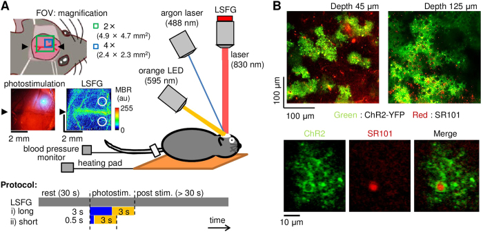
Figure 1
A ) Experimental setup (original drawing). To open ChR2 channels specifically expressed in the astrocytes, an argon laser was induced through an electromagnetic shutter, while spatiotemporal CBF were ...
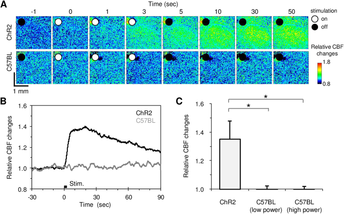
Figure 2
Photostimulation induced CBF changes.
A ) Baseline and activation-induced CBF images captured with LSFG. A robust increase in CBF was evoked only in the ChR2 mouse (top) and not in the C57BL/6 mouse (bottom). White and black circles in th...
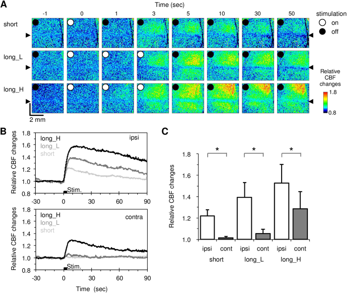
Figure 3
Comparison of the photostimulation-induced CBF changes in the stimulated ipsilateral and unstimulated contralateral cortices in ChR2 mice (Nu2009=u200910).
A ) Spatiotemporal dynamics of the CBF responses to short irradiation (top), long blue laser irradiation with low power (long_L, middle), and long blue laser irradiation with high power (long_H, botto...
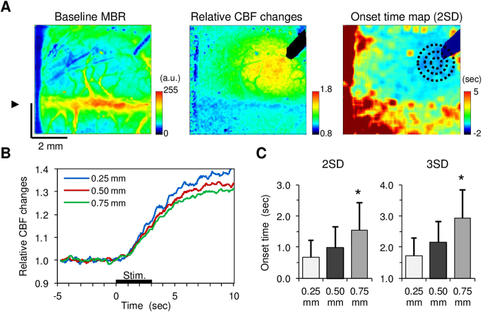
Figure 4
Spatiotemporal dynamics of CBF responses to transcranial photostimulation in ChR2 mice (Nu2009=u200910).
A ) A representative image of the mean baseline MBR averaged for 10-sec pre-stimulus periods ( left ). The arrowhead indicates the location of the midline. Broad CBF changes were evoked with photostim...
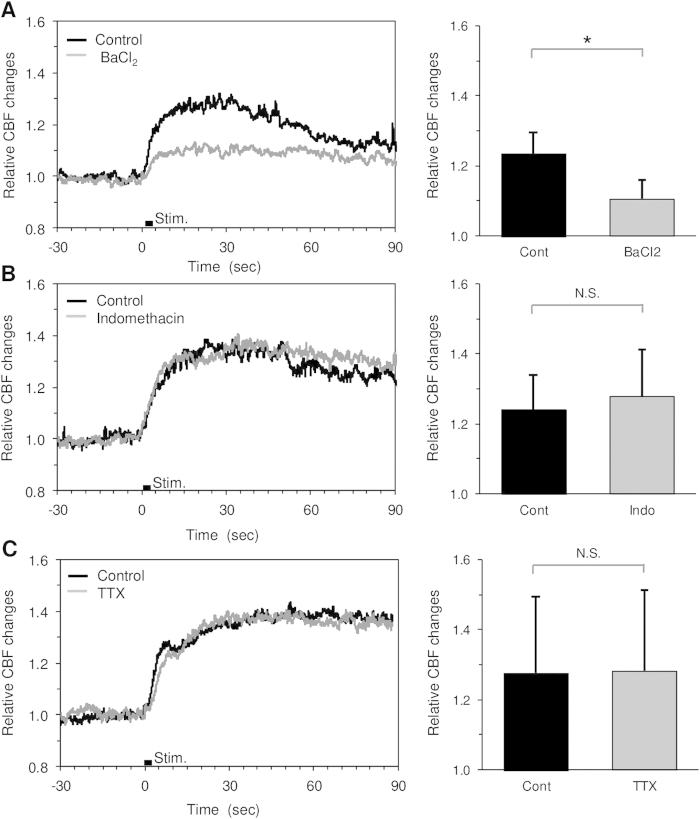
Figure 5
CBF responses to direct photostimulation measured before and after pharmacological treatment in ChR2 mice.
A ) Topical administration of BaCl 2 (0.1u20130.5u2009mM) significantly reduced the CBF responses (Nu2009=u20096). B ), C ) Neither significant difference in the CBF response was detected for topical ...
Figure images are served from the NIH/NLM PubMed Central Open Access Subset or Europe PMC; copyright remains with the publishers and authors.
💬 Discussion
0 commentsNo comments yet. Be the first to start a discussion!
Leave a Comment