Abstract
Annexins are Ca(2+)-binding, membrane-fusogenic proteins with diverse but poorly understood functions. Here, we show that during cell cycle progression annexin 11 translocates from the nucleus to the spindle poles in metaphase and to the spindle midzone in anaphase. Annexin 11 is recruited to the midbody in late telophase, where it forms part of the detergent-resistant matrix that also contains CHO1. To investigate the significance of these observations, we used RNA interference to deplete cells of annexin 11. A combination of confocal and video time-lapse microscopy revealed that cells lacking annexin 11 fail to establish a functional midbody. Instead, daughter cells remain connected by intercellular bridges that contain bundled microtubules and cytoplasmic organelles but exclude normal midbody components such as MKLP1 and Aurora B. Annexin 11-depleted cells failed to complete cytokinesis and died by apoptosis. These findings demonstrate an essential role for annexin 11 in the terminal phase of cytokinesis.
🔬 Techniques
🧬 Organisms
💻 Software
✨ Fluorophores
🧪 Sample Preparation
🔬 Cell Lines
🏭 Microscope Brands
🧪 Reagent Suppliers
📷 Detectors
💻 Software Details
🏛️ Research Organizations (ROR)
Affiliated research institutions:
📋 Methods
Cell lines and culture conditions Human A431, HeLa, and HEp2 cells were grown in DME (GIBCO BRL) supplemented with 10% (vol/vol) FCS, 100 U/ml penicillin, 100 μg/ml streptomycin, and 292 μg/ml l -glutamine at 37°C in 5% CO 2 humidified incubators. Cells were kept subconfluent, and the medium was changed every 2 to 3 d. For microscopy, cells were plated onto sterilized coverslips or grown in 35-mm glass bottom microwell dishes (MatTek).
Antibodies
Antibodies used for immunofluorescence were L-19 goat polyclonal anti–human annexin 11 (Santa Cruz Biotechnology, Inc.), mouse monoclonal anti–mouse annexin 11 (BD Biosciences), mouse monoclonal anti–chicken α-tubulin (Zymed Laboratories), rabbit polyclonal anti–human α-tubulin (gift of K. Matter, Institute of Ophthalmology, University College London, London, UK), E18 rabbit polyclonal anti–chicken CHO1 (gift of R. Kuriyama, University of Minnesota, Minneapolis, MN; Kuriyama et al., 2002 ), mouse monoclonal anti–rat Aim-1 (BD Biosciences), N-19 rabbit polyclonal anti-MKLP1 (Santa Cruz Biotechnology, Inc.), E-15 goat polyclonal anti-ARK-2 (Santa Cruz Biotechnology, Inc.), and M-20 goat polyclonal anti–mouse lamin B (Santa Cruz Biotechnology, Inc.). Secondary antibodies were donkey anti-goat-Alexa Fluor 488 (Molecular Probes), goat anti–mouse-Alexa Fluor 488 (Molecular Probes), donkey anti–mouse-Alexa Fluor 488 (Molecular Probes), donkey anti–rabbit-Cy5, donkey anti–mouse-Cy5, and donkey anti–mouse-TRITC. Antibodies used for Western blotting were as for CHO1, L-19 goat polyclonal anti–human annexin 11 (Santa Cruz Biotechnology, Inc.), sheep polyclonal anti–chicken annexin 11 (raised against full-length recombinant chicken annexin 11), mouse monoclonal anti–chicken α-tubulin (Zymed Laboratories), mouse monoclonal anti–β-tubulin (Sigma-Aldrich), and rabbit polyclonal anti–human PARP (Santa Cruz Biotechnology, Inc.). Antisera to annexins 1 and 5 have been described previously ( Hawkins et al., 1999 ).
Show full methods section
Cell lines and culture conditions Human A431, HeLa, and HEp2 cells were grown in DME (GIBCO BRL) supplemented with 10% (vol/vol) FCS, 100 U/ml penicillin, 100 μg/ml streptomycin, and 292 μg/ml l -glutamine at 37°C in 5% CO 2 humidified incubators. Cells were kept subconfluent, and the medium was changed every 2 to 3 d. For microscopy, cells were plated onto sterilized coverslips or grown in 35-mm glass bottom microwell dishes (MatTek).
Antibodies
Antibodies used for immunofluorescence were L-19 goat polyclonal anti–human annexin 11 (Santa Cruz Biotechnology, Inc.), mouse monoclonal anti–mouse annexin 11 (BD Biosciences), mouse monoclonal anti–chicken α-tubulin (Zymed Laboratories), rabbit polyclonal anti–human α-tubulin (gift of K. Matter, Institute of Ophthalmology, University College London, London, UK), E18 rabbit polyclonal anti–chicken CHO1 (gift of R. Kuriyama, University of Minnesota, Minneapolis, MN; Kuriyama et al., 2002 ), mouse monoclonal anti–rat Aim-1 (BD Biosciences), N-19 rabbit polyclonal anti-MKLP1 (Santa Cruz Biotechnology, Inc.), E-15 goat polyclonal anti-ARK-2 (Santa Cruz Biotechnology, Inc.), and M-20 goat polyclonal anti–mouse lamin B (Santa Cruz Biotechnology, Inc.). Secondary antibodies were donkey anti-goat-Alexa Fluor 488 (Molecular Probes), goat anti–mouse-Alexa Fluor 488 (Molecular Probes), donkey anti–mouse-Alexa Fluor 488 (Molecular Probes), donkey anti–rabbit-Cy5, donkey anti–mouse-Cy5, and donkey anti–mouse-TRITC. Antibodies used for Western blotting were as for CHO1, L-19 goat polyclonal anti–human annexin 11 (Santa Cruz Biotechnology, Inc.), sheep polyclonal anti–chicken annexin 11 (raised against full-length recombinant chicken annexin 11), mouse monoclonal anti–chicken α-tubulin (Zymed Laboratories), mouse monoclonal anti–β-tubulin (Sigma-Aldrich), and rabbit polyclonal anti–human PARP (Santa Cruz Biotechnology, Inc.). Antisera to annexins 1 and 5 have been described previously ( Hawkins et al., 1999 ).
Cell synchronization and isolation of midbodies
Cells were grown to 50% confluence and incubated in medium containing 4 mM thymidine (Sigma-Aldrich) overnight to achieve S phase block. Dishes were extensively washed in PBS and cultured for a further 6 h in medium containing 0.07 μg/ml nocodazole (Sigma-Aldrich) for growth arrest at prometaphase. Cells were extensively washed and allowed to recover in normal medium for 30–60 min or incubated for 45 min in the presence of 15 μg/ml BFA (Sigma-Aldrich). For midbody isolation, cells were synchronized at prometaphase and spindles were prepared as described previously ( Kuriyama et al., 1984 ).
Fluorescence microscopy
Cells grown on coverslips or in 35-mm glass bottom microwell dishes were washed in PBS, fixed/permeabilized on ice for 30 min in 2% PFA + 0.1% Triton X-100, washed four times in PBS, and blocked for 15 min with 1% BSA (Sigma-Aldrich). Fixed cells were incubated in a moist chamber with primary antibody dissolved in 1% BSA for 1 h, followed by four washes of 5–10 min each in PBS before incubation with the secondary antibody dissolved in 1% BSA for another hour. After four further washes in PBS, samples were either observed immediately in PBS or mounted onto slides with Moviol and sealed with nail polish. Stained cells were observed under a Radiance 2000 AGR-3 (Q) confocal (Bio-Rad Laboratories) attached to an inverted microscope (model Axiovert S100TV; Carl Zeiss MicroImaging, Inc.) using 60× or 100× oil immersion lenses, and images were processed using Metamorph 4.6r9.
Electron microscopy
A431 cells were cultured on thermanox coverslips (Agar Scientific) and, after annexin 11 depletion, were fixed, processed, and treated with tannic acid as described by Stinchcombe et al. (1995) . Coverslips were mounted on Epon stubs, the Epon polymerized overnight at 60°C, and the coverslips removed by heating. 70-nm sections were cut en face and stained with lead citrate before examination in a JEOL 1010 electron microscope.
Video time-lapse microscopy
A431 cells were placed in a 37°C preheated microscope chamber with 5% CO 2 supply and observed using phase illumination on an inverted microscope (model Axiovert S100M; Carl Zeiss MicroImaging, Inc.) using a 40× oil immersion lens. Images were acquired with Openlab (Improvision) and analyzed using Metamorph 4.6r9 (Universal Imaging Corp.). Immunoprecipitation, SDS-PAGE, and Western blotting A431 cell monolayers were washed in PBS, resuspended in 500 μl lysis buffer (50 mM Tris-Cl, pH 7.4, 50 mM NaCl, 30 mM sodium pyrophosphate, 50 mM sodium fluoride, 100 μM sodium orthovanadate, 0.2 mM CaCl 2 , 1% Triton X-100, 0.1% BSA, and 0.02% sodium azide) supplemented with 10 μl of protease inhibitor cocktail (Sigma-Aldrich) per 10 7 cells, and lysed by three rounds of sonication on ice. Lysates were precleared at 16,000 g for 30 min at 4°C, followed by incubation with 30 μl of 50% protein G–Sepharose 4 Fast Flow slurry (Amersham Biosciences) for 30 min, and then 5 min centrifugation at 16,000 g at 4°C. L-19 goat anti–human annexin 11 antibody (Santa Cruz Biotechnology, Inc.) was immobilized on protein G–Sepharose beads and added to the precleared lysates for 2 h at 4°C, centrifuged, and the supernatant discarded. Beads were washed four times in lysis buffer minus BSA and once in ice-cold PBS. After removal of the supernatant, the immunoprecipitates were resuspended in SDS-PAGE sample buffer, boiled for 5 min, and resolved by SDS-PAGE on discontinuous mini-gels. Proteins were electroblotted for 45 min at 400 mA onto Hybond-P PVDF Transfer Membrane (Amersham Biosciences). Membranes were treated with blocking solution (10% skimmed milk or 10% BSA in PBS-Tween [PBS + 0.05% Tween 20]) for 40 min at RT and incubated for 16 h at 4°C with primary antibody in PBS-Tween. After three PBS-Tween washes, membranes were incubated with secondary antibody for 90 min, also in PBS-Tween (1/10,000 dilution). IgG-HRP conjugates were used as secondary antibodies. Blots were developed after three further PBS-Tween washes using the ECL Plus Western blotting detection system (Amersham Biosciences) and visualized in a Fujifilm Intelligent Dark Box II coupled to a LAS-1000 CCD camera. Depletion of annexin 11 in HeLa and A431 cells by RNAi Preparation of siRNA oligonucleotides was performed as follows. A search was performed for AA(N19)TT sequences with ∼50% G/C-content in human annexin 11 cDNA. One such sequence was located at nt 727 in the coding sequence. Sense (5′-gACggCUUACggCAAggAUdTdT-3′) and antisense (5′-AUCCUUgCCgUAAgCCgUCdTdT-3′) siRNA oligonucleotides were purchased from Dharmacon Research, Inc. Oligonucleotides were diluted with 1 ml of RNase-free water to a concentration of 50 μM and annealed by combining 30 μl of each with 15 μl of 5× annealing buffer (Dharmacon Research, Inc.). The solution was incubated at 90°C for 1 min followed by 1 h at 37°C. The final concentration of the siRNA duplex was 20 μM. Transfection of cells with siRNA was performed as follows. Cells were seeded on 24-well plates in normal medium without antibiotics and transfected at 30 to 40% confluency as follows. For each well, 3 μl Oligofectamine reagent (Invitrogen) was dissolved in 12 μl Opti-MEM I medium (Invitrogen), and 3–5 μl of siRNA duplex was added to 50 μl of Opti-MEM I medium. The solutions were incubated for 10 min at RT, combined, and mixed by inversion. The mixture was incubated for 25 min at RT, and 30–32 μl of Opti-MEM I medium was added to bring the final volume to 100 μl. The complexes were added to the cells for 1 to 3 d before analysis by FACS ® , immunofluorescence, or Western blotting. In some experiments, to further increase the depletion of annexin 11, transfected cells were split after 3 d and retransfected as aforementioned.
Fluorescence-activated cell sorting
To determine the level of apoptosis caused by annexin 11 depletion, siRNA-treated cells were analyzed by FACS ® . Cells were collected, washed in PBS, fixed for 30 min in 70% ethanol at −20°C, centrifuged at 560 g for 5 min at 4°C, washed in PBS, and resuspended in 100 μl of 1 mg/ml RNase I-A (Sigma-Aldrich) solution in PBS for 30 min. To stain the DNA, 150 μl of propidium iodide solution (50 μg/ml propidium iodide [Sigma-Aldrich], 0.1% sodium citrate, and 0.1% Triton X-100) was added to this preparation. Samples were processed using a FACscan™ flowcytometer (BD Biosciences), and the results were analyzed with WinMDI 2.8. Online supplemental material Two supplemental figures are available showing the localization of annexin 11 and the Golgi marker GM130 in A431 cells undergoing cytokinesis and the localization of MKLP1 and Aurora B in annexin 11–depleted A431 cells undergoing cytokinesis. Videos showing control and annexin 11–depleted cells failing to complete cytokinesis are available as supplements to Fig. 7 . Note that in Video 2 the apoptosing cells detach from the plate after the final frame. Online supplemental material is available at http://www.jcb.org/cgi/content/full/jcb.200311054/DC1 .
Online supplemental material Two supplemental figures are available showing the localization of annexin 11 and the Golgi marker GM130 in A431 cells undergoing cytokinesis and the localization of MKLP1 and Aurora B in annexin 11–depleted A431 cells undergoing cytokinesis. Videos showing control and annexin 11–depleted cells failing to complete cytokinesis are available as supplements to Fig. 7 . Note that in Video 2 the apoptosing cells detach from the plate after the final frame. Online supplemental material is available at http://www.jcb.org/cgi/content/full/jcb.200311054/DC1 .
📊 Figures
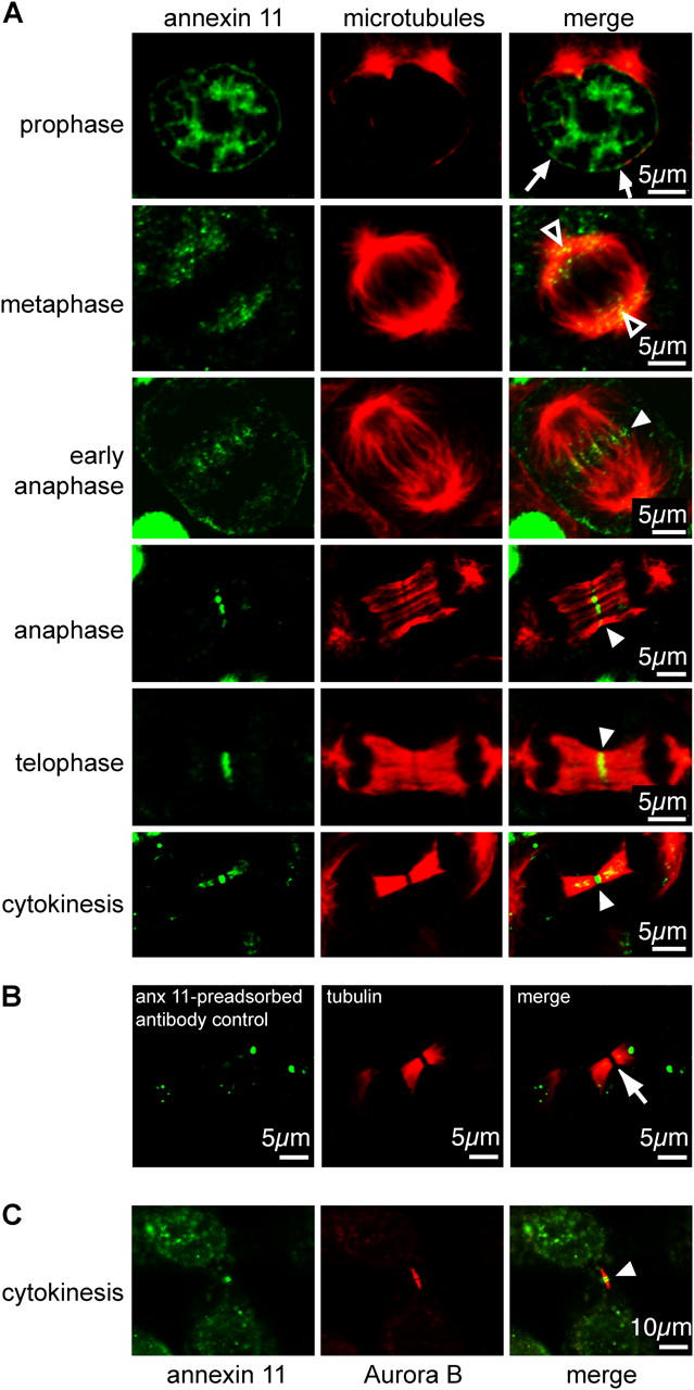
Figure 1.
Immunolocalization of annexin 11 at the different stages of the cell cycle. (A) A431 cells were fixed at different stages of cell cycle progression and stained for u03b1-tubulin and annexin 11. Annexi...
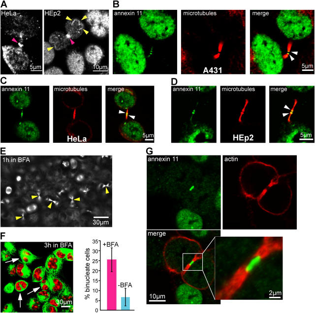
Figure 2.
Annexin 11 is a component of the midbody in different cell lines. (A) HeLa and HEp2 cells were fixed in cytokinesis and stained for annexin 11. The pink arrowheads indicate annexin 11 at the midbody, ...
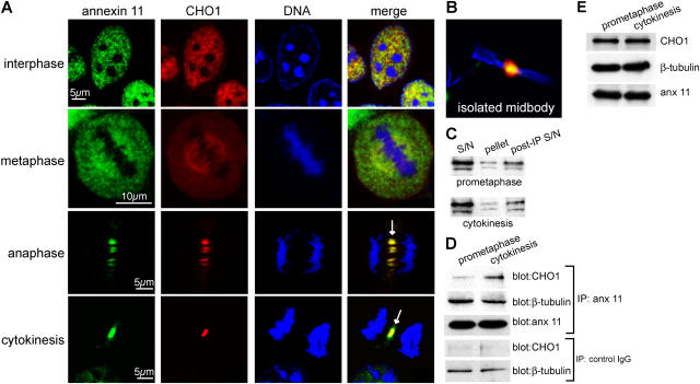
Figure 3.
Annexin 11 colocalizes and interacts with CHO1 at cytokinesis. (A) A431 cells were synchronized in mitosis and fixed and stained for annexin 11 and CHO1. DNA was labeled with propidium iodide. Cells i...
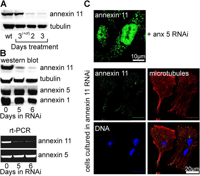
Figure 4.
Depletion of annexin 11 using siRNA. (A) A431 cells were treated with siRNA for annexin 11, with oligofectamine alone (+of) or with no additives (wt) for the periods indicated. Whole cell lysates were...
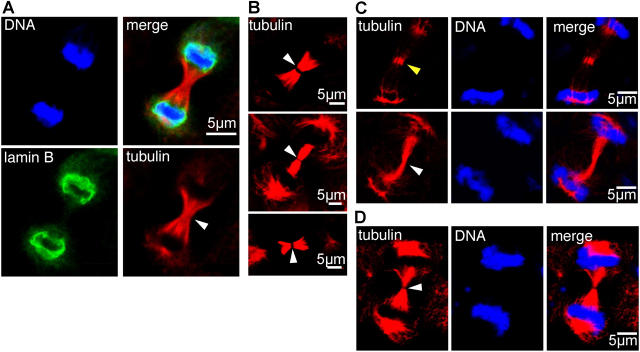
Figure 5.
Annexin 11 is required for midbody matrix formation. (A) HeLa cells treated with annexin 11 siRNA for 2 d were fixed and stained for lamin B and u03b1-tubulin. DNA was labeled using propidium iodide. ...
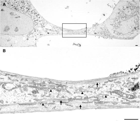
Figure 6.
Electron microscopy shows a complete absence of midbody formation in annexin 11u2013depleted cells. A431 cells were treated with annexin 11 siRNA for 3 d, and were then fixed and processed for electro...
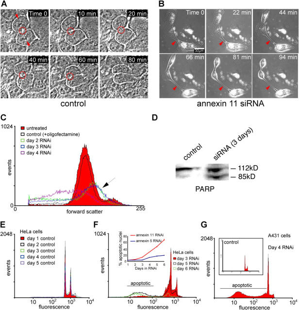
Figure 7.
Cytokinesis failure and apoptosis in cells treated with annexin 11 siRNA. (A) Control A431 cells undergoing cytokinesis were examined by video time-lapse microscopy (see Video 1). The two dividing cel...
Figure images are served from the NIH/NLM PubMed Central Open Access Subset or Europe PMC; copyright remains with the publishers and authors.
💬 Discussion
0 commentsNo comments yet. Be the first to start a discussion!
Leave a Comment