Abstract
The identities of toxic aggregate species in Huntington's disease pathogenesis remain ambiguous. While polyQ-expanded huntingtin (Htt) is known to accumulate in compact inclusion bodies inside neurons, this is widely thought to be a protective coping response that sequesters misfolded conformations or aggregated states of the mutated protein. To define the spatial distributions of fluorescently-labeled Htt-exon1 species in the cell model PC12m, we employed highly sensitive single-molecule super-resolution fluorescence imaging. In addition to inclusion bodies and the diffuse pool of monomers and oligomers, fibrillar aggregates -100 nm in diameter and up to -1-2 µm in length were observed for pathogenic polyQ tracts (46 and 97 repeats) after targeted photo-bleaching of the inclusion bodies. These short structures bear a striking resemblance to fibers described in vitro. Definition of the diverse Htt structures in cells will provide an avenue to link the impact of therapeutic agents to aggregate populations and morphologies.
🔬 Techniques
✨ Fluorophores
🧪 Sample Preparation
🔬 Cell Lines
🏭 Microscope Brands
🧪 Reagent Suppliers
🔴 Lasers
📷 Detectors
🎨 Filters
💻 Software Details
🏛️ Research Organizations (ROR)
Affiliated research institutions:
📋 Methods
PC12m cell model
We used PC12m cells, a sub-clone of the original PC12 neuronal model cell line developed by L. Greene (Columbia University). PC12m was selected for its enhanced attachment to plastics, facilitating culture work, in M. Montminy's laboratory (Salk Institute, La Jolla). PC12m cells are well suited for imaging experiments in culture due to their moderate thickness of 10–20 μm when adhered to the microscope slide. PC12 and its sub-clones, differentiated into sympathetic-neuron-like cells, have served as models for early pathogenic events in Huntington's Disease with fluorescent-protein fusions to huntingtin (e.g. 16 17 ).
Cell culture
PC12m cells were grown to confluency at 37°C in a 5% CO 2 , 96% relative humidity incubator in tissue-culture treated flasks (75 cm 2 , BD Biosciences) in cell culture medium (10% fetal bovine serum (FBS), 90% phenol red free DMEM, both Gibco). Cells were passaged with 1:5 dilution at least 3 times prior to experiments. Each passage consisted of a 5 min incubation with 2 ml of trypsin replacement (TrypLE Express, Gibco), addition of 8 ml cell media to deactivate the enzyme, a brief centrifugation (
📊 Figures
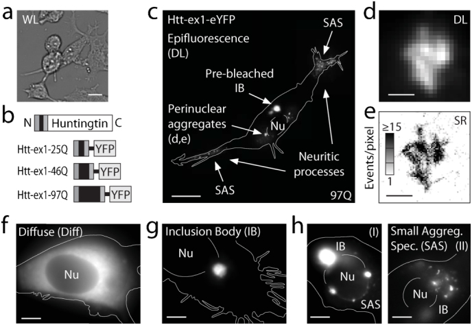
Figure 1
Overview of fluorescence microscopy of sub-cellular huntingtin distributions.
(a) Low-magnification white-light (WL) image of NGF-differentiated PC12m cell culture. (b) Schematic of full-length Htt (not to scale) and the Htt-ex1-xQ constructs fused at the C terminus to eYFP for...
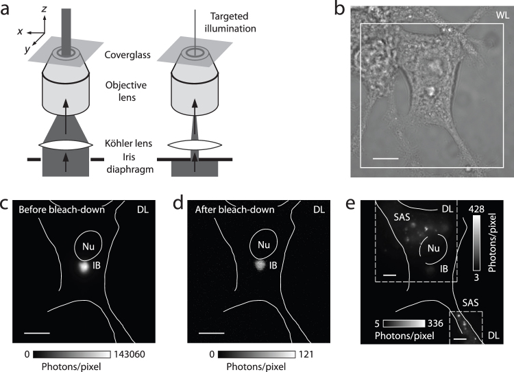
Figure 2
Targeted photo-bleaching of inclusion bodies and selective illumination of sub-cellular regions.
(a) Schematic of the procedure employed for bleaching a sample region containing a strongly fluorescent inclusion body (IB), to reduce its detrimental signal interference with nearby dimmer structures...
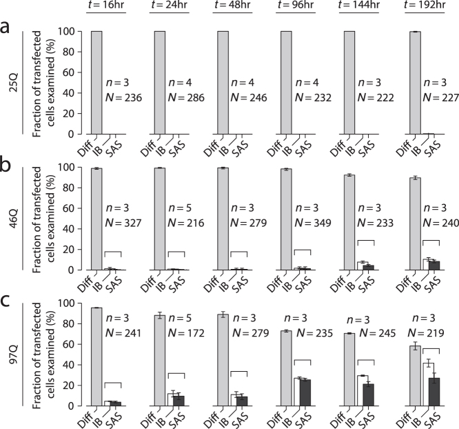
Figure 3
Phenotype counting analysis of cellular huntingtin distributions for three repeat-lengths (25,46,97 Q-repeats) by high-magnification examination of NGF-differentiated PC12m cells.
(a,b,c) Count of Htt-ex1-transfected cells (fixed) classified as the three phenotypes defined in Fig. 1fu2013h for the three Q-repeat lengths, at time-points t = 16u2013192u2005hours post-transfection...
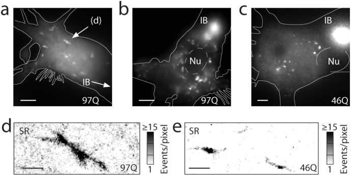
Figure 4
Sub-cellular distributions of huntingtin aggregates.
(a,b,c) Small aggregates dispersed throughout the cell bodies form for both 46Q (example e, from a different image plane in c) and 97Q (a,b,d) at time points beyond 16u2005hrs post-transfection. Their...
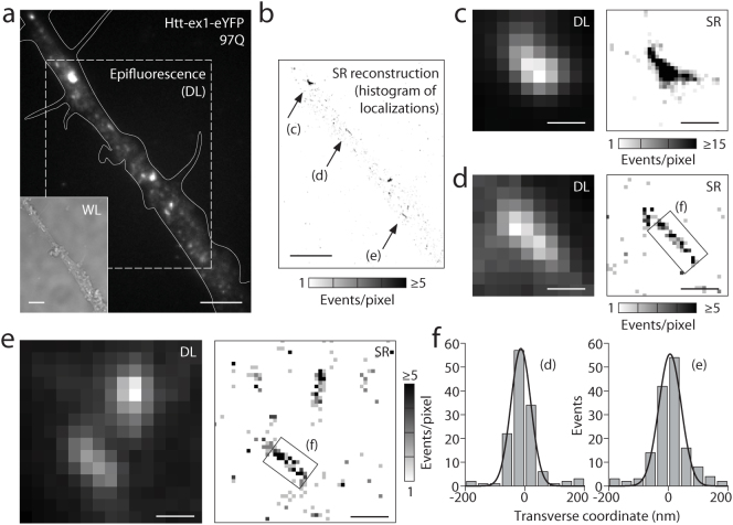
Figure 5
Aggregate species in neuritic processes imaged by super-resolution microscopy.
(a,b) Diffraction-limited and super-resolution image of a cellular process (large field). The cell was transfected with fluorescently labeled Htt-ex1-97Q for 24u2005hours before fixation. Scale bar: 5...
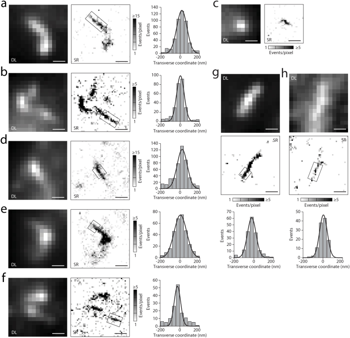
Figure 6
Small aggregates, imaged with super-resolution microscopy, based on the light-induced blinking of single eYFP fused to Htt-ex1.
(a) u2013 (h) (Left) Diffraction-limited (DL) images, (middle) super-resolution (SR) reconstructions of structures as histograms of localizations. The thin linear segments in the boxed regions were an...
Figure images are served from the NIH/NLM PubMed Central Open Access Subset or Europe PMC; copyright remains with the publishers and authors.
💬 Discussion
0 commentsNo comments yet. Be the first to start a discussion!
Leave a Comment