Abstract
This review summarizes the historical and more recent developments of multiphoton microscopy, as applied to dermatology. Multiphoton microscopy offers several advantages over competing microscopy techniques: there is an inherent axial sectioning, penetration depths that compete well with confocal microscopy on account of the use of near-infrared light, and many two-photon contrast mechanisms, such as second-harmonic generation, have no analogue in one-photon microscopy. While the penetration depths of photons into tissue are typically limited on the order of hundreds of microns, this is of less concern in dermatology, as the skin is thin and readily accessible. As a result, multiphoton microscopy in dermatology has generated a great deal of interest, much of which is summarized here. The review covers the interaction of light and tissue, as well as the various considerations that must be made when designing an instrument. The state of multiphoton microscopy in imaging skin cancer and various other diseases is also discussed, along with the investigation of aging and regeneration phenomena, and finally, the use of multiphoton microscopy to analyze the transdermal transport of drugs, cosmetics and other agents is summarized. The review concludes with a look at potential future research directions, especially those that are necessary to push these techniques into widespread clinical acceptance.
🔬 Techniques
✨ Fluorophores
🔬 Cell Lines
🏭 Microscope Brands
🧪 Reagent Suppliers
🏛️ Research Organizations (ROR)
Affiliated research institutions:
📊 Figures

Fig. 1
Spectral dependence of CPD damage production. Pixel dwell time: 30 u03bc s, pulse width at the focal plane: 164 fs. Reproduced with permission from Ref. 117 .

Fig. 2
Multiphoton imaging of collagen matrix remodeling induced by fibroblast contraction. Combined 2PEF (red) and SHG (green) images of fibroblasts within control and Y-27632 treated collagen gels. The ima...
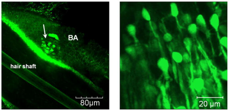
Fig. 3
Typical nestin-GFP expressing stem cells within the hair follicle bulge (white arrow). The cells have an oval-shaped body with a typical size of 7mm and dendritic-like arms. Images were obtained from ...

Fig. 4
Concentration of R123 as a function of UVB irradiation time (upper x-axes) or UVB dose (Jm u22122 , lower x-axes ) in the stratum corneum (a) and the viable epidermal strata (b-d) for lightly pigmente...
Figure images are served from the NIH/NLM PubMed Central Open Access Subset or Europe PMC; copyright remains with the publishers and authors.
💬 Discussion
0 commentsNo comments yet. Be the first to start a discussion!
Leave a Comment