Abstract
AbstractSingle molecule localization microscopy (SMLM) has become a powerful imaging tool for biomedical research, but it is mostly available in imaging facilities and a small number of laboratories due to its high cost. Here, we evaluate the possibility of replacing high-cost components on standard SMLM with appropriate low-cost alternatives and build a simple but high-performance super-resolution SMLM setup. Through numerical simulation and biological experiments, we demonstrate that our low-cost SMLM setup can yield similar localization precision and spatial resolution compared to the standard SMLM equipped with state-of-the-art components, but at a small fraction of their cost. Our low-cost SMLM setup can potentially serve as a routine laboratory microscope with high-performance super-resolution imaging capability.
🔬 Techniques
🧪 Sample Preparation
🔬 Cell Lines
🏭 Microscope Brands
🧪 Reagent Suppliers
📷 Detectors
🎨 Filters
💻 Software Details
🏛️ Research Organizations (ROR)
Affiliated research institutions:
📋 Methods
SNR definition for camera comparison The SNR is defined as follows: documentclass[12pt]{minimal} usepackage{amsmath} usepackage{wasysym} usepackage{amsfonts} usepackage{amssymb} usepackage{amsbsy} usepackage{mathrsfs} usepackage{upgreek} setlength{oddsidemargin}{-69pt} begin{document}$$begin{array}{c}SNR=Signalcdot QE/sqrt{Signalcdot QE+ExcessNoisecdot Signalcdot QE+ReadNois{e}^{2}}\ {begin{array}{l}ExcessNoise=1,EMCCD\ ExcessNoise=0,other,camerasend{array}end{array}$$end{document} S N R = S i g n a l ⋅ Q E / S i g n a l ⋅ Q E + E x c e s s N o i s e ⋅ S i g n a l ⋅ Q E + R e a d N o i s e 2 { E x c e s s N o i s e = 1 , E M C C D E x c e s s N o i s e = 0 , o t h e r c a m e r a s Please note that, only quantum efficiency (QE) and read noise are considered. The effect of dark current noise is ignored. For camera without cooling, the dark current noise can be up to several electrons per pixel per second under the room temperature, which needs to be significantly reduced via deep cooling if long exposure time (e.g., several seconds) is required. But it is not crucial in the context of SMLM imaging, because the acquisition speed is usually several tens of frames per second; and for each frame, the dark current noise is far less than 1 electron, which can be neglected. To validate this point, we measured the sum of the read noise and dark current noise of IMX265 under the experimental condition 20 , and found it is only 2.57 electrons.
Numerical simulation
To compare the performance of our low-cost SMLM setup with the standard SMLM setup, a series of image sets with different signal levels are numerically generated 21 . For each image, a single molecule is randomly distributed in the central pixel. The PSF was modeled with Gaussian function with a kernel width of 0.21 · λ / NA . The wavelength λ is set to be 700 nm to mimic the wavelength of a commonly used fluorophore (Alexa 647). The pixel size is set to be 160 nm for standard SMLM setup equipped with TIRF objective (100X oil, NA = 1.49) and EMCCD cameras (iXon 897, Andor) and 115 nm for our low-cost SMLM setup. The total photon number of the molecules is set to be from 1000 to 8000 to cover the signal range of the commonly used fluorescent dyes. The background for each pixel is set to be 1/50 th of total photon number per molecule. The noise is modeled with a Poisson model plus the excess noise for standard SMLM, and Poisson model plus the read noise with standard deviation of 2.57 electrons for our low-cost SMLM. For each signal level, 1000 images are generated and analyzed to calculate the localization precision (defined as the standard deviation of the localization error). Protocol to coat gold nanoparticles on the coverslip We first coat the glass bottom dish (FD3510-100, WPI) with poly-D-lysine (P7280, SIGMA) for 20 minutes, followed by 200 µL diluted 100 nm gold nanoparticle solution (1:60 with ddH 2 O, EM.GC100, BBI) for 3 hours. Finally, dishes are coated with another layer of poly-D-lysine for 20 minutes for better cell adherence.
Show full methods section
SNR definition for camera comparison The SNR is defined as follows: documentclass[12pt]{minimal} usepackage{amsmath} usepackage{wasysym} usepackage{amsfonts} usepackage{amssymb} usepackage{amsbsy} usepackage{mathrsfs} usepackage{upgreek} setlength{oddsidemargin}{-69pt} begin{document}$$begin{array}{c}SNR=Signalcdot QE/sqrt{Signalcdot QE+ExcessNoisecdot Signalcdot QE+ReadNois{e}^{2}}\ {begin{array}{l}ExcessNoise=1,EMCCD\ ExcessNoise=0,other,camerasend{array}end{array}$$end{document} S N R = S i g n a l ⋅ Q E / S i g n a l ⋅ Q E + E x c e s s N o i s e ⋅ S i g n a l ⋅ Q E + R e a d N o i s e 2 { E x c e s s N o i s e = 1 , E M C C D E x c e s s N o i s e = 0 , o t h e r c a m e r a s Please note that, only quantum efficiency (QE) and read noise are considered. The effect of dark current noise is ignored. For camera without cooling, the dark current noise can be up to several electrons per pixel per second under the room temperature, which needs to be significantly reduced via deep cooling if long exposure time (e.g., several seconds) is required. But it is not crucial in the context of SMLM imaging, because the acquisition speed is usually several tens of frames per second; and for each frame, the dark current noise is far less than 1 electron, which can be neglected. To validate this point, we measured the sum of the read noise and dark current noise of IMX265 under the experimental condition 20 , and found it is only 2.57 electrons.
Numerical simulation
To compare the performance of our low-cost SMLM setup with the standard SMLM setup, a series of image sets with different signal levels are numerically generated 21 . For each image, a single molecule is randomly distributed in the central pixel. The PSF was modeled with Gaussian function with a kernel width of 0.21 · λ / NA . The wavelength λ is set to be 700 nm to mimic the wavelength of a commonly used fluorophore (Alexa 647). The pixel size is set to be 160 nm for standard SMLM setup equipped with TIRF objective (100X oil, NA = 1.49) and EMCCD cameras (iXon 897, Andor) and 115 nm for our low-cost SMLM setup. The total photon number of the molecules is set to be from 1000 to 8000 to cover the signal range of the commonly used fluorescent dyes. The background for each pixel is set to be 1/50 th of total photon number per molecule. The noise is modeled with a Poisson model plus the excess noise for standard SMLM, and Poisson model plus the read noise with standard deviation of 2.57 electrons for our low-cost SMLM. For each signal level, 1000 images are generated and analyzed to calculate the localization precision (defined as the standard deviation of the localization error). Protocol to coat gold nanoparticles on the coverslip We first coat the glass bottom dish (FD3510-100, WPI) with poly-D-lysine (P7280, SIGMA) for 20 minutes, followed by 200 µL diluted 100 nm gold nanoparticle solution (1:60 with ddH 2 O, EM.GC100, BBI) for 3 hours. Finally, dishes are coated with another layer of poly-D-lysine for 20 minutes for better cell adherence.
Cell preparation and staining Primary mouse embryo fibroblast
(MEF) cells or MCF-10A cells were plated onto a PDL (poly-D-lysine) coated glass-bottom dish (FD3510, World Precision Instruments) at confluency of 50% and cultured overnight to let the cells attach to the dish. To stain microtubules, MEF cells were pre-extracted for 30 seconds in 0.5% Triton X-100 (Triton) in BRB80 buffer supplemented with 4 mM EGTA and fixed with Methanol (−20 °C) for 10 minutes. To stain acetylated histone H3, MCF-10A cells were fixed with 4% PFA for 15 minutes and permeabilized with 0.2% Triton for 10 minutes. After fixation, the cells were washed 3 times with PBS and blocked with 3% BSA for 1 hour, then incubated with primary antibody diluted in 3% BSA overnight at 4 °C (rabbit anti-acetyl-histone H3, EMD Milipore 06-599, 1:500). The cells were washed 3 times with PBS and incubated with lab-synthesized Alexa 647-conjugated secondary antibody (Donkey anti rabbit unconjugated antibody, Jackson ImmunoResearch, 711-005-152; Alexa 647 NHS Ester, ThermoFisher, Scientific, A20106) diluted in 3% BSA for 2 hours at room temperature, protected from light. Wash the cells with PBS 3 times and store in PBS until imaging. Immediately before imaging, the sample was switch to the STORM imaging buffer. For microtubule imaging, buffer contains 10% w/v glucose, 2% v/v β-me, 0.56 mg/mL glucose oxidase, 0.17 mg/mL catalase and 2 mM COT (cyclooctatetraene). For histone imaging, 60% TDE (2,2′-thiodiethanol) was used to dissolve the component above instead of water, and 2 mM COT was added to the buffer solution.
Protocol to coat gold nanoparticles on the coverslip We first coat the glass bottom dish (FD3510-100, WPI) with poly-D-lysine (P7280, SIGMA) for 20 minutes, followed by 200 µL diluted 100 nm gold nanoparticle solution (1:60 with ddH 2 O, EM.GC100, BBI) for 3 hours. Finally, dishes are coated with another layer of poly-D-lysine for 20 minutes for better cell adherence.
Electronic supplementary material Low cost LM supplementary
📊 Figures
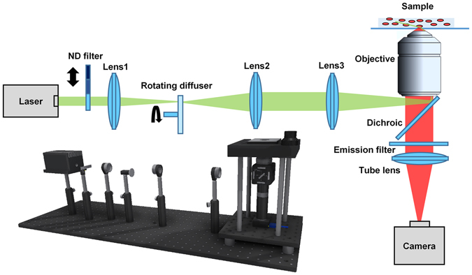
Figure 1
The schematic diagram of the low-cost SMLM setup. ND filter is a step variable metallic neutral density filters (NDL-10S-2, Thorlabs). Lens 1 and Lens 2 are achromatic lenses with a focal length of 10...
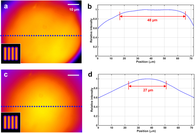
Figure 2
Uniformity of the illumination field in the low-cost SMLM by using different diffusers. ( a , c ) Spatial distribution of illumination intensity generated by the flat-top diffuser. ( b ) The correspon...
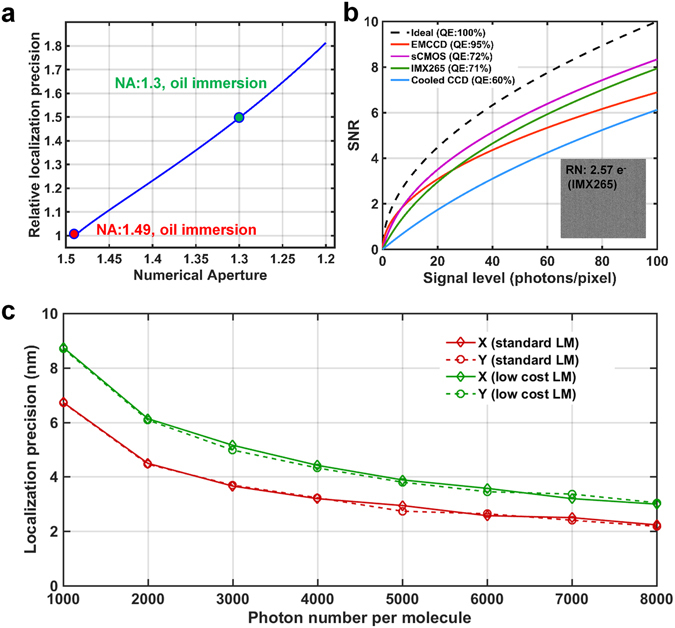
Figure 3
Comparison of localization performance by theoretical calculation and numerical simulation. ( a ) Comparison of relative localization precision for the objectives with different NAs. ( b ) Comparison ...
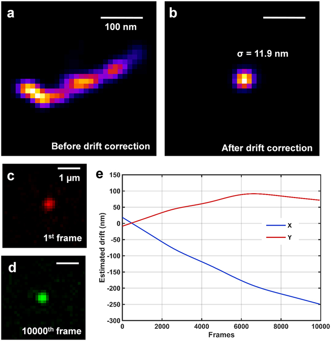
Figure 4
Performance of our low-cost SMLM for super-resolution imaging of nanoparticles. ( a , b ) Reconstructed super-resolution image of the nanoparticle ( a ) without drift correction and ( b ) with cross-c...
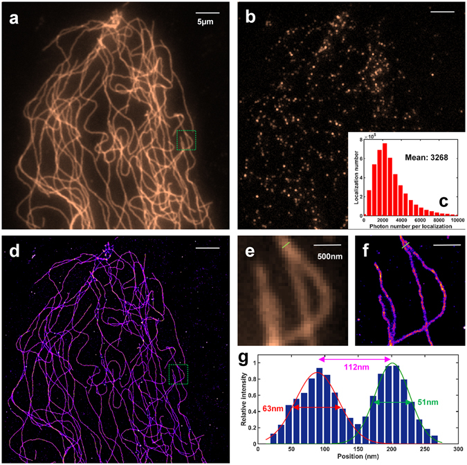
Figure 5
Performance of low-cost SMLM imaging of microtubules in MEF cells. ( a ) Conventional wide-field fluorescence image. ( b ) A single raw image under d STORM imaging condition. ( c ) Statistical distrib...
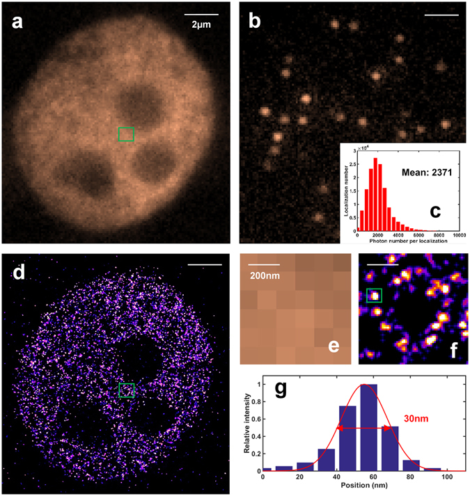
Figure 6
The performance of our low-cost SMLM imaging of acetylated H3 protein in MCF10A cells. ( a ) Conventional wide-field image. ( b ) A single raw image under d STORM imaging condition. ( c ) Statistical ...
Figure images are served from the NIH/NLM PubMed Central Open Access Subset or Europe PMC; copyright remains with the publishers and authors.
💬 Discussion
0 commentsNo comments yet. Be the first to start a discussion!
Leave a Comment