Abstract
The axoneme-the conserved core of eukaryotic cilia and flagella-contains highly specialized doublet microtubules (DMTs). A long-standing question is what protein(s) compose the junctions between two tubules in DMT. Here we identify a highly conserved flagellar-associated protein (FAP), FAP20, as an inner junction (IJ) component. The flagella of Chlamydomonas FAP20 mutants have normal length but beat with an abnormal symmetrical three-dimensional pattern. In addition, the mutant axonemes are liable to disintegrate during beating, implying that interdoublet connections may be weakened. Conventional electron microscopy shows that the mutant axonemes lack the IJ, and cryo-electron tomography combined with a structural labeling method reveals that the labeled FAP20 localizes at the IJ. The mutant axonemes also lack doublet-specific beak structures, which are localized in the proximal portion of the axoneme and may be involved in planar asymmetric flagellar bending. FAP20 itself, however, may not be a beak component, because uniform localization of FAP20 along the entire length of all nine DMTs is inconsistent with the beak's localization. FAP20 is the first confirmed component of the IJ. Our data also suggest that the IJ is important for both stabilizing the axoneme and scaffolding intra-B-tubular substructures required for a planar asymmetrical waveform.
🔬 Techniques
🧬 Organisms
💻 Software
✨ Fluorophores
🧪 Sample Preparation
🏭 Microscope Brands
🧪 Reagent Suppliers
📷 Detectors
💻 Software Details
🏛️ Research Organizations (ROR)
Affiliated research institutions:
📋 Methods
Strains and culture conditions
Chlamydomonas reinhardtii strains used for this study are listed in Supplemental Table S1. Cells were cultured on Tris-acetate-phosphate medium (TAP) plates with 1.5% agar or in liquid TAP with constant aeration at 22 or 25°C with a 12:12 h light:dark cycle. Gametes for dikaryon analyses were prepared by shaking cells in M-N (Sager and Granick minimum medium without nitrogen source) liquid medium for 4 h under illumination.
Isolation and mapping of FAP20 mutants
The first allele, dmj1-1 , was isolated from a library of motility mutants generated by insertional mutagenesis using a marker plasmid, pSI103 ( Sizova et al ., 2001 ). The mutation was not linked to the marker plasmid and thus was mapped to the FAP20 gene by AFLP analysis ( Kathir et al ., 2003 ). The second allele, dmj1-2 , was isolated from a library of motility mutants generated by ultraviolet (UV) mutagenesis. The third allele, RL-11 , was previously described ( Nakamura, 1981 ). Both dmj1-2 and RL-11 were identified as FAP20 mutants by sequencing of the FAP20 gene. The fourth allele, dmj1-3 , was screened from a phototaxis-defective mutant library created by insertional mutagenesis of a hygromycin B–resistant gene ( Berthold et al ., 2002 ). The mutation was mapped to the FAP20 gene by restriction enzyme site-directed amplification PCR ( Gonzalez-Ballester et al ., 2005 ). Oligonucleotide primers used for amplification and sequencing of the FAP20 gene are listed in Supplemental Table S2. Each mutant was backcrossed at least twice for the experiments reported in this study.
Show full methods section
Strains and culture conditions
Chlamydomonas reinhardtii strains used for this study are listed in Supplemental Table S1. Cells were cultured on Tris-acetate-phosphate medium (TAP) plates with 1.5% agar or in liquid TAP with constant aeration at 22 or 25°C with a 12:12 h light:dark cycle. Gametes for dikaryon analyses were prepared by shaking cells in M-N (Sager and Granick minimum medium without nitrogen source) liquid medium for 4 h under illumination.
Isolation and mapping of FAP20 mutants
The first allele, dmj1-1 , was isolated from a library of motility mutants generated by insertional mutagenesis using a marker plasmid, pSI103 ( Sizova et al ., 2001 ). The mutation was not linked to the marker plasmid and thus was mapped to the FAP20 gene by AFLP analysis ( Kathir et al ., 2003 ). The second allele, dmj1-2 , was isolated from a library of motility mutants generated by ultraviolet (UV) mutagenesis. The third allele, RL-11 , was previously described ( Nakamura, 1981 ). Both dmj1-2 and RL-11 were identified as FAP20 mutants by sequencing of the FAP20 gene. The fourth allele, dmj1-3 , was screened from a phototaxis-defective mutant library created by insertional mutagenesis of a hygromycin B–resistant gene ( Berthold et al ., 2002 ). The mutation was mapped to the FAP20 gene by restriction enzyme site-directed amplification PCR ( Gonzalez-Ballester et al ., 2005 ). Oligonucleotide primers used for amplification and sequencing of the FAP20 gene are listed in Supplemental Table S2. Each mutant was backcrossed at least twice for the experiments reported in this study.
Antibodies
The full-length FAP20 cDNA was amplified by reverse transcription-PCR with primers FAP20-BglNd-F and FAP20-Xho-R. The truncated FAP20 cDNA was amplified with primers FAP20-BglNd-F and FAP20-TR-Xho-R. The full-length product was digested with Nde I and Xho I and cloned into pET24a (Merck, Darmstadt, Germany) to produce FAP20full-His. Both the full-length and the truncated products were digested with Bgl II and Xho I and cloned between Bam HI and Xho I sites of pGEX6P2 (GE Healthcare, Little Chalfont, United Kingdom) to produce glutathione S -transferase (GST)–FAP20full and GST-FAP20ΔC20aa, respectively. Each of the three plasmids was transformed into BL21 (DE3). The expression of the recombinant proteins was induced by adding 1 mM isopropyl-β- d -thiogalactoside into a logarithmically growing culture. Almost all of the expressed proteins were contained within inclusion bodies. The inclusion bodies were purified as previously described ( Yanagisawa and Kamiya, 2004 ). FAP20full-His was used to immunize two rabbits. The antibodies were affinity purified using GST-FAP20full or GST-FAP20ΔC20aa blotted onto polyvinylidene difluoride membranes. Sequences of the primers used for the plasmid constructions are listed in Supplemental Table S2. Other antibodies used in this study are listed in Supplemental Table S3.
Isolation and biochemical fractionation of axonemes
Chlamydomonas cells were deflagellated with dibucaine-HCl (Wako Pure Chemical Industries, Tokyo, Japan), and axonemes were collected by centrifugation ( Witman et al ., 1978 ). The flagella were demembranated with 1% Nonidet P-40 in HMDEK buffer (30 mM 4-(2-hydroxyethyl)-1-piperazineethanesulfonic acid–NaOH, pH 7.2, 5 mM MgCl 2 , 1 mM dithiothreitol, 1 mM ethylene glycol tetraacetic acid, 50 mM CH 3 COOK, 1× protease inhibitor cocktail [Nacalai Tesque, Kyoto, Japan]). Biochemical fractionation of the axonemes was performed as previously described ( Witman et al ., 1972 ; Yanagisawa and Kamiya, 2004 ).
Plasmid constructions To make
FAP20 constructs with tags, an ∼4.5-kb fragment containing the FAP20 gene was amplified from Chlamydomonas genomic DNA using primers FAP20-gDNA-F and FAP20-gDNA-R. The PCR product was cloned into the Eco RV site of pBluescript II to create the construct pBS-FAP20. A unique Eco RV site was introduced before the stop codon by PCR amplification of the entire plasmid with primers FAP20-gDNA-C-ERV-F and FAP20-gDNA-C-ERV-R. The PCR product with 16-base cohesive ends was transformed into DH5α to make the construct pBS-FAP20-C-ERV. A GFP tag was cut out with Bam HI from pCrGFP ( Fuhrmann et al ., 1999 ) and blunted with Klenow. A 3xHA tag was cut out with Nru I and Sca I from p3xHA ( Silflow et al ., 2001 ). Each of the two tag fragments was ligated into the Eco RV site of pBS-FAP20-C-ERV. A BCCP tag was amplified from pGenD-LC2-BCCP ( Furuta et al ., 2009 ) using primers FAP20-BCCP-F and FAP20-BCCP-R and cloned into the Eco RV site of pBS-FAP20-C-ERV using In-Fusion HD Cloning Kit (Clontech, Mountain View, CA). Each of the three FAP20 constructs was linearized with Eco RI and cotransformed with pSI103 into the dmj1-1 cells by electroporation to produce the rescued strains dmj1-1 ::FAP20-GFP, dmj1-1 ::FAP20-3xHA, and dmj1-1 ::FAP20-BCCP. Sequences of the primers used for the plasmid constructions are listed in Supplemental Table S2.
Flagella length measurement
Cells were fixed with 2× Lugol's iodine and placed in 4°C until the end of the experiment. The cells were observed by an Axioplan phase contrast microscope (Carl Zeiss, Oberkochen, Germany). Still images were taken with a Phantom Miro-eX2 (Vision Research, Wayne, NJ), and the length of a single flagellum from each cell was measured using the ImageJ ( Schneider et al ., 2012 ) segmented line tool.
Assessment of flagellar motility
For waveform analyses, cells were observed by an Axioplan phase contrast microscope or a BX51 dark-field microscope (Olympus, Tokyo, Japan). To prevent photoshock, a red filter was used. The filter was removed to induce waveform change in wild-type cells. Movies for waveform analysis were taken with a Phantom Miro-eX2 or an EoSens MC1362 (Mikrotron, Unterschleissheim, Germany) at either 600 or 1000 fps. To trace the waveforms, the movies were imported into ImageJ and trimmed to 10 frames with equal time elapsed between frames of a given set. Shapes of the flagella were traced over a lowered opacity using Illustrator (Adobe, San Jose, CA). For measurement of swimming velocity, cells were observed by a dark-field microscope and movies were recorded as described at 30 fps. Swimming velocity of the cells was measured using the MTrack2 plug-in ( Klopfenstein and Vale, 2004 ) of ImageJ.
Immunofluorescence microscopy
Nucleoflagellar apparatus prepared as previously described ( Taillon and Jarvik, 1995 ) was fixed with 2% formaldehyde for 10 min at room temperature, followed by treatment with cold acetone and methanol (−20°C). Fixed samples were immmunostained as previously described ( Sanders and Salisbury, 1995 ). Samples were observed by an IX70 fluorescence microscope (Olympus) with an ORCA-R2 charge-coupled device (CCD) camera (Hamamatsu Photonics, Shizuoka, Japan).
Live-cell imaging of the GFP-tagged strain
Live cells of the dmj1-1 ::FAP20-GFP were observed by an IX70 microscope as described or an IX81 microscope (Olympus) equipped with a disk scanning unit and iXon electron-multiplying CCD camera (Andor Technology, Belfast, United Kingdom). Fluorescence intensities of flagella were measured using ImageJ. Reactivation and disintegration of isolated axonemes To analyze the stability of the dmj1-1 axoneme, isolated axonemes were reactivated in HMDEK buffer containing 0.1 mM ATP without proteases. Motility and disintegration of the axonemes were observed by a dark-field microscope and recorded using a MC681SPD high-sensitive CCD camera (Texas Instruments, Dallas, TX). To observe FAP20-GFP fluorescence on the DMTs, the isolated axoneme from dmj1-1 ::FAP20-GFP was adsorbed on a slide and treated with 1 μg/ml Type XXIV bacterial proteinase (Sigma-Aldrich, St. Louis, MO) and 1 mM ATP in HMDEK-PI (HMDEK without protease inhibitors) buffer for 30 s at room temperature. GFP fluorescence was observed as described.
Thin-section electron microscopy
Thin-section electron microscopy of the axonemes was performed as previously described ( Huang et al ., 1979 ). The cacodylate buffer in the method was substituted by 50 mM Na-phosphate, pH 7.0. The samples were observed by a JEM-3100FEF transmission electron microscope (JEOL, Tokyo, Japan) equipped with TemCam-F416 (TVIPS, Gauting, Germany). Images were recorded at 300 keV, with ∼3-μm defocus, at a magnification of ×82,100 and a pixel size of 1.9 Å. Cross-sectional images in which the PFs were clearly visible were selected and used for averaging of DMTs. Alignment and averaging of DMTs were conducted using custom Ruby-Helix scripts ( Metlagel et al ., 2007 ) and the align2d program of EMAN ( Ludtke et al ., 1999 ).
Assessment of the BCCP-tagged strain
Biotinylation of the FAP20-BCCP in the dmj1-1 ::FAP20-BCCP axonemes was confirmed by Western blotting with streptavidin–horseradish peroxidase (Thermo Fisher Scientific, Waltham, MA). Accessibility of the BCCP tag in the intact axoneme was assessed using fluorescence-labeled streptavidin. Demembranated axonemes were loaded to a flow cell and blocked with 1% bovine serum albumin (BSA) in HMDEK buffer. Then the flow cell was perfused with streptavidin–Alexa Fluor 546 (Life Technologies, Carlsbad, CA) diluted 1:1000 in the same buffer and washed with the buffer alone. Samples were observed by an IX70 fluorescence microscope as described. Enhanced streptavidin labeling of the axonemes Demembranated axonemes were incubated with 0.05 mg/ml streptavidin (Wako Pure Chemical Industries) for 15 min at 4°C in HMDEK buffer. Axonemes were washed five times with HMDEK buffer and incubated with 0.05 mg/ml biotinylated cytochrome C for 15 min at 4°C in the presence of 1 mg/ml BSA and 0.1 mg/ml unlabeled cytochrome C. Axonemes were washed five times with HMDEK again and incubated with 0.05 mg/ml streptavidin for 15 min at 4°C. Labeled axonemes were separated from unbound streptavidin by centrifugation and were resuspended in HMDEK buffer at a concentration of 0.02 mg/ml and mixed with equal amount of 15-nm colloidal gold suspension conjugated with BSA (Aurion, Wageningen, Netherlands). Home-made holey carbon grids were glow discharged and coated with 20-nm colloidal gold (BBInternational, Cardiff, United Kingdom). Suspended axonemes plus colloidal gold (5 μl) was loaded onto the grids and plunge frozen in liquid ethane at −180°C with an automated plunge-freezing device, EM GP (Leica Microsystems, Wetzlar, Germany). Images were acquired and processed as previously described ( Oda and Kikkawa, 2013 ) with little modification. The details are described in the Supplemental Methods.
📊 Figures
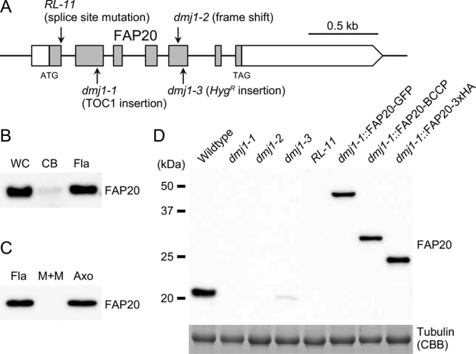
FIGURE 1:
Four mutant alleles of dmj1 . (A) The mutation sites of four different dmj1 alleles are indicated on the exon/intron structure of the FAP20 gene (Cre07.g351650.t1.3 in Phytozome v9.1; www.phytozome.ne...
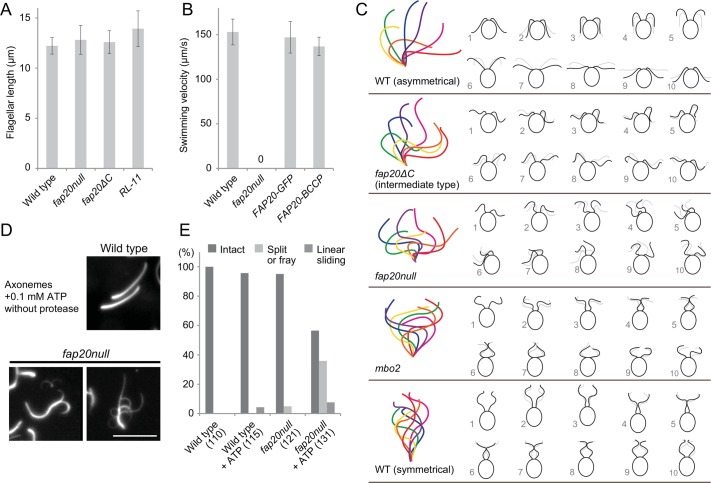
FIGURE 2:
FAP20 is essential for both asymmetrical waveform and stability of flagella. (A) FAP20 mutants produce wild-type length of the flagella ( n = 33). (B) Introduction of the tagged FAP20 constructs rescu...
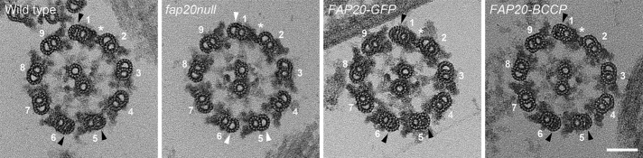
FIGURE 3:
Ultrastructural defects of the fap20null axoneme. Cross sections of Epon-embedded axonemes of wild type, fap20null , and rescued strains. The 1u20132 bridge (asterisks) was used to determine the posit...
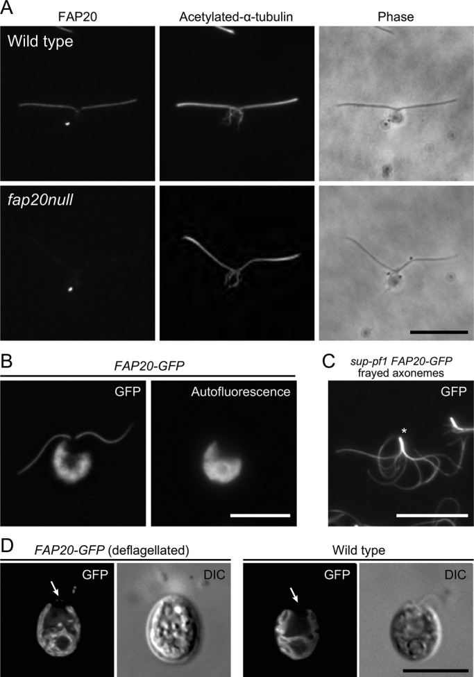
FIGURE 4:
FAP20 localizes along the entire length of flagellum and is present in all nine DMTs. (A) Immunofluorescence microscopy of nucleoflagellar apparatus (NFA) shows that FAP20 is evenly distributed along ...
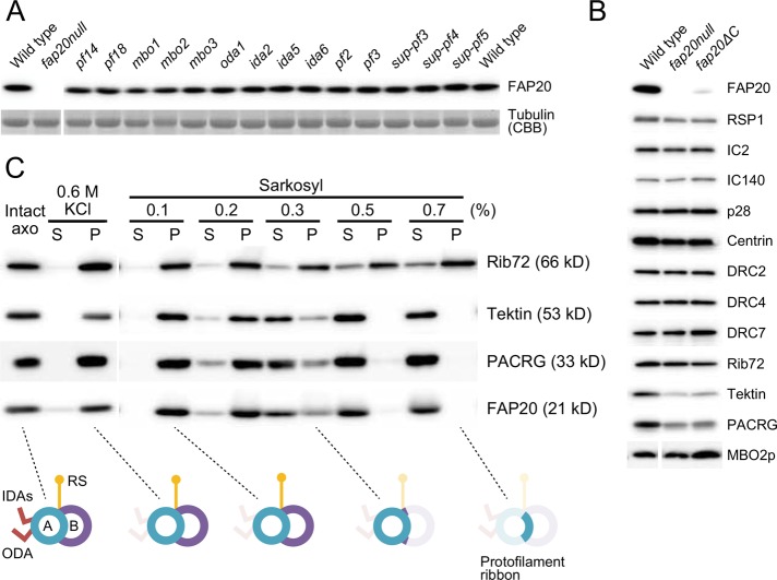
FIGURE 5:
FAP20 is tightly associated with the DMT. (A) Western blot analysis of axonemes from a variety of flagellar mutants with FAP20 antibody. Coomassie-stained bands of tubulins were used as a loading cont...
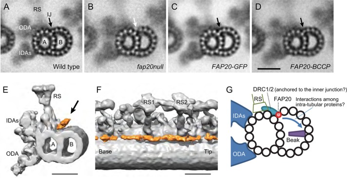
FIGURE 6:
FAP20 is an IJ protein. (Au2013D) The fap20null axoneme is defective in the IJ between A- and B-tubules. Images of DMTs in cross sections of Epon-embedded axonemes were averaged. DMT1, 5, and 6 were e...
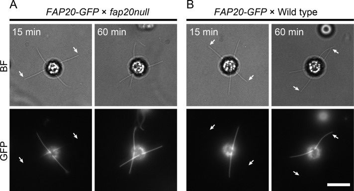
FIGURE 7:
FAP20 is assembled into the flagella from base to tip. FAP20-GFP gametes were mated with both fap20null (A) and wild-type (B) gametes and dikaryons observed for the emergence of FAP20-GFP in the unlab...
Figure images are served from the NIH/NLM PubMed Central Open Access Subset or Europe PMC; copyright remains with the publishers and authors.
💬 Discussion
0 commentsNo comments yet. Be the first to start a discussion!
Leave a Comment