Abstract
In vitro, dendritic cells (DCs) bind and transfer intact, infectious HIV to CD4 T cells without first becoming infected, a process known as trans-infection. trans-infection is accomplished by recruitment of HIV and its receptors to the site of DC-T cell contact and transfer of virions at a structure known as the infectious synapse. In this study, we used fluorescent microscopy to track individual HIV particles trafficking in DCs during virus uptake and trans-infection. Mature DCs rapidly concentrated HIV into an apparently intracellular compartment that lacked markers characteristic of early endosomes, lysosomes, or antigen-processing vesicles. Live cell microscopy demonstrated that the HIV-containing compartment was rapidly polarized toward the infectious synapse after contact with a T cell; however, the bulk of the concentrated virus remained in the DCs after T cell engagement. Individual virions were observed emerging from the compartment and fusing with the T cell membrane at the infectious synapse. The compartmentalized HIV, although engulfed by the cytoplasm, was fully accessible to HIV envelope-specific inhibitors and other membrane-impermeable probes that were delivered to the cell surface. These results demonstrate that HIV resides in an invaginated domain within DCs that is both contiguous with the plasma membrane and distinct from endocytic vesicles. We conclude that HIV virions are routed through this specialized compartment, which allows individual particles to be delivered to T cells during trans-infection.
🔬 Techniques
🔭 Microscopes
✨ Fluorophores
🧪 Sample Preparation
🔬 Cell Lines
🏭 Microscope Brands
🧪 Reagent Suppliers
📷 Detectors
🏛️ Research Organizations (ROR)
Affiliated research institutions:
📋 Methods
Cells and antibodies
Hos-CD4 (human osteosarcoma cell line stably expressing human CD4) and HEK293T cells were maintained in DMEM supplemented with 10% FBS. Jurkat, Jurkat LTR-GFP (kindly provided by Olaf Kutsch) and LuSIV (CEM T cells transduced with an HIV LTR-Luciferase reporter) (AIDS Reference and Reagent Program) cells were maintained in RPMI supplemented with 10% FBS. Monocyte-derived DCs were generated as described previously [27] . Briefly, CD14-positive monocytes were purified from PBMC by magnetic bead separation (Miltenyi Biotec) and cultured in RPMI (Life Technologies) supplemented with 10% FBS (Hyclone), 100 ng/ml IL-4 and 50 ng/ml GM-CSF (Gentaur Biosciences) for 5–7 days to generate immature DC. In a typical experiment, greater than 90% of the cells were CD14- HLA-DR+ DC-SIGN+ and 80%–90% displayed an immature phenotype as determined by low or no expression of CD80 and CD86. Activated DCs were generated by the addition of LPS (100 ng/ml, Sigma) for 12–24 h. Maturation was assessed by upregulation of CD86 and HLA-DR. Myeloid DCs were purified from PBMC using CD1c (BDCA-1) + dendritic cell magnetic bead selection kit according to the manufacturer (Miltenyi Biotech). Myeloid DCs were maintained overnight in GM-CSF (5 ng/ml) and activated with LPS (100 ng/ml). For immunofluorescent studies, monoclonal antibodies (mAbs) specific for DC-SIGN, CXCR4, CCR5 (R&D Systems), CD4 (Sigma), CD9, CD63, CD80, HLA-DM, HLA-(DR, DP, DQ), LAMP-1 (Pharmingen) were diluted to predetermined concentrations in PBS+10% normal donkey serum+.1% Triton X-100. Cy3- or Cy5-labeled Donkey anti-mouse antibodies (Jackson ImmunoResearch) were used as secondary reagents. Anti-HIV env antibody 2G12 (AIDS Reference and Reagent Program) and Pro-542 (sCD4-hIgG) (kindly provided by Norbert Shulke, Progenics, Inc.) were detected using anti-human IgG secondary reagents (Molecular Probes). For multi-color staining, mAbs were pre-labeled with appropriate Zenon reagents (Molecular Probes) and added after secondary antibody labeling. Flow cytometric analysis was performed using direct labeled antibodies to the specified antigen with appropriate isotype controls (BD Biosciences). Actin cytoskeleton was stained with fluorescent-phalloidin (Molecular Probes) and nuclei were stained with Hoechst dye (Sigma). Inhibitors Nocodazole (Sigma, 5 µM) or Latrunculin B (BioMol, 2.5 µM) were added to MDDCs 15 min. prior to HIV exposure to disrupt cytoskeletal structures and maintained in the cultures throughout the experiment. Inhibition of HIV trans -infection was performed as described using recombinant sCD4 (10 µg/ml) (AIDS Reference and Reagent Program) or the Progenics sCD4-hIgG (8 µg/ml) (Pro 542, Progenics, Inc.) fusion with similar results (data not shown).
Show full methods section
Cells and antibodies
Hos-CD4 (human osteosarcoma cell line stably expressing human CD4) and HEK293T cells were maintained in DMEM supplemented with 10% FBS. Jurkat, Jurkat LTR-GFP (kindly provided by Olaf Kutsch) and LuSIV (CEM T cells transduced with an HIV LTR-Luciferase reporter) (AIDS Reference and Reagent Program) cells were maintained in RPMI supplemented with 10% FBS. Monocyte-derived DCs were generated as described previously [27] . Briefly, CD14-positive monocytes were purified from PBMC by magnetic bead separation (Miltenyi Biotec) and cultured in RPMI (Life Technologies) supplemented with 10% FBS (Hyclone), 100 ng/ml IL-4 and 50 ng/ml GM-CSF (Gentaur Biosciences) for 5–7 days to generate immature DC. In a typical experiment, greater than 90% of the cells were CD14- HLA-DR+ DC-SIGN+ and 80%–90% displayed an immature phenotype as determined by low or no expression of CD80 and CD86. Activated DCs were generated by the addition of LPS (100 ng/ml, Sigma) for 12–24 h. Maturation was assessed by upregulation of CD86 and HLA-DR. Myeloid DCs were purified from PBMC using CD1c (BDCA-1) + dendritic cell magnetic bead selection kit according to the manufacturer (Miltenyi Biotech). Myeloid DCs were maintained overnight in GM-CSF (5 ng/ml) and activated with LPS (100 ng/ml). For immunofluorescent studies, monoclonal antibodies (mAbs) specific for DC-SIGN, CXCR4, CCR5 (R&D Systems), CD4 (Sigma), CD9, CD63, CD80, HLA-DM, HLA-(DR, DP, DQ), LAMP-1 (Pharmingen) were diluted to predetermined concentrations in PBS+10% normal donkey serum+.1% Triton X-100. Cy3- or Cy5-labeled Donkey anti-mouse antibodies (Jackson ImmunoResearch) were used as secondary reagents. Anti-HIV env antibody 2G12 (AIDS Reference and Reagent Program) and Pro-542 (sCD4-hIgG) (kindly provided by Norbert Shulke, Progenics, Inc.) were detected using anti-human IgG secondary reagents (Molecular Probes). For multi-color staining, mAbs were pre-labeled with appropriate Zenon reagents (Molecular Probes) and added after secondary antibody labeling. Flow cytometric analysis was performed using direct labeled antibodies to the specified antigen with appropriate isotype controls (BD Biosciences). Actin cytoskeleton was stained with fluorescent-phalloidin (Molecular Probes) and nuclei were stained with Hoechst dye (Sigma). Inhibitors Nocodazole (Sigma, 5 µM) or Latrunculin B (BioMol, 2.5 µM) were added to MDDCs 15 min. prior to HIV exposure to disrupt cytoskeletal structures and maintained in the cultures throughout the experiment. Inhibition of HIV trans -infection was performed as described using recombinant sCD4 (10 µg/ml) (AIDS Reference and Reagent Program) or the Progenics sCD4-hIgG (8 µg/ml) (Pro 542, Progenics, Inc.) fusion with similar results (data not shown).
Virus stocks GFP-Vpr labeled
HIV was produced by calcium phosphate co-transfection of HEK293T cells with an eGFP-Vpr expression construct, HIV env deficient proviral clone pLAI∂env and HXB2 envelope glycoprotein expression construct as previously described [20] . GFP-Vpr/S15-RFP was generated by including S15-mCherry, a myrystoylated fusion protein that associates with lipid bilayers in transfected cells and marks the HIV lipid envelope [21] . Transfected cells were washed 16 hours post-transfection, media was replaced again 8 hours later and supernatants containing labeled virus was collected the next morning, approximately 40 hours post-transfection. Cleared supernatant was passed through a .45 μ filter and frozen at −80°C. Stocks were assayed for infectivity and p24 concentration, and incorporation of GFP-Vpr was assessed by co-staining with Gag antibodies [20] . GFP-Vpr/S15-RFP was assessed by co-localization of GFP and RFP with Gag staining, and optimized so that greater than 95% of the GFP-positive particles were also RFP-positive. Single-round infectious, HIV luciferase stocks were generated by transfection of HEK293T cells with the env -deficient proviral vector plasmid NL-Luc-E - R - containing a firefly luciferase reporter gene or NL-Ren-E-R- containing renilla luciferase reporter (kindly provided by Dr. Nathaniel Landau) [28] along with an HIV-1 HXB2 envelope glycoprotein expression construct. HIV-1 trans -infection MDDCs (10 6 /ml) were incubated with HxB2 pseudotyped Luciferase or Renilla stocks (37°C, 2 h), washed twice and resuspended in culture medium. DCs (5×10 3 ) were then co-cultured with Hos-CD4 target cells (2×10 4 ) in 96-well plates and assayed 40 hours later using the Brite-Luc or Dual-Luc luciferase assay reagents (Promega) and reading the plates on a multi-well format luminometer (BioRad). Freshly thawed aliquots of HIV-Luciferase were included as normalization standards. Alternatively, MDDC or myDCs were incubated with HXB2 pseudotyped GFP-Vpr HIV, washed and co-cultured with LuSIV (HIV LTR-Luciferase) indicator cells for 40 hours and assayed as above. For inhibitor studies, DCs were incubated at the appropriate times with sCD4 (AIDS Reference and Reagent Program) at 4°C for 1 h, washed twice at 4°C and incubated further as indicated in the text.
Immunofluorescence
DCs were allowed to adhere to poly L-Lysine–treated coverslips, rinsed with PBS and fixed with 4% EM grade formaldehyde (Polysciences) in PBS. Antibodies were added in SB (PBS, 10% normal donkey serum [Jackson ImmunoResearch]) or SBTx (SB+0.1% Triton X-100) to remove cellular membranes for staining intracellular antigens for 20 mn at RT. Coverslips were rinsed extensively and stained with donkey anti-mouse secondary antibodies (Jackson ImmunoResearch) in SB. For live cell staining, cells were incubated at 4°C with the indicated probes for 30 to 60 mn, washed twice with cold PBS and fixed onto poly-L-lysine coverslips. Probes were then detected with the appropriate fluorescent reagents. Coverslips were mounted onto glass slides using Gel Mount (Biomedia) containing an anti-fade reagent. Dried slides were imaged on a Deltavision RT epifluorescent microscope system fitted with an automated stage (Applied Precision, Inc) and images were captured in z-series on a CCD digital camera. Out-of-focus light was digitally removed using the Softworks deconvolution software (Applied Precision, Inc). 3-D volume projections were generated using the Softworx analysis program. Images were exported as .tif files and figures were composed using Adobe Photoshop CS (Adobe, Inc). Accession numbers of proteins referenced (SwisProt) CD4: P01730 , CD209 (DC-SIGN): Q9NNX6 , CD86: P42081 , CD63: P08962 , CD9: P21926 , CD80: P33681 , CD81: P60033 , HLA-DR: P04229 , HLA-DP: P20036 , HLA-DQ: P01907 , HLA-DM: P28067 , EEA1: Q15075 , Transferrin Receptor: P02786 , LAMP-1: P11279 , ICAM-1: P05362 , LFA-1: P20701 , CXCR4: P61073 , CCR5: P51681 , HIV gp120: O70902 .
Supporting Information Figure S1 Accumulation of HIV in immature MDDCs. Unactivated MDDCs were plated onto coverslips, exposed to GFP-HIV (green) for 1 h, washed and fixed at 1 h (A–C), 4 h (D–F), or 24 h (G–I, J–L) after pulse. Cells were stained for Actin (red, left panels), DNA (blue), and CD81 (red, right 2 panels), and imaged and projected as 3-D volume renderings. Arrows denote overlap of the CD81 and HIV signals. (10 MB TIF) Click here for additional data file. Figure S2 The HIV compartment is a highly dynamic structure. Mature MDDCs were incubated with GFP-Vpr/S15-RFP–labeled HIV for 1 h at 37°C, washed, and plated onto a glass coverslip dish. Jurkat LTR-GFP T cells (marked by low GFP expression) were added, and cells were imaged at 2-min intervals immediately after identifying the DC–T cell interaction. Video shows merged light and fluorescent signals, rendered as whole-cell volume projections. Arrows denote a single HIV compartment that splits into two after 8 min and reforms by 14 min. Insets are magnified views of the concentrated GFP/RFP signals. No viral transmission was observed during this interaction. See also Video S4 . (6.6 MB TIF) Click here for additional data file. Figure S3 Peripheral blood myeloid DCs sequester HIV in the CD81-positive, surface accessible compartment. (A–D) BDCA-1-positive myeloid DCs were isolated from PBMCs and activated with LPS for 14 h. The matured myDCs were pulsed with GFP-HIV for 1 h, washed, and cultured an additional hour. The cells were then incubated at 4°C with 2G12 anti-HIV Env (gp120) mAb, washed, fixed, and immunostained for 2G12 (gp120) (orange) and CD81 (red). Arrows denote regions of HIV concentration. Images are 3-D renderings of the entire cell volumes. Bars, 5 μ. (2 MB TIF) Click here for additional data file. Video S1 Transmission of HIV to a Jurkat T cell at the infectious synapse. Mature MDDCs were incubated with GFP-Vpr/S15-RFP–labeled HIV for 1 h at 37°C, washed, and plated onto a glass coverslip dish. Jurkat LTR-GFP T cells (marked by low GFP expression) were added, and cells were imaged at 2-min intervals immediately after identifying the DC–T cell interaction. The movie shows merged light and fluorescent signals until just prior to transfer, then GFP/RFP fluorescent signals only. Arrows denote transmitted HIV particles. (12.2 MB MOV) Click here for additional data file. Video S2 Blood myeloid DCs transmit single particles from the HIV compartment. LPS matured myeloid DCs were incubated with labeled HIV as in Video S1 and plated onto a glass coverslip dish. Jurkat T cells labeled with CellTrace DDAO-SE (diffuse red signal) were added, and cells were imaged as above. Movie shows merged light and fluorescent signals until just prior to transfer, then GFP/RFP fluorescent signals only. Arrows denote transmitted HIV particles. (4.3 MB MOV) Click here for additional data file. Video S3 Surface transmission of HIV after loading at 4°C. Mature MDDCs were exposed to 20× concentrated GFP-Vpr labeled HIV for 2 h at 4°C. Cells were washed and plated on coverslip dishes with Jurkat LTR-GFP cells and imaged as above. Movie shows merged light and fluorescent signals until just prior to transfer, then the GFP signal only during particle transfer. Arrows denote transmitted HIV particles. (1.4 MB MOV) Click here for additional data file. Video S4 The HIV compartment is a highly dynamic structure. Mature MDDCs were incubated with GFP-Vpr/S15-RFP labeled HIV for 1 h at 37°C, washed, and plated onto a glass coverslip dish. Jurkat LTR-GFP T cells (marked by low GFP expression) were added, and cells were imaged at 2-min intervals immediately after identifying the DC–T cell interaction. The movie shows merged light and fluorescent signals, rendered as whole-cell volume projections. Arrows denote a single HIV compartment that splits into two after 8 min and reforms by 14 min. No viral transmission was observed during this interaction. (3.9 MB MOV) Click here for additional data file. Video S5 Compartmentalized HIV remains accessible to surface probes after extended culture. z-stack movie of the cell shown in Figure 8A . Mature MDDCs were exposed to HxB2-pseudotyped GFP-HIV for 1 h, washed, and cultured at 37°C for 24 h. Cells were then incubated at 4°C with a mouse anti-CD81 mAb, washed and fixed onto coverslips, and stained with anti-mouse secondary antibody (red). The movie was compiled using individual focal planes of the entire z-stack taken at .2-μ intervals. (849 KB MOV) Click here for additional data file.
📊 Figures
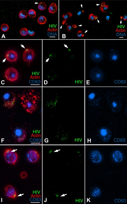
Figure 1
HIV concentration in mature MDDCs requires an intact actin cytoskeleton.
(A,B) Mature DCs were plated onto coverslips, pulsed with HxB2 pseudotyped GFP-Vpr HIV (green) for 30 min, washed and fixed (A) or incubated an additional hour and fixed (B). Fixed cells were stained ...
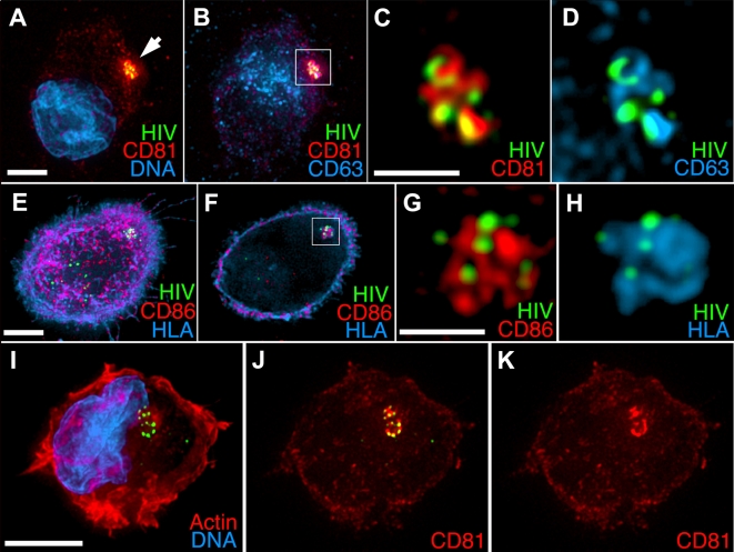
Figure 2
HIV and cell-surface proteins are sequestered into a distinct subcellular compartment.
(Au2013H) Mature MDDCs were pulsed with HxB2 pseudotyped GFP-Vpr HIV (green) for 1 h, washed, plated onto poly-L-Lysine coverslips, and fixed and stained for the indicated markers. (A) 3-Du2013rendere...
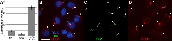
Figure 3
trans -infection and HIV sequestration in mature blood myeloid DCs.
BDCA-1u2013positive myDCs were purified from PBMCs and cultured for 14 h without or with LPS to induce maturation. Cells were incubated with HxB2 pseudotyped GFP-Vpr HIV (green) for 1 h, washed and cu...
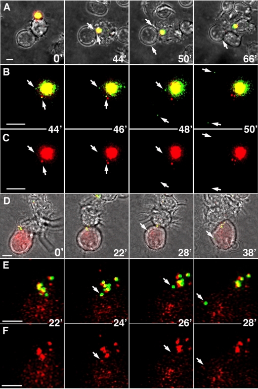
Figure 4
DCs transmit individual HIV particles to T cells at the infectious synapse.
(Au2013C) Mature MDDCs were incubated with GFP-Vpr/S15-RFP labeled HIV for 1 h at 37u00b0C, washed, and plated onto a glass coverslip dish. GFP-Vpr (green) marks the HIV core, and S15-RFP (red) is a m...
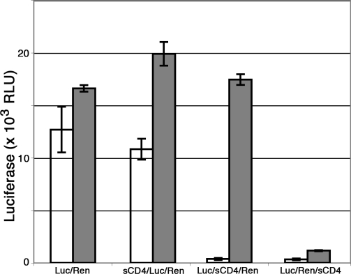
Figure 5
Inhibition of surface accessible HIV abolishes trans -infection.
Mature MDDCs were sequentially exposed to HIV-Firefly luciferase (Luc) and HIV-Renilla luciferase (Ren) at 37u00b0C and treated at 4u00b0C with soluble CD4 (sCD4, 10 u00b5g/ml) for 1 h either before, ...
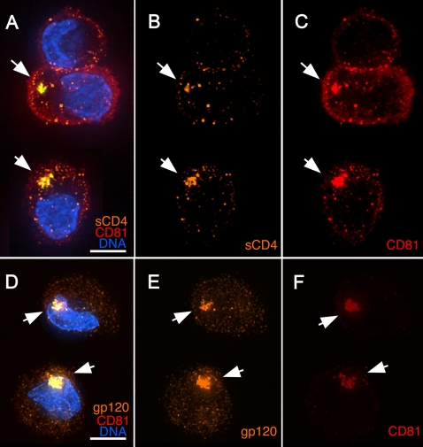
Figure 6
Internalized HIV is accessible to surface applied HIV-specific inhibitory antibodies.
(Au2013F) Mature MDDCs were pulsed with HxB2 pseudotyped GFP-Vpr HIV (green) for 1 h, washed, and cultured for 1 h at 37u00b0C. Cells were then placed on ice and incubated for 30 min with (Au2013C) CD...
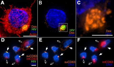
Figure 7
Internalized HIV is accessed by a surface-applied fluid phase marker.
(Au2013C) Mature MDDCs were pulsed with HIV as in Figure 6 and incubated with fluorescent dextran (orange) at 4u00b0C. Cells were plated onto coverslips, fixed, and stained for Actin (Red) and DNA (Bl...
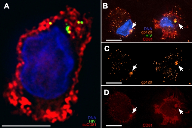
Figure 8
Compartmentalized HIV remains accessible to surface probes after extended culture.
(A) Mature MDDCs were exposed to HxB2 pseudotyped GFP-HIV for 1 h, washed, and cultured at 37u00b0C for 24 h. Cells were then incubated at 4u00b0C with a mouse anti-CD81 mAb, washed, fixed onto covers...
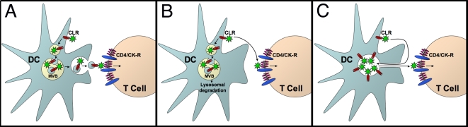
Figure 9
Models of trans -infection.
(A) MVB/exosomal transfer. HIV is endocytosed and concentrated in a non-degradative late endosome/multivesicular body (MVB). The MVB is subsequently transported to the T cell interface, where it fuses...
Figure images are served from the NIH/NLM PubMed Central Open Access Subset or Europe PMC; copyright remains with the publishers and authors.
💬 Discussion
0 commentsNo comments yet. Be the first to start a discussion!
Leave a Comment