Abstract
The intimate synapsis of homologous chromosome pairs (homologs) by synaptonemal complexes (SCs) is an essential feature of meiosis. In many organisms, synapsis and homologous recombination are interdependent: recombination promotes SC formation and SCs are required for crossing-over. Moreover, several studies indicate that initiation of SC assembly occurs at sites where crossovers will subsequently form. However, recent analyses in budding yeast and fruit fly imply a special role for centromeres in the initiation of SC formation. In addition, in budding yeast, persistent SC-dependent centromere-association facilitates the disjunction of chromosomes that have failed to become connected by crossovers. Here, we examine the interplay between SCs, recombination, and centromeres in a mammal. In mouse spermatocytes, centromeres do not serve as SC initiation sites and are invariably the last regions to synapse. However, centromeres are refractory to de-synapsis during diplonema and remain associated by short SC fragments. Since SC-dependent centromere association is lost before diakinesis, a direct role in homolog segregation seems unlikely. However, post-SC disassembly, we find evidence of inter-centromeric connections that could play a more direct role in promoting homolog biorientation and disjunction. A second class of persistent SC fragments is shown to be crossover-dependent. Super-resolution structured-illumination microscopy (SIM) reveals that these structures initially connect separate homolog axes and progressively diminish as chiasmata form. Thus, DNA crossing-over (which occurs during pachynema) and axis remodeling appear to be temporally distinct aspects of chiasma formation. SIM analysis of the synapsis and crossover-defective mutant Sycp1⁻/⁻ implies that SCs prevent unregulated fusion of homolog axes. We propose that SC fragments retained during diplonema stabilize nascent bivalents and help orchestrate local chromosome reorganization that promotes centromere and chiasma function.
🔬 Techniques
🔭 Microscopes
🧬 Organisms
💻 Software
✨ Fluorophores
🧪 Sample Preparation
🏭 Microscope Brands
🧪 Reagent Suppliers
📷 Detectors
💻 Software Details
🏛️ Research Organizations (ROR)
Affiliated research institutions:
📋 Methods
Ethics Statement
All experiments conformed to relevant regulatory standards and were approved by the U.C Davis Institutional Animal Care and Use Committee.
Mice
All mice were congenic with the C57BL/6J background. The Sycp1 and Spo11 knock-out lines were previously described [17] , [91] . Generation of the Rnf212 knock-out line will be described elsewhere (Reynolds et al., submitted). PCR genotyping of Rnf212 mice was performed using primers exon forward ( 5′-CGCTGGAATGAACGCAGGCGC-3′ ), exon reverse ( 5′-CAGGGGAGTGAAGCCACGGTC-3′ ), pH530 ( 5′-TCCATGGGCTTAAACCAGTGC-3′ ), and VM3 ( 5′-GCGCATGCTCCAGACTGCCTTG-3′ ). Primers, exon forward and exon reverse, generate a 290-bp fragment diagnostic of the Rnf212 wild-type allele; pH530 and VM3 detect the Rnf212 mutant allele as a 383-bp fragment. PCR conditions were 30 seconds at 94°C, 30 seconds at 60°C, and 1 minute at 72°C for 30 cycles. Cytology Testes were removed from 2–4 month old mice and processed for surface spreading as described [92] . Immunofluorescence staining was performed as described [93] using the following primary antibodies overnight at room temperature (dilutions in parentheses): rabbit anti-SYCP3 (sc-33195 Santa Cruz, 1∶300); mouse anti-SYCP3 (sc-74568 Santa Cruz, 1∶200); mouse anti-rat SYCP1 monoclonal antibody [94] (1∶400); CREST antiserum (generously provided by Shelby White, ARUP Laboratories; 1∶10000); mouse monoclonal anti-γH2AX (05-636 Millipore, 1∶500), rabbit anti-mouse RAD21L (a generously gift of K. Ishiguro and Y. Watanabe, University of Tokyo [45] (1∶200); guinea pig anti-SYCE1 (1∶2000), guinea pig anti-SYCE2 (1∶400) and guinea pig anti-TEX12 (1∶200) [95] , [96] . Slides were subsequently incubated with the following goat secondary antibodies for 1 hour at 37°C: anti-rabbit 488 (A11070 Molecular Probes, diluted 1∶10000), anti-rabbit 568 (A11036 Molecular Probes, diluted 1∶2000), anti-human 488 (A11013 Molecular Probes, 1∶2000), anti-mouse 594 (A11020 Molecular Probes, 1∶10000), anti-human DyLight 649 (109-495-088 Jackson Labs, 1∶200), and anti-guinea pig fluorescein isothiocyanate (106-096-006 FITC, Jackson Labs, 1∶200). Coverslips were mounted with ProLong Gold antifade reagent (Molecular Probes).
Show full methods section
Ethics Statement
All experiments conformed to relevant regulatory standards and were approved by the U.C Davis Institutional Animal Care and Use Committee.
Mice
All mice were congenic with the C57BL/6J background. The Sycp1 and Spo11 knock-out lines were previously described [17] , [91] . Generation of the Rnf212 knock-out line will be described elsewhere (Reynolds et al., submitted). PCR genotyping of Rnf212 mice was performed using primers exon forward ( 5′-CGCTGGAATGAACGCAGGCGC-3′ ), exon reverse ( 5′-CAGGGGAGTGAAGCCACGGTC-3′ ), pH530 ( 5′-TCCATGGGCTTAAACCAGTGC-3′ ), and VM3 ( 5′-GCGCATGCTCCAGACTGCCTTG-3′ ). Primers, exon forward and exon reverse, generate a 290-bp fragment diagnostic of the Rnf212 wild-type allele; pH530 and VM3 detect the Rnf212 mutant allele as a 383-bp fragment. PCR conditions were 30 seconds at 94°C, 30 seconds at 60°C, and 1 minute at 72°C for 30 cycles. Cytology Testes were removed from 2–4 month old mice and processed for surface spreading as described [92] . Immunofluorescence staining was performed as described [93] using the following primary antibodies overnight at room temperature (dilutions in parentheses): rabbit anti-SYCP3 (sc-33195 Santa Cruz, 1∶300); mouse anti-SYCP3 (sc-74568 Santa Cruz, 1∶200); mouse anti-rat SYCP1 monoclonal antibody [94] (1∶400); CREST antiserum (generously provided by Shelby White, ARUP Laboratories; 1∶10000); mouse monoclonal anti-γH2AX (05-636 Millipore, 1∶500), rabbit anti-mouse RAD21L (a generously gift of K. Ishiguro and Y. Watanabe, University of Tokyo [45] (1∶200); guinea pig anti-SYCE1 (1∶2000), guinea pig anti-SYCE2 (1∶400) and guinea pig anti-TEX12 (1∶200) [95] , [96] . Slides were subsequently incubated with the following goat secondary antibodies for 1 hour at 37°C: anti-rabbit 488 (A11070 Molecular Probes, diluted 1∶10000), anti-rabbit 568 (A11036 Molecular Probes, diluted 1∶2000), anti-human 488 (A11013 Molecular Probes, 1∶2000), anti-mouse 594 (A11020 Molecular Probes, 1∶10000), anti-human DyLight 649 (109-495-088 Jackson Labs, 1∶200), and anti-guinea pig fluorescein isothiocyanate (106-096-006 FITC, Jackson Labs, 1∶200). Coverslips were mounted with ProLong Gold antifade reagent (Molecular Probes).
Imaging
Immunolabeled chromosome spreads were imaged using a Zeiss AxioPlan II microscope with 63× Plan Apochromat 1.4 objective and EXFO X-Cite metal halide light source. Images were captured by a Hamamatsu ORCA-ER CCD camera. Image processing and measurements were performed using Volocity (Perkin Elmer) and Photoshop (Adobe) software packages. Any pair of CREST foci that was ≤0.6 µm apart was classified as associated; convergent SYCP1 staining defined synapsed centromeres. To account for overlapping CREST foci, total numbers of CREST foci were counted for all nuclei. In nearly all cases, overlapping pairs of CREST foci could be discerned as larger, more intense, bi-lobed staining structures. Only nuclei for which all centromeres could be accounted for were used to determine levels of centromere association/synapsis. SIM analysis was performed using a Nikon N-SIM super-resolution microscope system and NIS-Elements 2 image processing software.
Supporting Information Figure S1 Synapsis does not initiate at centromeres in the absence of recombination. Spermatocytes from Spo11 −/− knock-outs show a general defect in homolog pairing and synapsis, but a fraction of spermatocytes show significant levels of SC formation, which frequently involves non-homologous chromosomes ( [91] , [97] ). We analyzed zygotene-like Spo11 −/− nuclei to determine whether initial stretches of SC were associated with centromeres (A, B, E and F). For 69 SC stretches (from 10 nuclei), only 6 included the centromeres. Therefore, synapsis does not preferentially initiate between centromeres in the absence of recombination. Levels of centromere association were also determined and plotted as a function of the synapsis level of individual nuclei (C, D and F). Consistent with our analysis of wild-type spermatocytes, high levels of centromere association were only observed in nuclei with high levels of synapsis. This observation supports the inference that polymerization of SC is the major driver of centromere association during meiotic prophase in mouse. (A–D) Representative spermatocyte nuclei from a Spo11 −/− knock out immunolabled for SYCP3 (green), SYCP1 (red), and CREST (white). (E) Magnification of the chromosome indicated by an arrow in A. Synapsis appears to have nucleated between the non-centromeric terminus of a short chromosome and an internal region of a long chromosome. (F) Levels of centromere association as a function of synapsis level in Spo11 −/− spermatocytes. Scale bars = 10 µm for panels A–D; 1 µm for E. (TIF) Click here for additional data file. Figure S2 Synapsis does not initiate at centromeres in the absence of mammalian Zip3 ortholog, Rnf212.
Analysis of initial
SC stretches in zygotene-stage Rnf212 −/− spermatocytes shows that SC formation does not initiate between centromeres (A and B). Only 1 out of 158 SYCP1 stretches was associated with a CREST signal (11 nuclei analyzed). Moreover, centromeres remain among the last regions to synapse (FC–F). Representative early- (A,B) and late-zygotene (C,D) stage spermatocyte nuclei from a Rnf212 −/− knock out immunolabled for SYCP3 (green), SYCP1 (red), and CREST (white). (E and F) Magnification of the chromosome indicated by an arrow in C, highlighting the late synapsis of centromeres. Scale bars = 10 µm for panels A–D; 1 µm for E and F. (TIF) Click here for additional data file.
📊 Figures
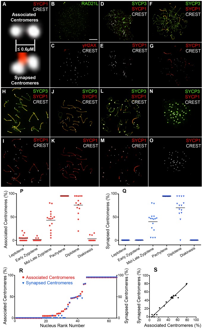
Figure 1
Centromere association and synapsis during meiotic prophase I.
(A) Criteria for assigning u201cassociatedu201d and u201csynapsedu201d centromeres. Magnified images from nuclei stained with CREST (white) and SYCP3 (red) are shown. (B and C) Surface spread pre-lept...
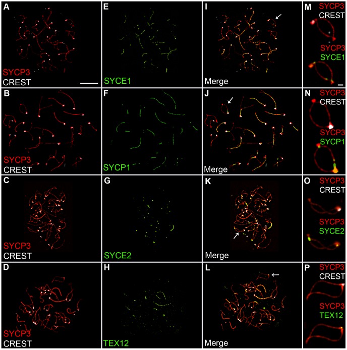
Figure 2
Localization of synaptonemal complex central element components during diplonema.
Diplotene stage spermatocyte nuclei, immunolabeled for SYCP3 (red), CREST (white) and various SC central-element proteins (green). (Mu2013P) Magnifications of bivalent chromosomes (indicated by arrows...
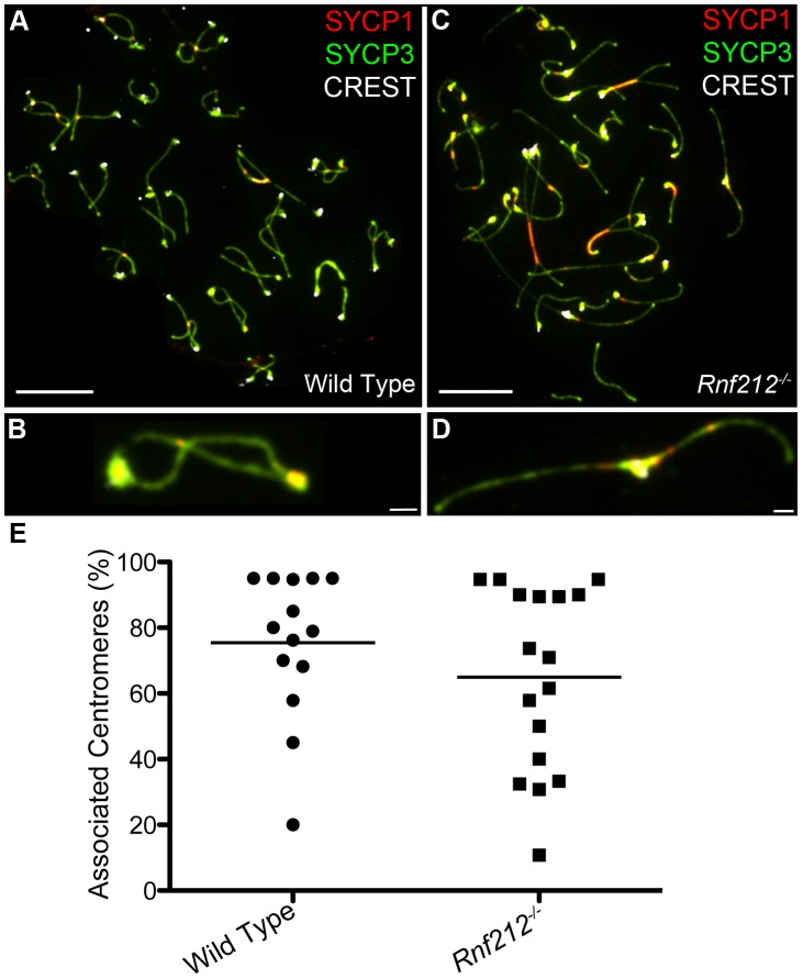
Figure 3
Centromere association in the absence of crossing-over.
Representative diplotene-stage spermatocytes from wild-type (A and B) and Rnf212 u2212/u2212 (C and D) mice, immunostained for SYCP3 (green), SYCP1 (red) and CREST (white). Selected homolog pairs are ...
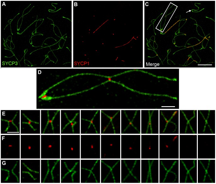
Figure 4
Axis remodeling revealed by structured illumination microscopy of diplotene-stage spermatocytes.
All panels show chromosomes from diplotene-stage nuclei immunolabled for SYCP3 (green) and SYCP1 (red). (Au2013C) A representative diplotene-stage nucleus. The arrow highlights the X-Y chromosome pair...
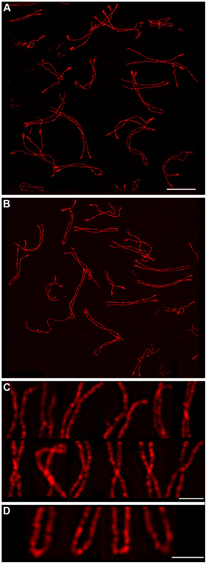
Figure 5
Chiasma-like structures and terminal fusions in the absence of synapsis and crossing-over.
(A and B) Representative diplotene-like nuclei from the Sycp1 u2212/u2212 knock-out immunostained for SYCP3. (C) Gallery of selected examples of axial association sites showing a variety of axis morph...
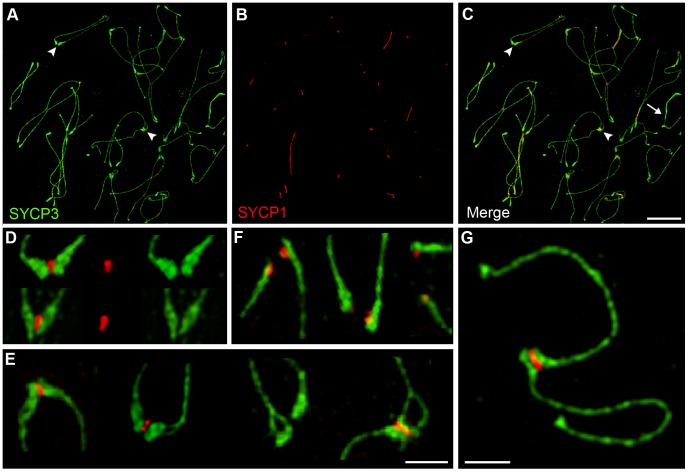
Figure 6
Centromere-associated SC fragments during diplonema.
(Au2013C) A representative diplotene-stage nucleus immunostained for SYCP3 (green) and SYCP1 (red). Arrowheads indicate paddle-like terminal structures with and without axis splitting. The arrow in C ...
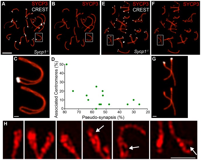
Figure 7
Centromere association and morphology of centromere regions in the absence of synapsis.
Spread spermatocyte nuclei from the Sycp1 u2212/u2212 knock-out immunostained for SYCP3 (red) and CREST (white). (A and B) Pachytene-like nucleus showing extensive coalignment or pseudo-synapsis of ho...
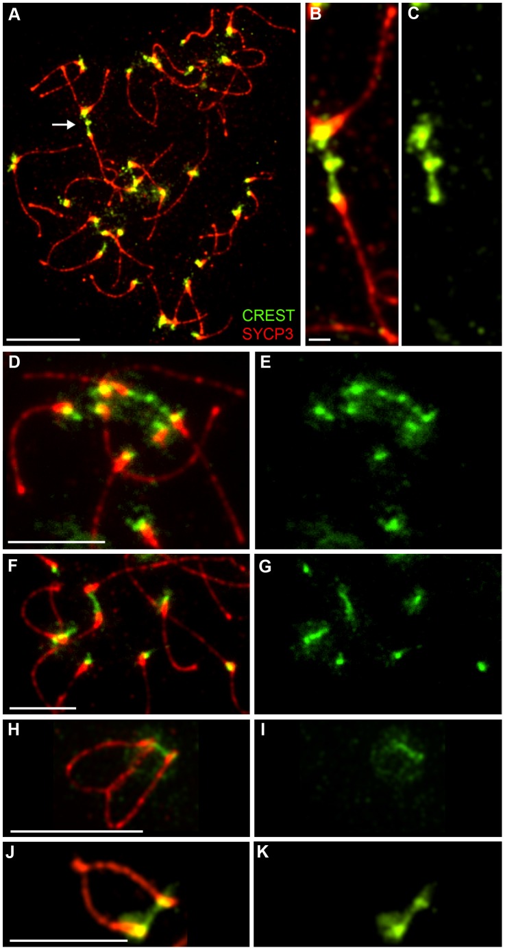
Figure 8
Identification of post-synapsis inter-centromeric CREST-staining bridges.
(A) Selected image of a late diplotene/early diakinesis spermatocyte from an Rnf212 u2212/u2212 mouse, immunostained for SYCP3 (red) and CREST (green). The arrow in panel A highlights the chromosomes ...
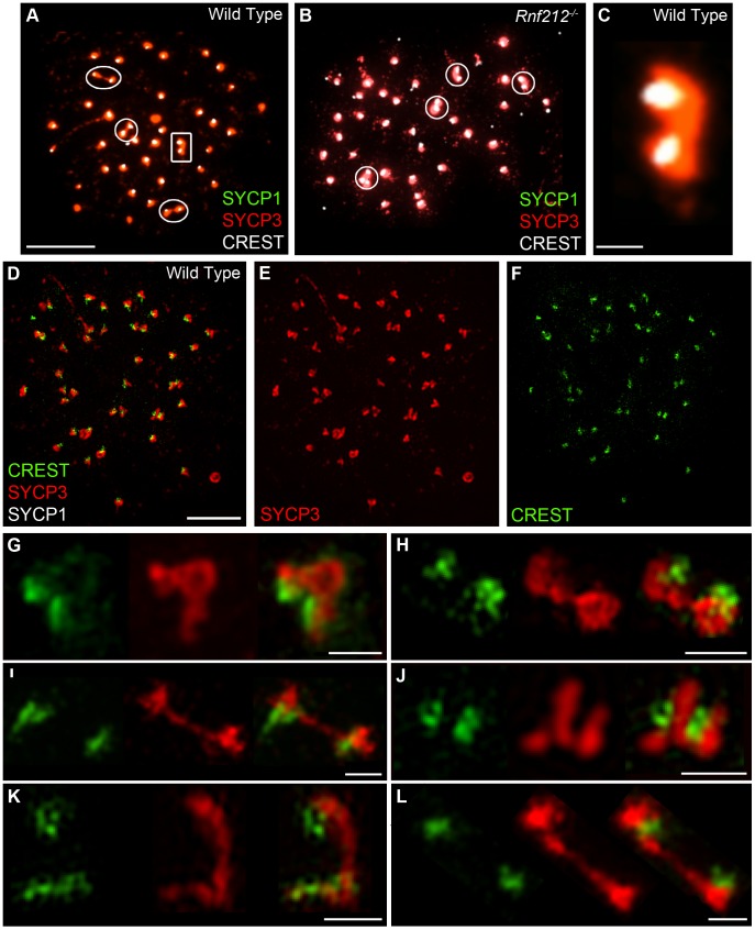
Figure 9
Identification of inter-centromeric SYCP3-staining bridges in diakinesis/metaphase I spermatocytes.
(Au2013C) Selected diakinesis/metaphase-I stage spermatoytes from wild-type and Rnf212 u2212/u2212 mice, immunostained for SYCP3 (red), SYCP1 (green), and CREST (white). Circles highlight pairs of CRE...
Figure images are served from the NIH/NLM PubMed Central Open Access Subset or Europe PMC; copyright remains with the publishers and authors.
💬 Discussion
0 commentsNo comments yet. Be the first to start a discussion!
Leave a Comment