Abstract
Localized mRNA provides spatial and temporal protein expression essential to cell development and physiology. To explore the mechanisms involved, considerable effort has been spent in establishing new and improved methods for visualizing mRNA. Here, we discuss how these techniques have extended our understanding of intracellular mRNA localization in a variety of organisms. In addition to increased ease and specificity of detection in fixed tissue, in situ hybridization methods now enable examination of mRNA distribution at the ultrastructural level with electron microscopy. Most significantly, methods for following the movement of mRNA in living cells are now in widespread use. These include the introduction of labeled transcripts by microinjection, hybridization based methods using labeled antisense probes and complementary transgenic methods for tagging endogenous mRNAs using bacteriophage components. These technical innovations are now being coupled with super-resolution light microscopy methods and promise to revolutionize our understanding of the dynamics and complexity of the molecular mechanism of mRNA localization.
🔬 Techniques
🧬 Organisms
✨ Fluorophores
🧪 Sample Preparation
🔬 Cell Lines
📷 Detectors
💻 Software Details
🏛️ Research Organizations (ROR)
Affiliated research institutions:
📊 Figures
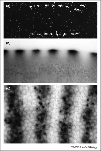
Figure 1
Detecting RNA in fixed cells. (a) ISH on 5-u03bcm-thick wax sections of Drosophila syncytial blastoderm embryos using a tritiated probe against fushi tarazu pair-rule transcripts that are expressed in...
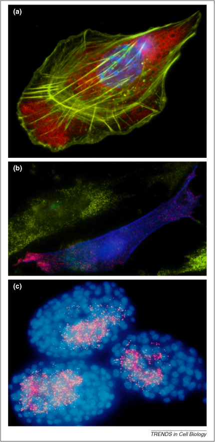
Figure 2
Detecting RNA in fixed cells by fluorescence. (a) Indirect labeling of RNA through its association with ZBP1. Double indirect immuno-fluorescence of a fixed primary mouse embryo fibroblast shows that ...
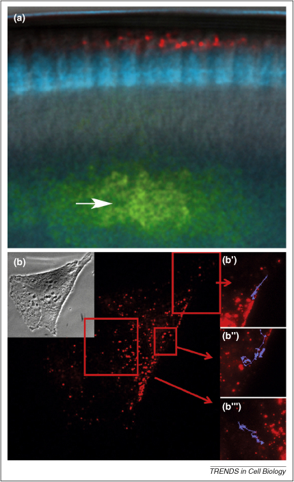
Figure 3
Detection of RNA in live tissue. (a) Localization of multiple RNAs via injection. Drosophila syncytial blastoderm expressing nuclear GFP (blue) first injected with an Alexa Fluor 546-labeled runt RNA ...
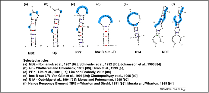
Figure 4
Different options for endogenous tagging of mRNA s in vivo . UNAfold (by Stewart and Zuker ( http://dinamelt.bioinfo.rpi.edu/download.php ) predicted secondary structures for RNA stemu2013loop sequenc...
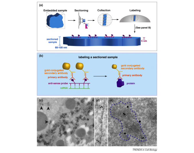
Figure I
ISH-IEM on ultrathin frozen sections.
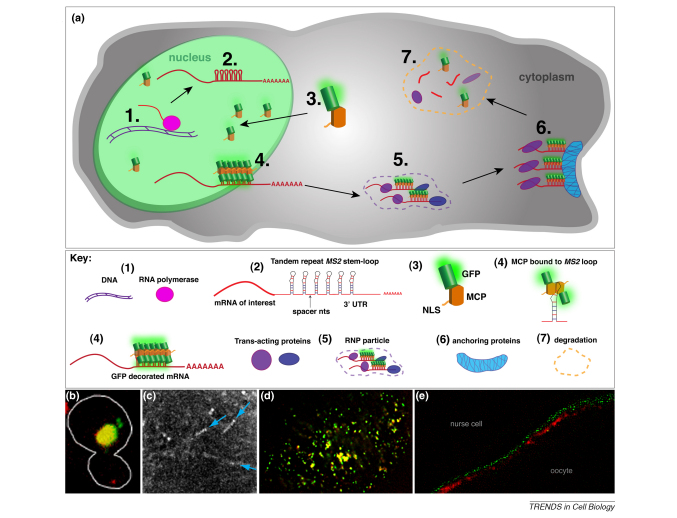
Figure II
MS2 -MCP labeling the life cycle of endogenous mRNA.
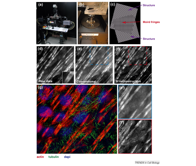
Figure III
Super-resolution imaging by SI on the Sedat OMX.
Figure images are served from the NIH/NLM PubMed Central Open Access Subset or Europe PMC; copyright remains with the publishers and authors.
💬 Discussion
0 commentsNo comments yet. Be the first to start a discussion!
Leave a Comment