Abstract
We studied two aspects of vimentin intermediate filament dynamics-transport of filaments and subunit exchange. We observed transport of long filaments in the periphery of cells using live-cell structured illumination microscopy. We studied filament transport elsewhere in cells using a photoconvertible-vimentin probe and total internal reflection microscopy. We found that filaments were rapidly transported along linear tracks in both anterograde and retrograde directions. Filament transport was microtubule dependent but independent of microtubule polymerization and/or an interaction with the plus end-binding protein APC. We also studied subunit exchange in filaments by long-term imaging after photoconversion. We found that converted vimentin remained in small clusters along the length of filaments rather than redistributing uniformly throughout the network, even in cells that divided after photoconversion. These data show that vimentin filaments do not depolymerize into individual subunits; they recompose by severing and reannealing. Together these results show that vimentin filaments are very dynamic and that their transport is required for network maintenance.
🔬 Techniques
🔭 Microscopes
💻 Software
✨ Fluorophores
🧪 Sample Preparation
🔬 Cell Lines
🏭 Microscope Brands
🧪 Reagent Suppliers
📷 Detectors
🔎 Objectives
💻 Software Details
💾 Data Repositories
🏛️ Research Organizations (ROR)
Affiliated research institutions:
📋 Methods
DNA constructs PA-GFP-vimentin. PA-GFP plus linker and human vimentin were amplified by PCR from two separate donor vectors. The amplicons were then inserted into pQCXIP (Clontech, Mountain View, CA) using Clontech In-Fusion cloning kit as recommend by the manufacturer. Next PA-GFP was PCR amplified from this vector. This amplicon and the PA-GFP-vimentin vector were digested with Eco RI, and the digestion products were ligated together. The resulting vector had two copies of PA-GFP separated by TCCGGACTCAGATCTCGAGCTCAAGCTTCGAATTCC placed on the N-terminus of vimentin with the linker TCCGGACTCAGATCTCGAGCTCAAGCTTCGAATTCTGCAGTCGACGGTACCGCGGGCCCGGGATCCATG between the last tag and vimentin . mEos3.2-vimentin. Human vimentin was cloned into a pEGFP-C1 vector (Clontech) in which enhanced GFP had been replaced by mEos3.2, creating the linker TCCGGACTCAGATCTGGCAGCGGTGGAGGCAGCGCATCCGGCGGAAGCGGAAGC between mEos3.2 and vimentin . The mEos3.2-vimentin portion of this vector was then amplified by PCR. Next pQCXIP and the PCR amplicon were digested with Age I and Bam HI, and the products were ligated together to produce mEos3.2-vimentin in a viral vector. EB3-GFP was a gift from the Akhmanova lab ( Stepanova et al. , 2003 ). mTagRFP-EB3 was cloned as reported previously ( Robert et al. , 2014 ).
Cell lines
All cells were maintained at 37°C in 5% CO 2 . h-TERT RPE cells were maintained in DMEM plus 1 mM sodium pyruvate plus 10% fetal bovine serum (FBS); SW13 cells in Leibovitz's L-15 plus 10% FBS; NIH-3T3, MEF (primary cells at passage 4), and HEK 293-FT in DMEM plus 10% FBS; and CAD cells in 1:1 DMEM/F-12 plus 10% FBS. CAD cells were switched to DMEM/F-12 (without FBS) 16–24 h before imaging to induce differentiation and neurite outgrowth. mEmerald-vimentin RPE cells used in SIM experiments expressed mEmerald-vimentin under the endogenous vimentin promotor. mEmerald-vimentin and mTagRFPT-tubulin coexpressing RPE used in the two-color TIRF-SIM experiments were the mEmerald RPE cells also genome edited to express mTagRFPT-tubulin under the endogenous tubulin promotor. All genome-edited cells were a gift of the Danuser lab.
Show full methods section
DNA constructs PA-GFP-vimentin. PA-GFP plus linker and human vimentin were amplified by PCR from two separate donor vectors. The amplicons were then inserted into pQCXIP (Clontech, Mountain View, CA) using Clontech In-Fusion cloning kit as recommend by the manufacturer. Next PA-GFP was PCR amplified from this vector. This amplicon and the PA-GFP-vimentin vector were digested with Eco RI, and the digestion products were ligated together. The resulting vector had two copies of PA-GFP separated by TCCGGACTCAGATCTCGAGCTCAAGCTTCGAATTCC placed on the N-terminus of vimentin with the linker TCCGGACTCAGATCTCGAGCTCAAGCTTCGAATTCTGCAGTCGACGGTACCGCGGGCCCGGGATCCATG between the last tag and vimentin . mEos3.2-vimentin. Human vimentin was cloned into a pEGFP-C1 vector (Clontech) in which enhanced GFP had been replaced by mEos3.2, creating the linker TCCGGACTCAGATCTGGCAGCGGTGGAGGCAGCGCATCCGGCGGAAGCGGAAGC between mEos3.2 and vimentin . The mEos3.2-vimentin portion of this vector was then amplified by PCR. Next pQCXIP and the PCR amplicon were digested with Age I and Bam HI, and the products were ligated together to produce mEos3.2-vimentin in a viral vector. EB3-GFP was a gift from the Akhmanova lab ( Stepanova et al. , 2003 ). mTagRFP-EB3 was cloned as reported previously ( Robert et al. , 2014 ).
Cell lines
All cells were maintained at 37°C in 5% CO 2 . h-TERT RPE cells were maintained in DMEM plus 1 mM sodium pyruvate plus 10% fetal bovine serum (FBS); SW13 cells in Leibovitz's L-15 plus 10% FBS; NIH-3T3, MEF (primary cells at passage 4), and HEK 293-FT in DMEM plus 10% FBS; and CAD cells in 1:1 DMEM/F-12 plus 10% FBS. CAD cells were switched to DMEM/F-12 (without FBS) 16–24 h before imaging to induce differentiation and neurite outgrowth. mEmerald-vimentin RPE cells used in SIM experiments expressed mEmerald-vimentin under the endogenous vimentin promotor. mEmerald-vimentin and mTagRFPT-tubulin coexpressing RPE used in the two-color TIRF-SIM experiments were the mEmerald RPE cells also genome edited to express mTagRFPT-tubulin under the endogenous tubulin promotor. All genome-edited cells were a gift of the Danuser lab.
Vimentin cell lines
(PA-GFP-vimentin and mEos3.2-vimentin) were created by viral transduction of the wild-type cell lines listed, except MEF and HEK 293-FT. Virus was produced by transfection of HEK 293-FT cells with PA-GFP-vimentin or mEos3.2-vimentin and helper plasmids pVSVG (Clontech) and pCL-Eco (Imgenex, San Diego, CA). Supernatant from transfected cells plus 8 μg/ml Polybrene was filtered and applied to target cell lines for 6–8 h for two consecutive days. Target cells were then selected using puromycin at 0.5 μg/ml (SW13), 3 μg/ml puromycin (CAD and NIH-3T3), or 2 μg/ml (RPE) for 3–5 d. MEF cells were transfected with Eos3.2-vimentin using the Lipofectamine 2000 (Invitrogen) transfection agent per manufacturer's instructions.
APC RNA interference knockdown
A duplex scrambled sequence control and two APC siRNA duplex target sequences (5′-rCrGrA rCrArA rGrArG rCrUrA rGrArA rGrArU rArArU rUrCC A-3′ + 5′-rUrGrG rArArU rUrArU rCrUrU rCrUrA rGrCrU rCrUrU rGrUrC rGrArA-3′ and 5′-rUrGrA rCrArA rUrArA rArGrC rArGrA rGrGrA rArGrG rUrGA T-3′ + 5′-rArUrC rArCrC rUrUrC rCrUrC rUrGrC rUrUrU rArUrU rGrUrC rArUrC-3′) were ordered from Integrated DNA Technologies (Coralville, IA). RPE cells were transfected for two consecutive days for 14–16 h with either a combination of 10 nM of each APC sequence (APC condition) or 20 nM of scrambled sequence (control condition) using HiPerfect (Invitrogen, Carlsbad, CA). Cells were imaged in FluroBrite DMEM (Invitrogen) plus 10% FBS (17 cells in each group across two experiments; also see TIRFM and wide-field fluorescence microscopy ) and harvested for Western blotting on the same day, 48 h after the last transfection. Knockdown efficiency was assessed using Western blot analysis using the APC-M2 antibody at 1:5000 (generously gifted to us by Kristi Neufeld, University of Kansas, Lawrence, KS) and Coomassie stain as loading control.
Structured illumination microscopy
Cells were plated on glass coverslips 14–16 h before imaging in FluoroBrite DMEM supplemented with 10% FBS and 1 mM sodium pyruvate. Live-cell TIRF-SIM images were collected on a Nikon Ti-E inverted microscope (Nikon Instruments, Melville, NY) with a SIM illuminator and SIM enclosure equipped with an Apo TIRF 100×/numerical aperture (NA) 1.49 oil objective and electron-multiplying charge-coupled device (EMCCD) camera (iXon DU897; Andor, Belfast, United Kingdom) at the Nikon Center at the Northwestern University Feinberg School of Medicine Center for Advanced Microscopy. Cells were maintained at 37°C during imaging using a Tokai-Hit stage-top incubator (Tokai-Hit, Fujinomiya City, Japan). Images were collected at 1-s intervals and reconstructed using Nikon Elements software. For nocodazole experiments, FluoroBrite medium supplemented as before ± 10 μM nocodazole (Sigma-Aldrich, St. Louis, MO) was placed on cells, and cells were incubated at 4°C for 30 min, warmed to 37°C for 30 min, and then imaged at 37°C. Filament ends were tracked (14 filaments for 7–20 s across four cells) in reconstructed images using the Manual Tracking plug-in in Fiji (ImageJ processing package). Two-color TIRF-SIM experiments were conducted at the Advanced Imaging Center at the Janelia Research Campus. Cells were prepared as described for the other SIM experiments and kept at 37°C by regulated hot air blown into a plastic enclosure around the microscope stand. Briefly, this system offered the speed necessary to capture transport dynamics in two channels by using a spatial light modulator in place of a rotating diffraction grating. Details of the system have been described ( Kner et al. , 2009 ). TIRFM and wide-field fluorescence microscopy Live-cell TIRFM images were collected on a Nikon Eclipse U2000 inverted microscope equipped with a Plan-Apo TIRF 100×/1.45 NA objective and a Hamamatsu CMOS Orca Flash 4.0 camera (Hamamatsu Photonics, Hamamatsu, Japan), controlled by MetaMorph 7.7.7.0 software (Molecular Devices, Downingtown, PA). Cells were maintained at 37°C plus 5% CO 2 during imaging using a Tokai-Hit stage-top incubator (Tokai-Hit, Fujinomiya, Japan) and Okolab gas mixer (Okolab, Naples, Italy). The angle of a 561-nm laser was manually adjusted until near total internal reflection was reached, as judged by imaging of photoconverted mEos3.2-vimentin–expressing cells. To photoconvert, cells were exposed to UV light from a Hg + light source for 10 s through a pinhole in the light path. Time-lapse sequences were acquired at 20-s intervals for 3 min using the 561-nm laser. For all experiments except the serum starvation experiment, cells were plated on glass coverslips ∼16 h before imaging. For nocodazole experiments, cells were treated as described in Structured illumination microscopy (23 control and 19 nocodazole-treated cells). For vinblastine experiments, the medium for cells was switched to 37°C FluoroBrite DMEM plus 10% FBS ± 10 nM vinblastine (Sigma-Aldrich) for 10 min before imaging (16 control and 13 vinblastine-treated cells). For APC experiments, cells were treated as described in APC RNA interference knockdown and imaged in DMEM plus 10% FBS (17 cells in each group). For serum starvation experiments, cells were split to coverslips (in their maintenance medium), left unchanged (control, 10 cells), changed to serum-free medium 12–16 h later, and incubated in the serum-free medium for 72 h before imaging (serum-starved group, 19 cells) or imaged 2–10 min after the serum-free medium was replaced by that containing 10% FBS (serum stimulation group, 15 cells). For TSA experiments, cells were incubated in DMEM plus 10% FBS ± 500 nM TSA for ∼16 h (28 control, 28 TSA treated). For filament motility, time-lapse sequences were analyzed as described later. For display purposes (but not for analysis), TIRFM images of mEos3.2-vimentin RPE were modified using the Log function in Fiji imaging software, and gamma was adjusted to between 1.3 and 1.4. Similarly, images of mEos3.2-vimentin SW13 cells were logged, but gamma remained at 1.0. RPE cells were transfected using X-Treme Gene (Roche, Indianapolis, IN) with mTagRFPt-EB3. mTagRFPt-EB3–transfected RPE cells were also imaged at near-TIRF using the 561-nm laser at 2-s intervals for 16 s. These cells were treated ± 10 nM vinblastine as described for mEos3.2-vimentin cells. To display comets, image sequences were color coded using Temporal Color Code in Fiji, which assigns a single color to each frame and superimposes all colored frames. CAD cells were transfected with Lipofectamine 2000 with GFP-EB3. GFP-EB3–transfected CAD cells were imaged using a Plan Apo 100×/1.40 NA objective, Hg + light source, and Pixis 1024B CCD camera (Princeton Instruments, Roper Industries, Sarasota, FL) at intervals of 1 s for 1 min. An EB3 kymograph was created of a 5- pixel-wide section of GFP-EB3–expressing CAD neurite using the Reslice command in Fiji.
Confocal microscopy
Confocal images were collected on a Nikon Eclipse U2000 inverted microscope equipped with a Yokogawa CSU10 spinning-disk confocal head (Yokogawa Electric Corporation, Sugar Land, TX), a Plan Apo 100×/1.45 NA objective, an Agilent MLC 400 laser set (including 488- and 561-nm lasers; Agilent Technologies, Wood Dale, IL), 89 North Heliophor pumped phosphor light engine at 405 nm (Chroma Technology, Bellows Falls, VT) to drive photoactivation/photoconversion, and an Evolve EMCCD (Photometrics, Tucson, AZ) driven by Nikon Elements software. Cells were maintained at 37°C with 5% CO 2 as described earlier. Sections of PA-GFP-vimentin or mEos3.2-vimentin CAD cell neurites were photoconverted with 405-nm light restricted by a pinhole in the light path. Time-lapse sequences in the photoactivated (488 nm, PA-GFP-vimentin CAD) or photoconverted (561 nm, mEos3.2-vimentin CAD) channel were acquired for 3 min at 4 frames/min. Sequences from 32 neurites were visually inspected to determine the direction of movement of photoactivated/photoconverted filaments. For display only, images of PA-GFP-vimentin CAD were adjusted using the Log function in Fiji imaging software. mTagRFPt-tubulin RPE cells were treated ± nocodazole or vinblastine as described for mEos3.2-vimentin cells and then fixed with 0.5% glutaraldehyde plus 0.1% Triton for 5 min. Confocal images of these cells were gathered to record microtubule distribution. Similarly, RPE fixed with 0.5% glutaraldehyde plus 0.1% Triton and immunolabeled with 1:800 anti-vimentin antibody PCK-594P (Covance, Princeton, NJ), mEmerald-vimentin RPE, and mEos3.2-vimentin RPE were imaged on the confocal system to record vimentin distributions in these cells.
Quantification of filaments
Custom software was created in Matlab to process images for quantification of filament transport. Photomicrographs were first flattened with nonlinear mapping to boost the filament in the dimmer region. A Gaussian filter was then applied to reduce noise from the background. Multiple-scale steerable filtering was used to enhance the line features in the image. The non–maximum suppression image of the steerable filtering results were used to segment all possible filaments on their centerline locations. Based on geometrical and imaging features of filaments—the length, curvature, intensity, and steerable filtering response value—filaments were identified out of the whole set. From this, an output filament network was made by creating a binary image of the filament network with 1-pixel-wide filaments. For each image, the total length of filaments was counted by summing the identified pixels. Filament transport was defined as this amount outside the initial region of photoconversion. To account for variations in the initial number of photoconverted filaments between cells, filament transport for each frame was normalized to the sum intensity measured in the region of photoconversion in the first frame. These normalized values were then plotted against time, and slopes were calculated. Finally, these values were normalized to control average, groups were compared using Welch's t test analysis, and bar graphs were plotted using Prism software (GraphPad Software, La Jolla, CA).
📊 Figures
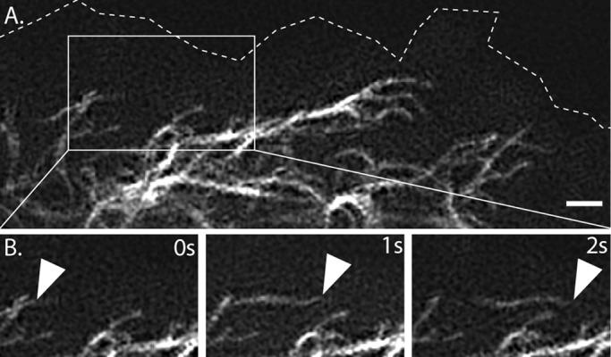
FIGURE 1:
Rapid motility of mature filaments at cell periphery revealed by TIRF-SIM. (A) Edge of Emerald-vimentinu2013expressing RPE cell. (B) Time-lapse at 1-s intervals shows rapid transport of filaments in t...
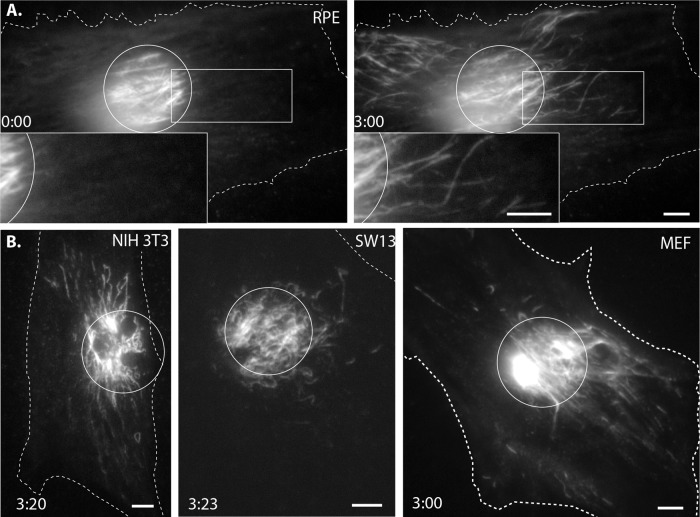
FIGURE 2:
Photoconvertible-vimentin reveals the rapid transport of mature vimentin intermediate filament (IF) in multiple cell types. (A) Left, Eos3.2-vimentin RPE cell immediately after a circular region was e...
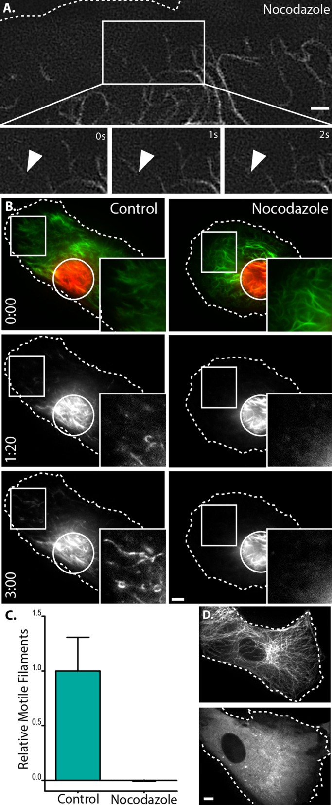
FIGURE 3:
Vimentin IF transport depends on microtubules. (A) Two-u00addimensional TIRF-SIM at the cell periphery of an emerald-vimentinu2013expressing RPE cell treated with nocodazole (top) shows that filaments...
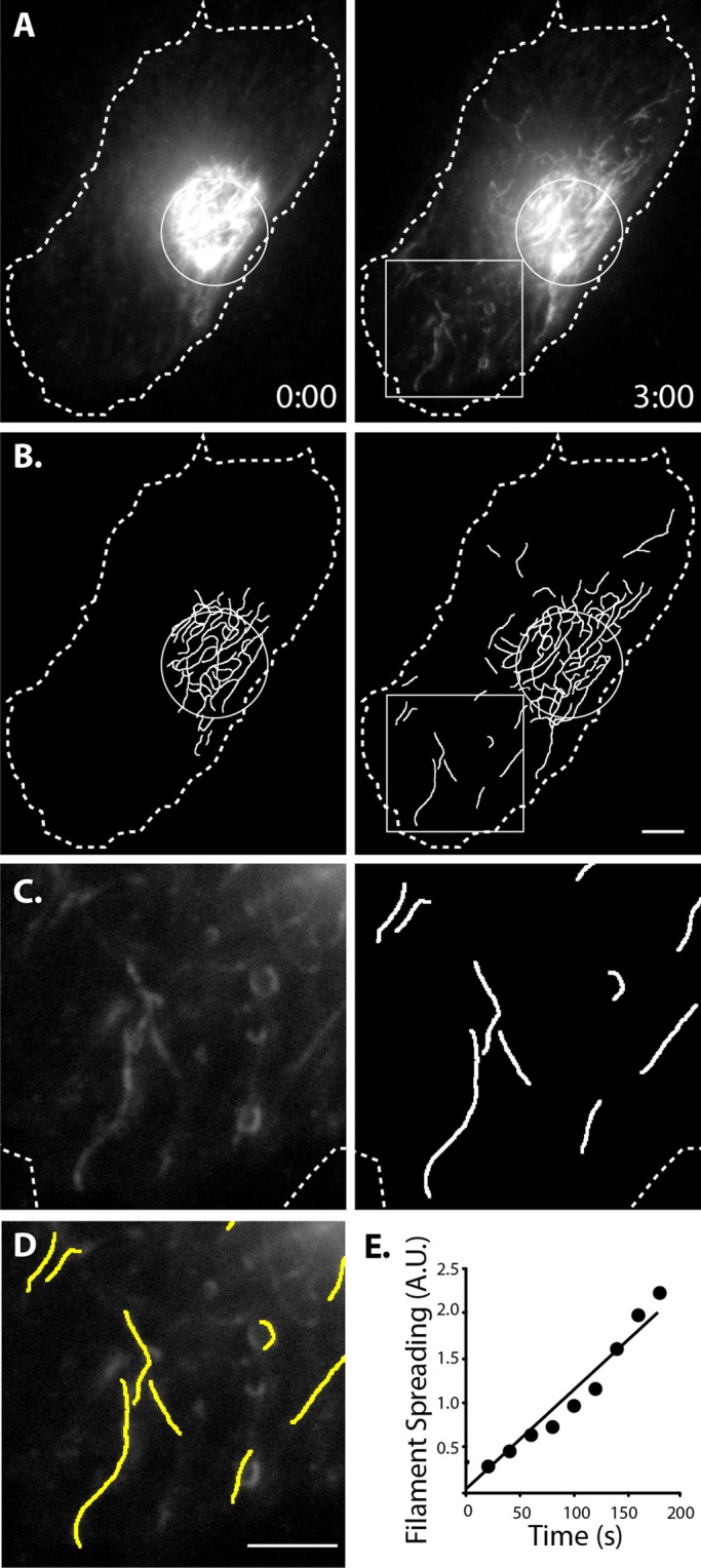
FIGURE 4:
Method to quantify vimentin filament motility. (A) Control cell 0 and 3 min after photoconversion. (B) Filaments detected using custom software to detect linear segments. (C) Enlargement of boxed regi...
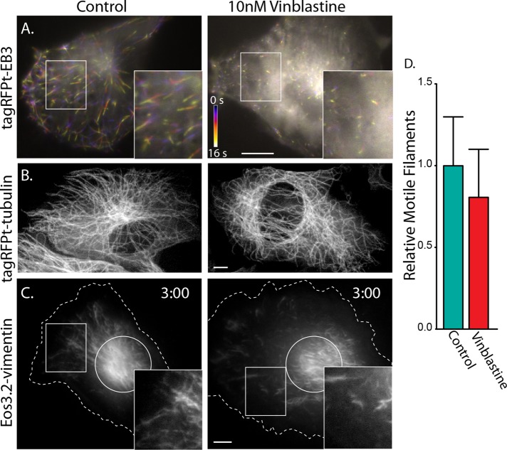
FIGURE 5:
Blocking microtubule dynamics does not affect vimentin IF transport. (A) mtagRFPT-EB3u2013labeled growing microtubule plus ends. Frames from time-lapse sequences were individually pseudocolored and su...
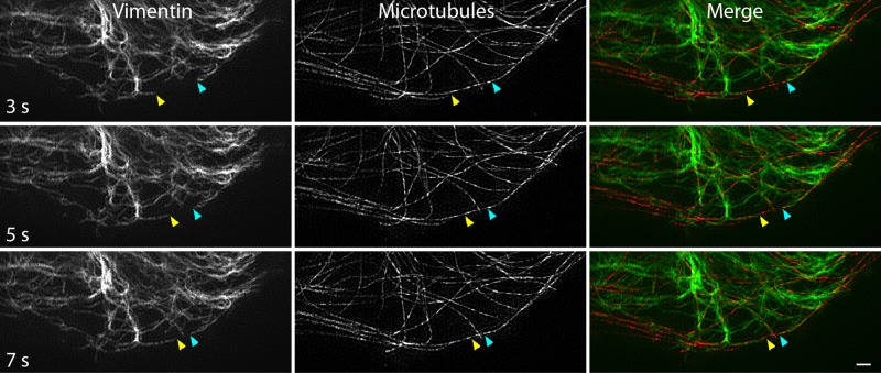
FIGURE 6:
Vimentin filament transport along microtubules revealed by live-cell, two-color TIRF-SIM. Frames from time-lapse imaging show vimentin filaments (left) moving in the periphery of a cell. Arrowheads in...
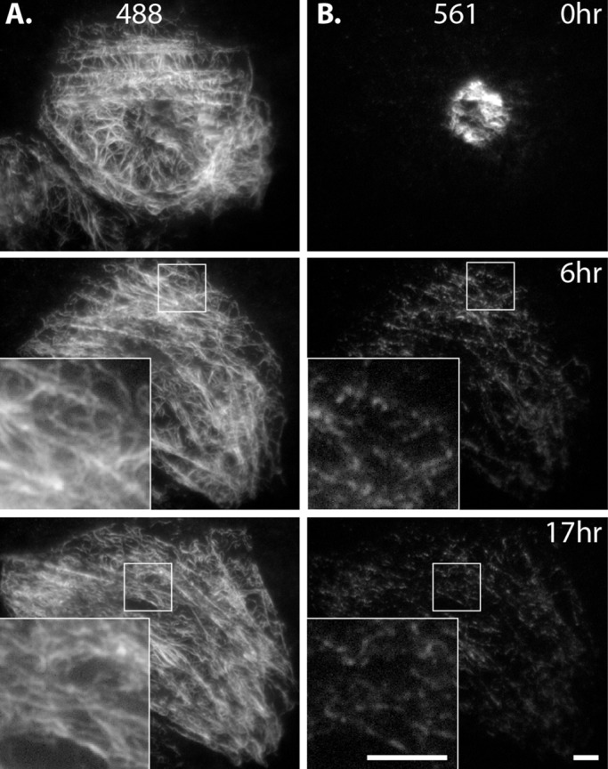
FIGURE 7:
Long-term imaging after photoconversion reveals extensive severing and reannealing of filaments. Eos3.2-vimentinu2013expressing SW13 cell immediately after photoconversion and 6 and 17 h later. (A) Th...
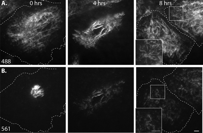
FIGURE 8:
Filaments remain in polymerized form throughout cell division in SW13 cells. Eos3.2-vimentinu2013expressing SW13 cell immediately after photoconversion (left), rounding and lifting from the coverslip ...
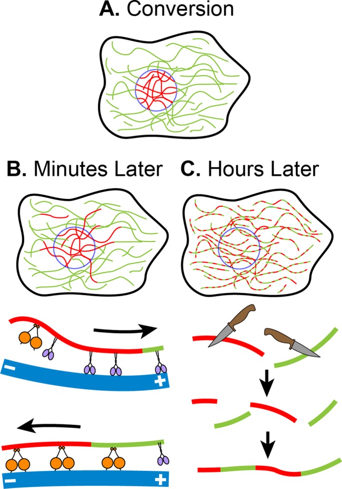
FIGURE 9:
Model of vimentin filament dynamics. (A) Filament dynamics can be followed by producing fiduciary marks on them using photoconversion. (B) Within only minutes, filaments can be seen to be transported ...
Figure images are served from the NIH/NLM PubMed Central Open Access Subset or Europe PMC; copyright remains with the publishers and authors.
💬 Discussion
0 commentsNo comments yet. Be the first to start a discussion!
Leave a Comment