Abstract
We used electron tomography to examine microtubules (MTs) growing from pure tubulin in vitro as well as two classes of MTs growing in cells from six species. The tips of all these growing MTs display bent protofilaments (PFs) that curve away from the MT axis, in contrast with previously reported MTs growing in vitro whose tips are either blunt or sheetlike. Neither high pressure nor freezing is responsible for the PF curvatures we see. The curvatures of PFs on growing and shortening MTs are similar; all are most curved at their tips, suggesting that guanosine triphosphate-tubulin in solution is bent and must straighten to be incorporated into the MT wall. Variations in curvature suggest that PFs are flexible in their plane of bending but rigid to bending out of that plane. Modeling by Brownian dynamics suggests that PF straightening for MT growth can be achieved by thermal motions, providing a simple mechanism with which to understand tubulin polymerization.
🔬 Techniques
🔭 Microscopes
🧬 Organisms
💻 Software
✨ Fluorophores
🧪 Sample Preparation
🏭 Microscope Brands
🧪 Reagent Suppliers
💻 Software Details
💾 Data Repositories
🏛️ Research Organizations (ROR)
Affiliated research institutions:
📋 Methods
Cell growth and the preparation of samples for microscopy
Mammalian cells (strain PtK 2 ) were plated on sapphire disks in McCoy’s medium at 37°C with 5% CO 2 and then cryoimmobilized by freezing under high pressure ( McDonald et al., 2007 ). Schizosaccharomyces pombe was grown in rich medium (YES) at 25°C, collected by vacuum filtration onto EMD Millipore filters, and transferred by spatula to a planchette for high-pressure freezing ( Giddings et al., 2017 ). For plunge freezing, S. pombe cells (strain CDC25-22) were grown at permissive temperature on nutrient agar covered with a thin Mylar film. Log-phase cells were frozen by rapid immersion of the Mylar into liquid ethane ( Tanaka and Kanbe, 1986 ). Saccharomyces cerevisiae was grown in rich medium and then collected and frozen as described previously ( Giddings et al., 2001 ). Arabidopsis was grown, and its root tips were harvested and then high-pressure frozen as described previously ( Austin et al., 2005 ). Chlamydomonas (strain 137c) was grown in liquid culture at 25°C on a 12-h light/dark cycle to enrich for mitotic cells ( Umen and Goodenough, 2001 ). The cells were collected by centrifugation after shifting to the dark and then high-pressure frozen essentially as described previously ( Preble et al., 2001 ; O’Toole and Dutcher, 2014 ). Hermaphrodites of C. elegans were high-pressure frozen in M9 worm buffer containing 20% BSA as described previously ( Muller-Reichert et al., 2008 ). All samples were fixed by freeze substitution at −90°C in acetone containing either glutaraldehyde and uranyl acetate or OsO 4 and then embedded in plastic as previously described ( Muller-Reichert et al., 2008 ). Serial sections ∼250 nm thick were cut using an Ultracut microtome (Leica Biosystems), picked up on slot grids coated with a film of Formvar, and poststained with lead citrate and uranyl acetate. The sections were then strewn with particles of 15 nm colloidal gold to serve as markers for alignment of serial tilts. Identification of cells whose MTs were growing Cells were identified as suitable based on their stage in anaphase B (a time when spindles elongate) and the orientation of their spindle axes approximately parallel to their plane of sectioning. In all these cell types, anaphase B begins near the end of anaphase A, so cells with well-separated chromosomes were selected. In PtK cells, the number of MTs in the interzone stays approximately constant as the MTs slide and elongate (Fig. S1; McIntosh et al., 1975 ). Thus, all MTs in the anaphase B interzones of these cells were elongating at the time of freezing. The same appears to be true for Chlamydomonas , although the data are less complete. In Arabidopsis , MT number stays high until near the end of anaphase B, whereupon it decreases as the cell plate forms ( Austin et al., 2005 ). Cells for study were therefore selected to be still in the spindle elongation phase of mitosis. In both yeasts, the number of interzonal MTs decreases during anaphase B, so some MTs must depolymerize as others elongate ( Ding et al., 1993 ; Winey et al., 1995 ). Growing MTs were identified as those whose plus ends were still overlapping; other interzone MT ends were ignored. In blastomeres of C. elegans , there are very few MTs in the anaphase interzone ( Muller-Reichert et al., 2008 ), so this organism was not used for studies of elongating IPMTs, only for examination of KMTs.
Show full methods section
Cell growth and the preparation of samples for microscopy
Mammalian cells (strain PtK 2 ) were plated on sapphire disks in McCoy’s medium at 37°C with 5% CO 2 and then cryoimmobilized by freezing under high pressure ( McDonald et al., 2007 ). Schizosaccharomyces pombe was grown in rich medium (YES) at 25°C, collected by vacuum filtration onto EMD Millipore filters, and transferred by spatula to a planchette for high-pressure freezing ( Giddings et al., 2017 ). For plunge freezing, S. pombe cells (strain CDC25-22) were grown at permissive temperature on nutrient agar covered with a thin Mylar film. Log-phase cells were frozen by rapid immersion of the Mylar into liquid ethane ( Tanaka and Kanbe, 1986 ). Saccharomyces cerevisiae was grown in rich medium and then collected and frozen as described previously ( Giddings et al., 2001 ). Arabidopsis was grown, and its root tips were harvested and then high-pressure frozen as described previously ( Austin et al., 2005 ). Chlamydomonas (strain 137c) was grown in liquid culture at 25°C on a 12-h light/dark cycle to enrich for mitotic cells ( Umen and Goodenough, 2001 ). The cells were collected by centrifugation after shifting to the dark and then high-pressure frozen essentially as described previously ( Preble et al., 2001 ; O’Toole and Dutcher, 2014 ). Hermaphrodites of C. elegans were high-pressure frozen in M9 worm buffer containing 20% BSA as described previously ( Muller-Reichert et al., 2008 ). All samples were fixed by freeze substitution at −90°C in acetone containing either glutaraldehyde and uranyl acetate or OsO 4 and then embedded in plastic as previously described ( Muller-Reichert et al., 2008 ). Serial sections ∼250 nm thick were cut using an Ultracut microtome (Leica Biosystems), picked up on slot grids coated with a film of Formvar, and poststained with lead citrate and uranyl acetate. The sections were then strewn with particles of 15 nm colloidal gold to serve as markers for alignment of serial tilts. Identification of cells whose MTs were growing Cells were identified as suitable based on their stage in anaphase B (a time when spindles elongate) and the orientation of their spindle axes approximately parallel to their plane of sectioning. In all these cell types, anaphase B begins near the end of anaphase A, so cells with well-separated chromosomes were selected. In PtK cells, the number of MTs in the interzone stays approximately constant as the MTs slide and elongate (Fig. S1; McIntosh et al., 1975 ). Thus, all MTs in the anaphase B interzones of these cells were elongating at the time of freezing. The same appears to be true for Chlamydomonas , although the data are less complete. In Arabidopsis , MT number stays high until near the end of anaphase B, whereupon it decreases as the cell plate forms ( Austin et al., 2005 ). Cells for study were therefore selected to be still in the spindle elongation phase of mitosis. In both yeasts, the number of interzonal MTs decreases during anaphase B, so some MTs must depolymerize as others elongate ( Ding et al., 1993 ; Winey et al., 1995 ). Growing MTs were identified as those whose plus ends were still overlapping; other interzone MT ends were ignored. In blastomeres of C. elegans , there are very few MTs in the anaphase interzone ( Muller-Reichert et al., 2008 ), so this organism was not used for studies of elongating IPMTs, only for examination of KMTs.
Identification of KMTs
KMTs were identified in PtK 2 cells, Chlamydomonas , and C. elegans as previously described ( McIntosh et al., 2013 ). We did not include fission yeast KMTs in our analysis in part because there is no flux of spindle MTs toward the spindle poles, so these MTs are not growing in metaphase, in part because chromosome in fission yeast cells are hard to see by EM, and in part because the number of examples of fission yeast anaphase spindles combined with the low numbers of MTs per kinetochore meant that we did not have large enough numbers of MTs for a serious analysis of their shapes. Preparation of electron tomograms and characterization of MT ends in vivo Sections of suitable cells were imaged as tilt series ranging over ±60° about two orthogonal axes using the SerialEM image acquisition software ( Mastronarde, 1997 ); 3D reconstruction was accomplished by back projection ( Kremer et al., 1996 ). Electron tomograms of regions near the spindle midplane contained many overlapping MTs whose growing plus ends were easy to identify. Graphic models of these MT trajectories were made using the 3dmod program from the IMOD software package ( Kremer et al., 1996 ). A subroutine in IMOD (mtrotlong) extracted volumes that contained each MT end. Additional controls for the impact of freezing and high pressure on PF shape As an additional control for the impact of freezing on MT structure, we have examined the plus ends of KMTs in metaphase PtK 2 cells that were chemically fixed with glutaraldehyde and then high-pressure frozen and further fixed by freeze-substitution before embedding, sectioning, and examination by ET in collaboration with the laboratory of J. DeLuca (Colorado State University, Fort Collins, CO). In this case, 264 PFs from 74 MTs showed a mean length of 43 ± 14 nm and a mean curvature of 14° ± 7° per dimer, further indicating that curving PFs are not a freezing artifact. As final evidence that high pressure is not the cause for curving PFs in vivo, we have examined KMTs in one anaphase PtK 2 cell that was plunge frozen, a rare case in which ice crystal damage did not obscure structural detail in a mitotic mammalian cell (all anaphase KMTs are shown in Fig. S4 [A, D, G, and J], and the anaphase numbers for PtK 2 cells are shown as KMT-PtK-ana in Table 2 ). The similarities among these numbers support the contention that neither high pressure nor freezing is a factor in PF shape. Growth and imaging of MTs in vitro For the study of MT polymerization in vitro, pure porcine tubulin purchased from Cytoskeleton (t238p) was polymerized onto the MTs of axonemes prepared from Chlamydomonas flagella ( Nicastro et al., 2006 ), a gift from M.E. Porter (University of Minnesota, Minneapolis, MN). Isolated axonemes were diluted into a buffer containing 80 mM Pipes, pH 6.9, supplemented with 1 mM GTP, EGTA, and MgCl 2 (BRB80) and then were applied as a 2–3-µl drop to a C-flat holy carbon-coated electron microscope grid (Electron Microscope Sciences) that had recently been glow discharged. Axonemes were given 30 s to attach to the carbon film, and then excess fluid was blotted away. 20 µM tubulin in BRB80 at 0°C was usually supplemented with a low titer of 10 nm colloidal gold, and then 5 µl of this mixture was immediately added to the grid, which was then drawn up into the prewarmed and hydrated chamber of a plunge-freezing device (Vitrobot; Thermo Fisher Scientific). Samples were incubated for 3–6 min at 37°C and 90% relative humidity. Both these chamber values were checked by independent measurement. Humidity was confirmed, but the temperature stated on the instrument’s console was ∼2°C higher than the temperature at the grid, as measured with a calibrated thermocouple. After sufficient time for MT growth, the grid was blotted with filter paper, plunged into liquid ethane, and then transferred to liquid nitrogen and kept under this liquid as it was transferred to a cryotransfer holder (910; Gatan) and inserted into a Tecnai F20 or F30 electron microscope from Thermo Fisher Scientific for examination at less than −170°C. Shortening MTs were made by first growing MTs with the above procedure and then inducing depolymerization by isothermal dilution. Polymerization buffer without tubulin was warmed to 37°C, and then an aliquot of 25 µl was added to the 5-µl drop of tubulin-containing buffer on the grid. Because two or more drops fell from the grid during this addition, the final tubulin concentration was
📊 Figures
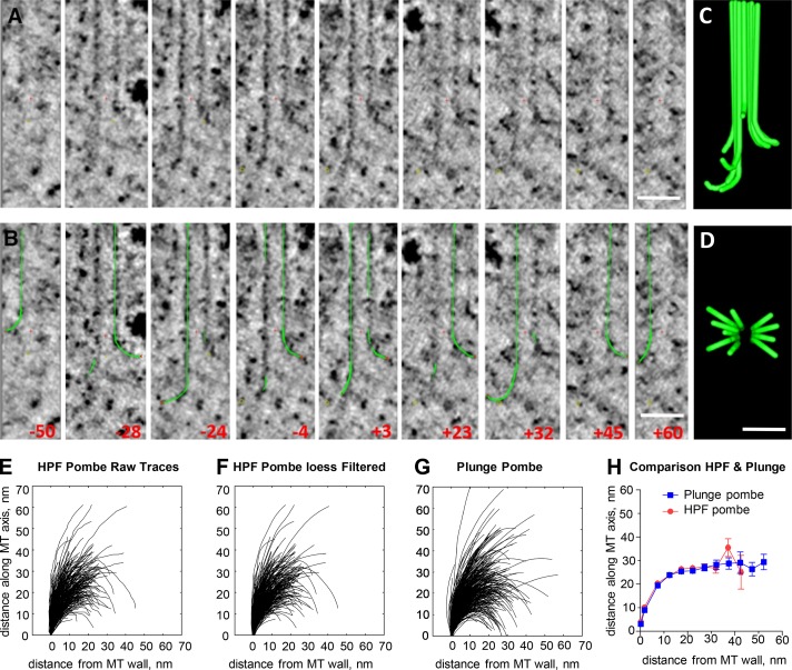
Figure 1.
Angular sampling of the 3D structure of a growing MT end. (A) Nine slices through the end of an MT from the anaphase B interzone in a PtK 2 cell. All images contain the MT axis, but the orientation of...
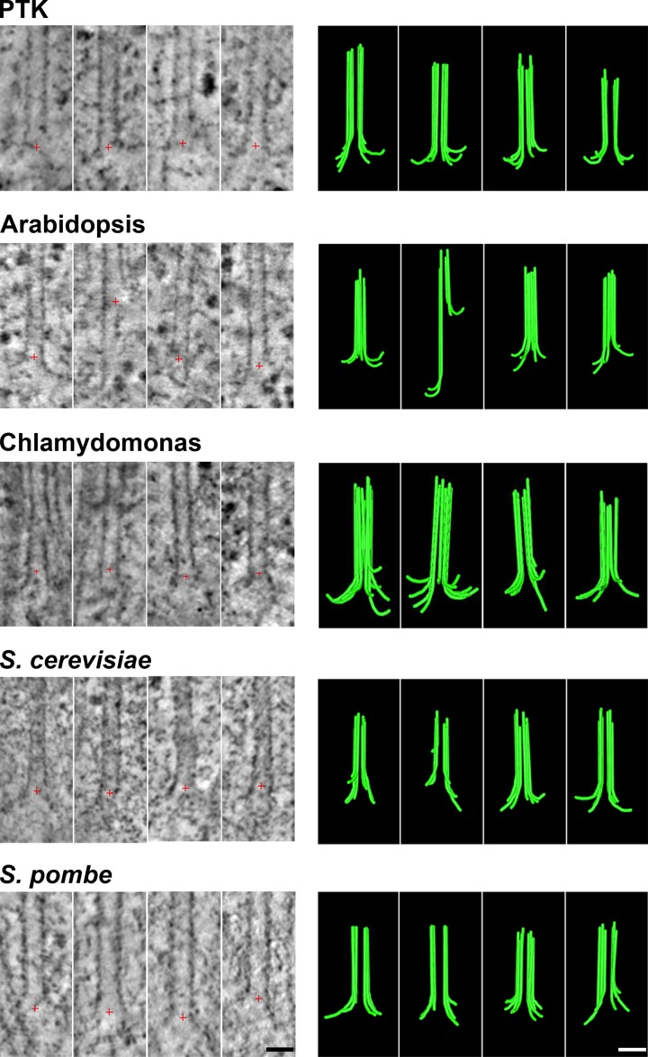
Figure 2.
Images and models of MTs growing in vivo. For each of five species names, the left column displays tomographic slices that contain the MT axis and show one or more PFs flaring out from the MT wall. Re...
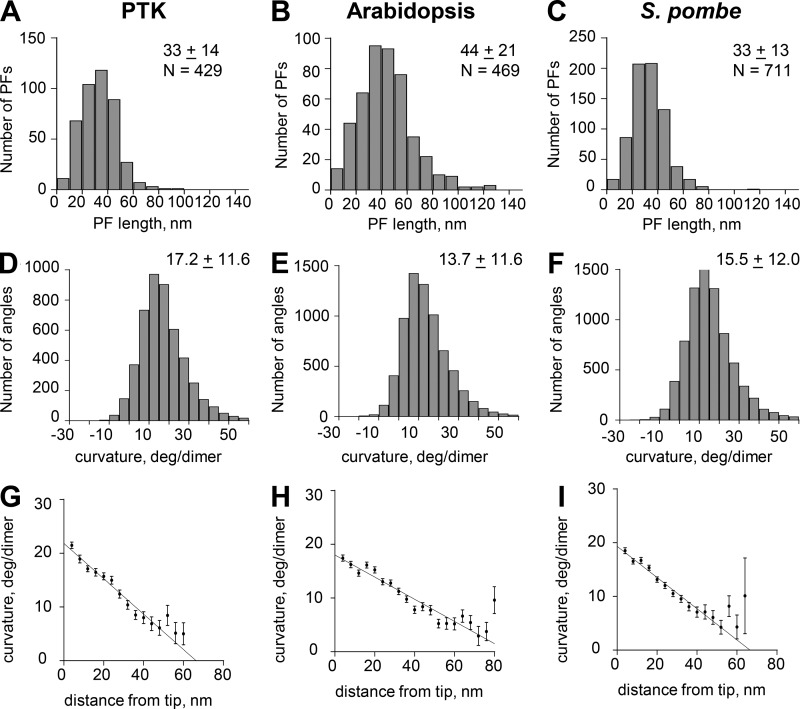
Figure 3.
Graphs depicting aspects of PF shape from three of the species studied. (Au2013C) Distributions of lengths for PFs from the species indicated. N, numbers of PFs traced. Other values are mean lengths u...
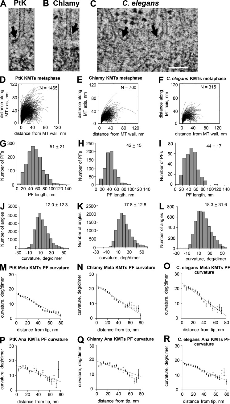
Figure 4.
Pictures and graphs describing the plus ends of metaphase KMTs. (Au2013C) Slices from tomograms of the plus ends of metaphase KMTs from the three species named. Arrows indicate flaring PFs. Bar, 100 n...
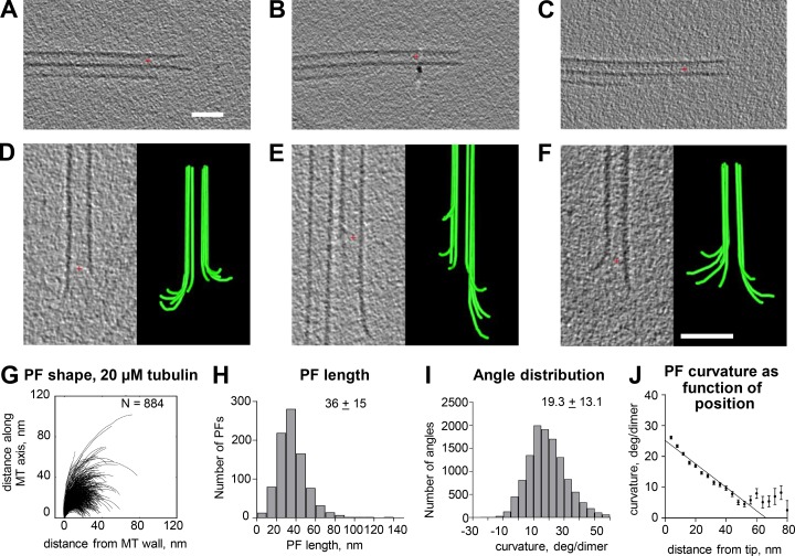
Figure 5.
Shapes of plus ends of MTs elongating in vitro. (Au2013C) Slices through tomograms of three examples of the axonemal doublet MTs that served as seeds to nucleate the polymerization of purified porcine...
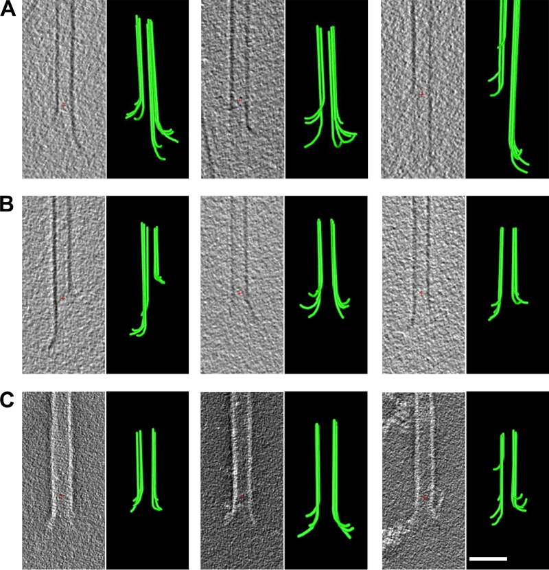
Figure 6.
Shapes of plus ends of in vitro MTs elongating or prepared under stabilizing conditions. (A) Tomographic slices and models of MT ends plunge frozen while elongating in GMPCPP. (B) Tomographic slices a...
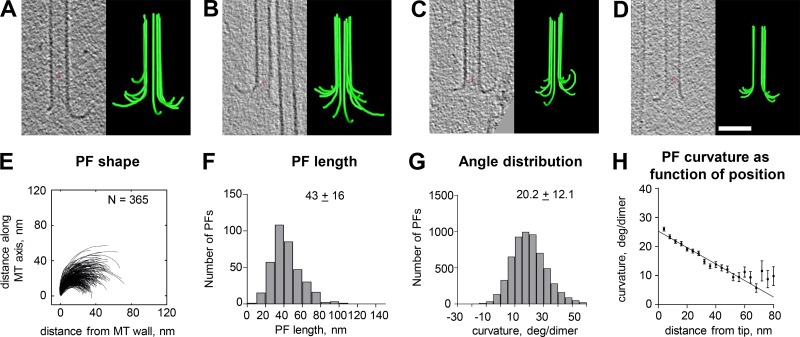
Figure 7.
Shapes of plus ends of MTs shortening in vitro. (Au2013D) Tomographic slices and models showing flaring PFs of MTs frozen u223c20 s after isothermal dilution. Red crosses mark the origins of the coord...
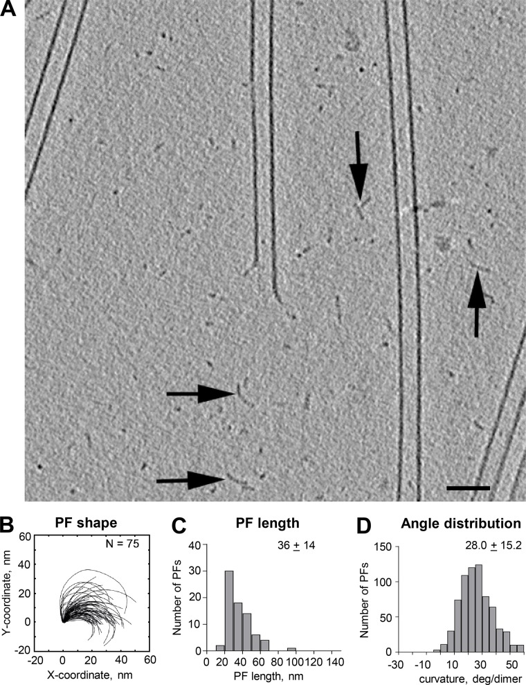
Figure 8.
Oligomers formed during tubulin polymerization. (A) 20-nm slice from a cryotomogram of an MT elongating in 20 u00b5M tubulin for 6 min before plunge freezing. A large enough area of background is show...
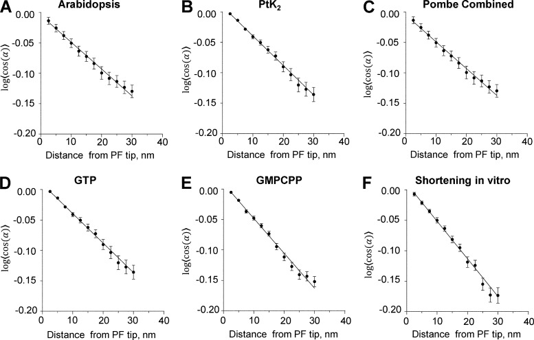
Figure 9.
Data describing PF flexibility. (Au2013F) Plots of the logarithms of the cosines of mean differences between each measured angle between adjacent line segments and the means of all angles at that dist...
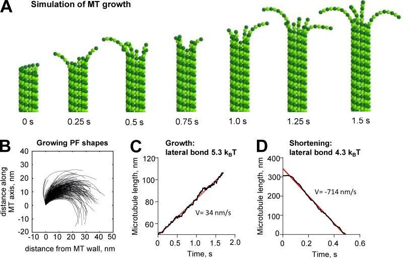
Figure 10.
MT growth with curved PFs illustrated with a Brownian dynamics model. (A) MT growth in a model with curved GTP-tubulin. (B) Shapes of simulated PFs on the growing MT tip. (C) Dependence of MT length o...
Figure images are served from the NIH/NLM PubMed Central Open Access Subset or Europe PMC; copyright remains with the publishers and authors.
💬 Discussion
0 commentsNo comments yet. Be the first to start a discussion!
Leave a Comment