Abstract
Neutrophils form the first line of host defense against bacterial pathogens. They are rapidly mobilized to sites of infection where they help marshal host defenses and remove bacteria by phagocytosis. While splenic neutrophils promote marginal zone B cell antibody production in response to administered T cell independent antigens, whether neutrophils shape humoral immunity in other lymphoid organs is controversial. Here we investigate the neutrophil influx following the local injection of Staphylococcus aureus adjacent to the inguinal lymph node and determine neutrophil impact on the lymph node humoral response. Using intravital microscopy we show that local immunization or infection recruits neutrophils from the blood to lymph nodes in waves. The second wave occurs temporally with neutrophils mobilized from the bone marrow. Within lymph nodes neutrophils infiltrate the medulla and interfollicular areas, but avoid crossing follicle borders. In vivo neutrophils form transient and long-lived interactions with B cells and plasma cells, and their depletion augments production of antigen-specific IgG and IgM in the lymph node. In vitro activated neutrophils establish synapse- and nanotube-like interactions with B cells and reduce B cell IgM production in a TGF-β1 dependent manner. Our data reveal that neutrophils mobilized from the bone marrow in response to a local bacterial challenge dampen the early humoral response in the lymph node.
🔬 Techniques
🔭 Microscopes
🧬 Organisms
✨ Fluorophores
🧪 Sample Preparation
🔬 Cell Lines
🏭 Microscope Brands
🧪 Reagent Suppliers
🔴 Lasers
📷 Detectors
💻 Software Details
📋 Protocols
💾 Data Repositories
🏛️ Research Organizations (ROR)
Affiliated research institutions:
📋 Methods
Mice
All animals were bred and housed under pathogen-free conditions and used according to the guidelines of the Animal Care and Use Committee (NIH). The LysM-GFP mice were provided by Dr. Hyeseon Cho and Dr. Ron Germain (NIAID) with permission from Dr. Thomas Graf (Center for Genomic Regulation, Barcelona, Spain). The BLIMP1-YFP mice were provided by Dr. David Fooksman (Skirball Institute of Biomolecular Medicine, New York, USA) with permission from Dr. Dimitris Skokos (The Rockefeller University, New York, USA). The C57BL/6 GFP-Lifeact mice were provided by Dr. Roland Wedlich-Soldne (Martinsried, Germany). DsRed mice (Jax Stock 006051) were received from Dr. Taha Bat and Dr. Cynthia Dunbar (NHLBI). CD45.1 C57BL/6 mice were purchased from the Jackson Laboratory (Bar Harbor, ME). DsRed and BLIMP1-YFP bone marrow chimeric mice were generated in animal facility within Comparative Medicine Branch (NIH/NIAID) as described in S1 Text . All targeted mouse genes are listed in S1 Table . All experiments were performed using sex and age matched animals, typically between 6 to 10 weeks old. S . aureus variants S . aureus LAC clone of strain USA300 (pulsed-field type USA300) was obtained from NARSA (Network on Antimicrobial Resistance in Staphylococcus aureus ). LAC-GFP clone was generated as a derivative of the USA300 LAC clone constitutively expressing genome-encoded GFP. For the integration of the gfp gene, whose DNA sequence was optimized for AT-rich Gram-positive bacteria ( gfpopt ), on the chromosome of LAC clone, first overlap extension PCR was used to create blaZ-gfpopt . The constitutively active beta-lactamase promoter was amplified from S . aureus N315 genomic DNA with primers BlaZFw (ATGCGGATCCCTAACAATAGAAATATAAAACAAAAGC) and BlaZ-GfpRv (AATTCTTCTCCTTTTGACATAATAAACCCTCCGATATTAC) and gfp-opt was amplified from plasmid pSW4-GFPopt [ 54 ] with BlaZ-GFPFw (GTAATATCGGAGGGTTTATTATGTCAAAAGGAGAAGAATT) and GFPRv (ATGCCTGCAGTTACTTATATAATTCATCCAT). A fusion product of the two PCR fragments was amplified with primers BlaZFw and GFPRv and cloned into plasmid pLL29 [ 55 ] BamHI and SalI restriction sites, resulting in plasmid (pLL29- blaZ-gfpopt ). Plasmid pLL29- blaZ-gfpopt was phage-transduced into LAC clone as described previously [ 55 ]. The integration of pLL29- blaZ-gfpopt into the ϕ11 attachment site of LAC clone was confirmed using primers scv4 (ACCCAGTTTGTAATTCCAGGAG) paired with scv10 (TATACCTCGATGATGTGCATAC) and primer scv8 (GCACATAATTGCTCACAGCCA) paired with scv9 (GCTGATCTAACAATCCAATCCA).
Show full methods section
Mice
All animals were bred and housed under pathogen-free conditions and used according to the guidelines of the Animal Care and Use Committee (NIH). The LysM-GFP mice were provided by Dr. Hyeseon Cho and Dr. Ron Germain (NIAID) with permission from Dr. Thomas Graf (Center for Genomic Regulation, Barcelona, Spain). The BLIMP1-YFP mice were provided by Dr. David Fooksman (Skirball Institute of Biomolecular Medicine, New York, USA) with permission from Dr. Dimitris Skokos (The Rockefeller University, New York, USA). The C57BL/6 GFP-Lifeact mice were provided by Dr. Roland Wedlich-Soldne (Martinsried, Germany). DsRed mice (Jax Stock 006051) were received from Dr. Taha Bat and Dr. Cynthia Dunbar (NHLBI). CD45.1 C57BL/6 mice were purchased from the Jackson Laboratory (Bar Harbor, ME). DsRed and BLIMP1-YFP bone marrow chimeric mice were generated in animal facility within Comparative Medicine Branch (NIH/NIAID) as described in S1 Text . All targeted mouse genes are listed in S1 Table . All experiments were performed using sex and age matched animals, typically between 6 to 10 weeks old. S . aureus variants S . aureus LAC clone of strain USA300 (pulsed-field type USA300) was obtained from NARSA (Network on Antimicrobial Resistance in Staphylococcus aureus ). LAC-GFP clone was generated as a derivative of the USA300 LAC clone constitutively expressing genome-encoded GFP. For the integration of the gfp gene, whose DNA sequence was optimized for AT-rich Gram-positive bacteria ( gfpopt ), on the chromosome of LAC clone, first overlap extension PCR was used to create blaZ-gfpopt . The constitutively active beta-lactamase promoter was amplified from S . aureus N315 genomic DNA with primers BlaZFw (ATGCGGATCCCTAACAATAGAAATATAAAACAAAAGC) and BlaZ-GfpRv (AATTCTTCTCCTTTTGACATAATAAACCCTCCGATATTAC) and gfp-opt was amplified from plasmid pSW4-GFPopt [ 54 ] with BlaZ-GFPFw (GTAATATCGGAGGGTTTATTATGTCAAAAGGAGAAGAATT) and GFPRv (ATGCCTGCAGTTACTTATATAATTCATCCAT). A fusion product of the two PCR fragments was amplified with primers BlaZFw and GFPRv and cloned into plasmid pLL29 [ 55 ] BamHI and SalI restriction sites, resulting in plasmid (pLL29- blaZ-gfpopt ). Plasmid pLL29- blaZ-gfpopt was phage-transduced into LAC clone as described previously [ 55 ]. The integration of pLL29- blaZ-gfpopt into the ϕ11 attachment site of LAC clone was confirmed using primers scv4 (ACCCAGTTTGTAATTCCAGGAG) paired with scv10 (TATACCTCGATGATGTGCATAC) and primer scv8 (GCACATAATTGCTCACAGCCA) paired with scv9 (GCTGATCTAACAATCCAATCCA).
Expression of GFPOPT in USA300
LAC clone was confirmed by fluorescence microscopy (excitation/emission at 470 ± 20 nm/ 515 nm, respectively). LAC S . aureus USA300 LAC spa (an isogenic protein A mutant) was a kind gift from Prof. A. Prince (Columbia University, New York, NY). Bacteria culture and CFU counts Glycerol stocks of S . aureus USA300-derivative LAC-GFP were grown to mid-exponential growth phase (for min of 2 h) in 50 ml of TSB at 37 °C with shaking at 180 rpm. Bacteria were harvested and washed and resuspended in sterile PBS prior to injections. CFU counts from infected LNs were performed as described [ 56 ]. Shortly, mice were euthanized and LNs were harvested. One LN of each mouse was placed into a 2-ml tube containing 1 ml of sterile PBS with 500 mg of 2 mm borosilicate glass beads (Sigma). The LNs were homogenized in a Fast Prep bead beater (Thermo Savant) at 6 m/s for 20 s. The homogenates were diluted in PBS, plated onto TSB plates, and incubated overnight at 37°C for CFU counting.
Antigens and immunization
CFA containing heat killed M . tuberculosis (H37Ra), 0.85 mL paraffin oil and 0.15 mL mannide monooleate (Sigma Aldrich) was injected in amount of 50 μl per iLN. S . aureus Bioparticles were purchased from Life Sciences (Molecular Probes, Cat #. S2851, S23371 , S23372 ). The reagent consists of heat-killed bacteria Wood 46 strain without protein A, unlabeled or fluorescently labeled with either Alexa Fluor 488 or Alexa Fluor 594. Alexa Fluor dyes of S . aureus Bioparticle conjugates are bound to the surface of bacterial cells, but not internalized. Prior to use, the Bioparticles were coated with S . aureus opsonizing reagent that contains rabbit polyclonal IgG antibodies specific for S . aureus and RIA-grade bovine serum albumin to block nonspecific binding (Molecular Probes, Life Sciences, Cat #. S2860). Opsonized S . aureus bioparticles were injected in amount of 2 x 10 7 bacterial particles per mouse (1 x 10 7 per iLN). NP-KLH (Biosearch Technologies) precipitated in Alum (Thermo Scientific) was given at amount of 25 μg per iLN. Sheep red blood cells (SRBC, Lonza) were administered in amount of 1 x 10 8 cells per iLN. S . aureus USA300-derivative LAC-GFP was injected at 1–3 x 10 5 CFU per mouse (0.5–1.5 x 10 5 CFU per iLN) in 200 μL of sterile PBS. All antigens were injected subcutaneously within 1 cm of the adjacent iLN ( S1A Fig ). Kinetics of neutrophil recruitment Neutrophil influx to the iLN was monitored using TP-LSM or confocal microscopy between 0 and 24 h; and in the iLN, blood, draining LNs and spleen using flow cytometry between 0 to 24 h, or days 1 to 5 after immunization. Whole blood was collected from mouse tails (tail snip), and iLN cells or splenocytes were isolated from euthanized mice. LysM-GFP cells were analyzed directly, and C57BL/6 cells were immunostained using fluorochrome conjugated anti-Ly6G/ (clone RB6-8C5),-B220,-CD11c,-GL7,-Fas,-CD4,-CD8,-CD169, and—F4/80 antibody (eBioscience). Flow cytometry was performed using FACS Canto II flow cytometer with FACS Diva 6.2 software (BD Biosciences).
Cell isolation and adoptive transfer
Immune cell subsets were isolated using magnetic negative selection system with dynabeads M-280 streptavidin and magnetic particle concentrator (Invitrogen) as previously described [ 57 ]. BM derived neutrophils were isolated from mouse femur and tibia using anti B220,-CD38,-CD138,-CD11c,-CD4 and-CD8; and B cells were isolated from mouse spleen by using anti CD11c,-Gr-1,-CD4 and-CD8 biotinylated antibody (BD Pharmingen). Isolated cells were cultured in complete lymphocyte medium (DMEM supplemented with 10% FBS, 25 mM HEPES, 50 μM β-ME, 1% Pen/Strep/L-Glu and 1% Sodium Pyruvate) in humidified CO 2 incubator. Cells were allowed to recover for 30 min, and labeled with CellTracker™ Blue, CMAC, CellTracker™ Green, CFDMA or CellTracker™ Red, CMTPX (Molecular Probes, Invitrogen) according to the manufacturer’s protocol. Labeled cells were administered intravenously into recipient mice (5 x 10 6 cells per mouse) via tail vein injection, and imaged between 16 and 24 h later.
Confocal microscopy Isolated mouse
LNs were sliced into 250 μm sections using Leica VT1000 S Vibrating Blade Microtome (Leica Microsystems). Live cell imaging of immunostained sections was performed using Leica SP8 inverted 5 channel confocal microscope equipped with a motorized stage and 2 HyD ultra-sensitive detectors (Leica Microsystems). Images of whole LNs were tiled using Leica Application Suite (Leica Microsystems) and processed using Imaris (Bitplane) software. For live cell imaging, BM-isolated neutrophils and LN-isolated B cells were cultured for 2 h on ICAM-1 + VCAM-1 + KC (Recombinant Mouse ICAM-1/CD54 Fc Chimera, CF; Recombinant Mouse VCAM-1/CD106 Fc Chimera, Recombinant Mouse CXCL1/KC CF; R&D Systems) coated glass-bottom dishes (No 1.5 coverglass; MatTek). Live cells were stained in complete medium with fluorescently labeled anti-Ly6G and anti-B220 correspondingly (BD Pharmingen). Confocal imaging was performed using Leica SP8 equipped with incubation chamber (CO 2 , 37°C) for live cell imaging (Pecon). Images were processed using Imaris (Bitplane) software. Detailed description of confocal microscopy setup is provided in supporting information ( S2 Text ).
Epifluorescent microscopy
Immunized mice were injected intravenously with 1% EB solution in PBS (Evans blue dye, Sigma Aldrich) at 1 ml/kg. Mice were euthanized, iLNs exposed on a skin flip, kept moisturized with PBS and imaged immediately after exposure. Fluorescent and bright field images of intact mouse iLNs were collected using motorized stereomicroscope Leica M205 (Leica Microsystems) equipped with 1x objective. GFP/YFP were excited at 488 nm and EB at 561 nm. Images were processed using Leica Application Suite (Leica Microsystems) and Imaris (Bitplane) software. Intravital two-photon laser scanning microscopy (TP-LSM) All imaging experiments were performed at Biological Imaging Section (NIH, NIAID) using Leica SP5 inverted confocal microscope (Leica Microsystems) equipped with dual Mai Tai lasers as previously described [ 57 ]. Mouse surgery for imaging the iLN was performed according to the Cold Spring Harbor protocol [ 58 ] modified for the inverted microscope setup. For imaging neutrophil recruitments from the BM mice were injected intravenously with KC+AMD3100 (AMD 3100 octahydrochloride; Recombinant Mouse CXCL1/KC CF; R&D Systems). Mouse calvarium BM was imaged as described [ 42 ], using upright microscope setup and a custom-made stage with the head holder (NIH Division of Scientific Equipment and Instrumentation Services). Post-acquisition image processing was performed using ImageJ (National Institutes of Health), Imaris (Bitplane) and Huygens (Scientific Volume Imaging) software. Detailed description of the imaging technique is provided in supporting information ( S3 Text ).
Neutrophil depletion and ELISA with LN derived B cells
Neutrophils were depleted in vivo as described [ 59 ] using anti Ly6G functional grade antibody 1A8 (eBioscience/BioLegend). Briefly, animals were injected intraperitoneally with 100 μg of isotype control rat immunoglobulin G (eBioscience/BioLegend) or 1A8 antibody at days -1, 0 and 1 of immunization. Efficiency of depletion was monitored by flow cytometry analysis or by TP-LSM, and typically represented > 90% in blood, spleen and draining LNs. At days 5 to 7 of immunization (6 to 8 of depletion) mice were sacrificed and the iLNs harvested. B cells isolated from a single iLN were cultured in complete lymphocyte medium for 72 h, and the supernatants were collected. Antibody concentration in the supernatants was measured with commercial ELISA kits (Mouse IgG total ELISA Ready-SET-Go, Mouse IgM total ELISA Ready-SET-Go; eBioscience) according to the manufacturer's protocol. LAC-specific IgG and IgM were measured using plates coated with bacterial lysates. Total lysates from LAC and LAC spa were prepared as previously described [ 60 ] with the following modifications. Bacteria were grown as above, pellets were resuspended in 1 ml of sterile PBS and incubated 30 minutes at 37 °C in the presence of Halt protease inhibitor single use cocktail (Thermo Scientific) and lysostaphin. The digested lysates were transferred to 2-ml Lysing Matrix B vials (MPbio) and homogenized in a Fast Prep bead beater (Thermo Savant) at 6 m/s for 20 s. Protein concentrations were determined with the Quant-iT assay kit (Life Technologies). IgG and IgM specific for NP-KLH were measured by ELISA using plates coated with NP-KLH. For antigen-specific ELISAs plates were coated at protein concentrations 10 μg/mL (LAC or LAC spa lysates) and 1 μg/mL (NP-KLH) in PBS overnight and blocked with 1% BSA in PBS. Neutrophil and B cell activation in vitro Neutrophils were isolated from the BM and cultured in for 24 h in presence of 2 μg/mL LPS (from E. coli, Serotype R515 (Re), TLR grade, ENZO Life Sciences) or 1 x 10 6 particles/mL S . aureus bioparticles. Supernatants were added to freshly isolated LN B cells. Isolated iLN B cells were cultured in complete lymphocyte medium at initial concentration between 5 x 10 5 to 2 x 10 6 cells/mL. B cells and neutrophils were added to the cultures in ratio 10 B cells: 1 neutrophil, for 5 days in presence of LPS or S . aureus bioparticles. Alternatively, supernatants from either activated or non-activated neutrophil cultures (25% of culture medium) were added to B cells. Final concentration of LPS was 2 μg/mL, and S . aureus 1 x 10 6 particles/mL in all cultures. Neutralizing anti TGF-β1 (TGF-beta 1/1.2 Polyclonal antibody; R&D Systems) were added at final concentration 1 μg/mL, and TGF-β1 (50 ng/mL). TGF-β1 in neutrophil and B cell cultures was measure using commercial ELISA kit (eBioscience). IgM and IgA levels in the supernatants were measured with commercial ELISA kits (eBioscience) according to the manufacturer's protocol.
Ethics statement
The animal experiments and protocols were performed according to the regulations of NIAID Division of Intramural Research Animal Care and Use Committee (DIR ACUC). Animal Study Proposal LIR 16 entitled “Analysis of Innate Immune Function in Mice” that covers this work was approved by the NIAID DIR ACUC on Dec 1 st , 2011 as the initiation date, and has been reviewed annually. The NIAID DIR ACUC as a part of the NIAID DIR Animal Care and Use Program, as part of the NIH Intramural Research Program (IRP), complies with all applicable provisions of the Animal Welfare Act and other Federal statutes and regulations relating to animals; and is guided by the "U.S.
Government Principles for the Utilization and Care of Vertebrate Animals
Used in Testing, Research, and Training". The policies, procedures and guidelines for the NIAID IRP are explicitly detailed in NIH Policy Manual 3040–2, “Animal Care and Use in the Intramural Program” (PM 3040–2) and the NIH Animal Research Advisory Committee Guidelines (ARAC Guidelines) that are posted on the NIH Office of Animal Care and Use public website at: http://oacu.od.nih.gov .
Statistical analysis
The statistical significance was evaluated by subjecting the data to a Student's t-test using GraphPad software. Values are presented as means ± SD or means ± SEM as indicated. *, P
📊 Figures
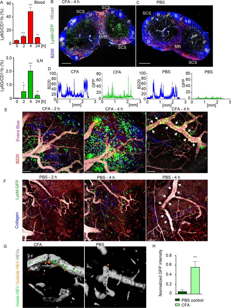
Fig 1
Neutrophils arrive from HEVs to occupy IFZ, MR and SCS in immunized iLN.
Mice were injected subcutaneously with CFA. C57BL/6 mice were subjected to flow cytometry analysis of whole blood and LN cell populations; LysM-GFP expression was imaged using confocal microscopy or T...
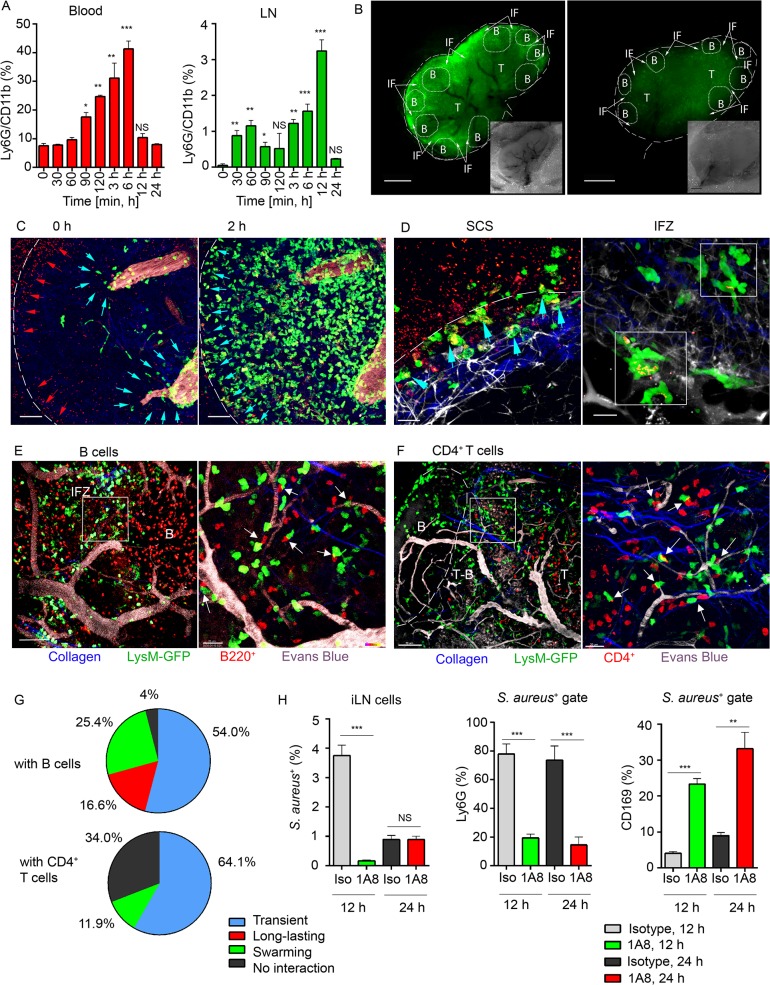
Fig 2
Local immunization with S . aureus bioparticles recruits neutrophils to the LN.
S . aureus bioparticles were opsonized and injected subcutaneously near the iLN. Neutrophil recruitment to the blood and lymphoid organs was analyzed by flow cytometry in C57BL/6 mice, or imaged using...
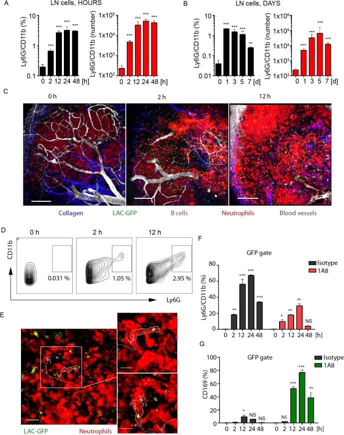
Fig 3
Local LAC-GFP infection recruits neutrophils to the iLN.
C57BL/6 or dsRed BM chimeric mice were injected subcutaneously near the iLN with LAC-GFP in amount of 1 x 10 5 CFU per iLN or given PBS as a control. Neutrophil recruitment to the iLN was analyzed by ...
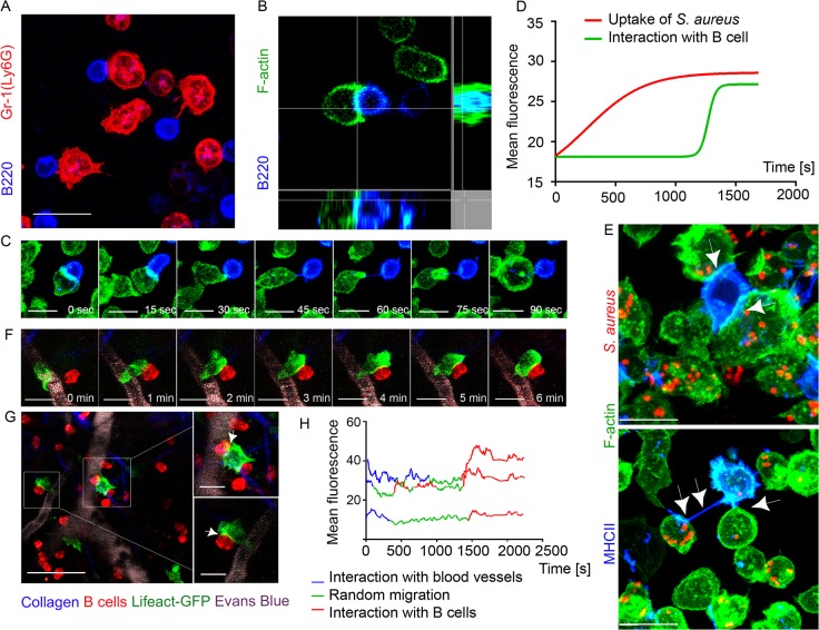
Fig 4
F-actin accumulates at interaction sites between neutrophils and B cells.
Lifeact-GFP neutrophils and B cells were co-cultured on ICAM-1+VCAM-1+KC coated surface and imaged using confocal microscopy. Lifeact-GFP neutrophils and dsRed B cells were adoptively transferred into...
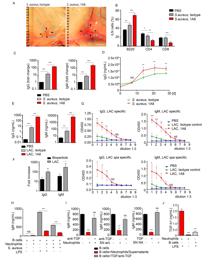
Fig 5
Neutrophils limit the humoral response following local immunization or infection.
C57BL/6 mice were given PBS as a baseline control, immunized with S . aureus bioparticles, or infected with LAC-GFP. Isotype control or 1A8 antibody was injected intraperitoneally at 100 u03bcg/mouse ...
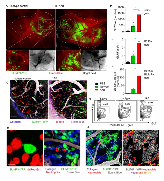
Fig 6
Neutrophil depletion results in increased population of BLIMP1-YFP+ and GC B cells in immunized iLN.
BLIMP1-YFP BM reconstituted mice were immunized with S . aureus subcutaneously, and injected with isotype control antibody or depleted of neutrophils using 1A8 antibody at days -1, 0 and 1 of immuniza...
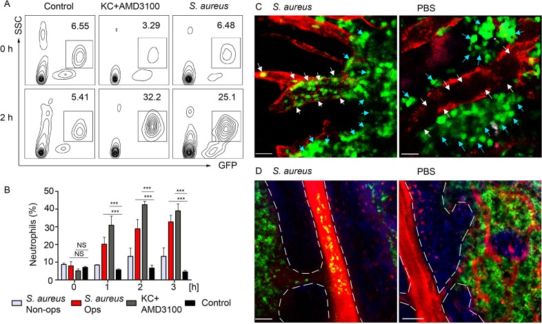
Fig 7
BM neutrophils are mobilized to the blood stream after local immunization.
LysM-GFP mice were injected with S . aureus bioparticles near the iLN. Neutrophil mobilization was analyzed using flow cytometry of the whole blood or TP-LSM of the calvarium bone marrow. Anti VE-cadh...
Figure images are served from the NIH/NLM PubMed Central Open Access Subset or Europe PMC; copyright remains with the publishers and authors.
💬 Discussion
0 commentsNo comments yet. Be the first to start a discussion!
Leave a Comment