Abstract
AbstractThe development, refinement, and use of techniques that allow high-throughput imaging of whole brains with cellular resolution will help us understand the complex functions of the brain. Such techniques are crucial for the analysis of complete neuronal morphology—anatomical and functional—connectivity, and repeated molecular phenotyping. CLARITY is a recently introduced technique that produces structurally intact, yet optically transparent tissue, which may be labeled and imaged without sectioning. However, the utility of this technique depends on several procedural variables during the process in which the light-scattering lipids in a tissue are replaced by a transparent hydrogel matrix. Here, we systematically varied a number of factors (including temperature, hydrogel composition, and polymerization conditions) to provide an optimized, highly replicable CLARITY procedure for clearing mouse brains. We found that for these preparations optimal tissue clearing requires electrophoresis (and cannot be achieved with passive clearing alone) for 5 d with a combination of 37 and 55°C temperature. Although this protocol is optimized for brains, we also show that it can be used to clear and analyze a variety of organs. Brain or other tissue prepared using this protocol is suitable for high-throughput imaging with confocal or single-plane illumination microscopy.
🔬 Techniques
🔭 Microscopes
💻 Software
✨ Fluorophores
🧪 Sample Preparation
🏭 Microscope Brands
🧪 Reagent Suppliers
💻 Software Details
🏛️ Research Organizations (ROR)
Affiliated research institutions:
📋 Methods
All of the necessary components and suppliers for performing the procedure can be found in Table 1 . Table 1 List of equipment and reagents Incubating orbital shaker VWR 97027-346 Vibrating tissue slicer Leica VT 1200S Recirculating water bath VWR 89203-010 Electrophoresis power supply Bio-Rad PowerPac HC Light sheet microscope LaVision Biotec UltraMicroscope Laser-scanning confocal microscope Zeiss LSM 710 Wide-field epifluorescent microscope Nikon Eclipse 80i Vacuum canister McMaster Carr 2204K7 SDS Sigma L3771 Boric acid Sigma B6768 Paraformaldehyde Sigma 441244 Acrylamide solution (40%) Bio-Rad 161-0140 Bis-acrylamide solution (2%) Bio-Rad 161-0142 VA-044 WAKO 877-714-1920 Glycerol Sigma G5516 Triton-X 100 Sigma T8787 Sodium azide Sigma S2002 10 N NaOH Sigma 656054 Sodium phosphate monobasic anhydrous Sigma S8282 FocusClear CelExplorer Labs FC-102 Nitrogen gas Praxair Propidium iodide Life Technologies P3566 Clear tubing (PVC, chemical resistant) McMaster Carr 5103K36 Clear tubing (PVC, chemical resistant) McMaster Carr 5103K34 Barbed tube fittings (polypropylene) McMaster Carr 53415K207 Barbed tube fittings (polypropylene) McMaster Carr 5463K245 Barbed tube fittings (polypropylene) McMaster Carr 5463K2458 Barbed tube fittings (polypropylene) McMaster Carr 5463K2457 Barbed tube fittings (polypropylene) McMaster Carr 5117K51 Manifold (polypropylene) McMaster Carr 5364K231 Manifold plugs (polypropylene) McMaster Carr 4515K209 Quick disconnect barbed plug McMaster Carr 5154K69 Quick disconnect barbed socket McMaster Carr 51545K63 Filter case McMaster Carr 4448K35 Filter cartridge McMaster Carr 4422K61 3M Scotch-weld Epoxy DP270 McMaster Carr 7467A17 Platinum wire Alfa Aesar 10286 Nalgene 60 ml wide-mouth jar VWR 36319-547 Cell strainer Fisher 22363548 Paraffin embedding cassette VWR 18000-000 Mice Adult (6-8 week) WT female mice in a hybrid (F1) genetic background were used for most studies (C57BL/6NTac × 129S6/SvEvTac). In some studies, we also used male and female activity-regulated cytoskeleton-associated protein-targeted recombination (ArcTRAP) in active populations of mice ( Guenthner et al., 2013 ; Jax, B6.129(Cg)- Arc tm1.1(cre/ERT2)Luo /J) crossed with a fluorescent reporter strain. In the ArcTRAP mice, the tamoxifen-inducible CreER T2 protein is driven by endogenous promoter/enhancer elements of the activity-dependent immediate early gene Arc ( Lyford et al., 1995 ). ArcTRAP mice were crossed with a reporter mouse (Jax, B6.Cg- Gt(ROSA)26Sor tm14(CAG-tdTomato)Hze / J) in which loxP sequences flank a STOP cassette upstream of a tdTomato reporter. In this way, following systemic tamoxifen injection, cells that are sufficiently active will show Cre-mediated recombination, thereby removing the STOP cassette and allowing expression of tdTomato. A 10 mg/ml solution of 4-hydroxytamoxifen was prepared by first dissolving the drug in 100% ethanol and then suspending the solution in sunflower oil. Mice were injected with a dose of 20 mg/ml. Genotypes were determined by PCR analysis of tail DNA samples as previously described ( Guenthner et al., 2013 ). Mice were bred in-house and group housed (3-5 mice per cage) on a 12 h light/dark cycle with food and water available ad libitum . Behavioral experiments were conducted during the light phase of the cycle. All procedures were conducted in accordance with the policies of the institutional Animal Care and Use Committee and conformed to the federal guidelines on the care and use of laboratory animals.
Show full methods section
All of the necessary components and suppliers for performing the procedure can be found in Table 1 . Table 1 List of equipment and reagents Incubating orbital shaker VWR 97027-346 Vibrating tissue slicer Leica VT 1200S Recirculating water bath VWR 89203-010 Electrophoresis power supply Bio-Rad PowerPac HC Light sheet microscope LaVision Biotec UltraMicroscope Laser-scanning confocal microscope Zeiss LSM 710 Wide-field epifluorescent microscope Nikon Eclipse 80i Vacuum canister McMaster Carr 2204K7 SDS Sigma L3771 Boric acid Sigma B6768 Paraformaldehyde Sigma 441244 Acrylamide solution (40%) Bio-Rad 161-0140 Bis-acrylamide solution (2%) Bio-Rad 161-0142 VA-044 WAKO 877-714-1920 Glycerol Sigma G5516 Triton-X 100 Sigma T8787 Sodium azide Sigma S2002 10 N NaOH Sigma 656054 Sodium phosphate monobasic anhydrous Sigma S8282 FocusClear CelExplorer Labs FC-102 Nitrogen gas Praxair Propidium iodide Life Technologies P3566 Clear tubing (PVC, chemical resistant) McMaster Carr 5103K36 Clear tubing (PVC, chemical resistant) McMaster Carr 5103K34 Barbed tube fittings (polypropylene) McMaster Carr 53415K207 Barbed tube fittings (polypropylene) McMaster Carr 5463K245 Barbed tube fittings (polypropylene) McMaster Carr 5463K2458 Barbed tube fittings (polypropylene) McMaster Carr 5463K2457 Barbed tube fittings (polypropylene) McMaster Carr 5117K51 Manifold (polypropylene) McMaster Carr 5364K231 Manifold plugs (polypropylene) McMaster Carr 4515K209 Quick disconnect barbed plug McMaster Carr 5154K69 Quick disconnect barbed socket McMaster Carr 51545K63 Filter case McMaster Carr 4448K35 Filter cartridge McMaster Carr 4422K61 3M Scotch-weld Epoxy DP270 McMaster Carr 7467A17 Platinum wire Alfa Aesar 10286 Nalgene 60 ml wide-mouth jar VWR 36319-547 Cell strainer Fisher 22363548 Paraffin embedding cassette VWR 18000-000 Mice Adult (6-8 week) WT female mice in a hybrid (F1) genetic background were used for most studies (C57BL/6NTac × 129S6/SvEvTac). In some studies, we also used male and female activity-regulated cytoskeleton-associated protein-targeted recombination (ArcTRAP) in active populations of mice ( Guenthner et al., 2013 ; Jax, B6.129(Cg)- Arc tm1.1(cre/ERT2)Luo /J) crossed with a fluorescent reporter strain. In the ArcTRAP mice, the tamoxifen-inducible CreER T2 protein is driven by endogenous promoter/enhancer elements of the activity-dependent immediate early gene Arc ( Lyford et al., 1995 ). ArcTRAP mice were crossed with a reporter mouse (Jax, B6.Cg- Gt(ROSA)26Sor tm14(CAG-tdTomato)Hze / J) in which loxP sequences flank a STOP cassette upstream of a tdTomato reporter. In this way, following systemic tamoxifen injection, cells that are sufficiently active will show Cre-mediated recombination, thereby removing the STOP cassette and allowing expression of tdTomato. A 10 mg/ml solution of 4-hydroxytamoxifen was prepared by first dissolving the drug in 100% ethanol and then suspending the solution in sunflower oil. Mice were injected with a dose of 20 mg/ml. Genotypes were determined by PCR analysis of tail DNA samples as previously described ( Guenthner et al., 2013 ). Mice were bred in-house and group housed (3-5 mice per cage) on a 12 h light/dark cycle with food and water available ad libitum . Behavioral experiments were conducted during the light phase of the cycle. All procedures were conducted in accordance with the policies of the institutional Animal Care and Use Committee and conformed to the federal guidelines on the care and use of laboratory animals.
Hydrogel solution preparation
The hydrogel solution used to perfuse the tissue is composed of four key reagents: acrylamide, bis-acrylamide, formaldehyde, and a thermal initiator (VA-044) in PBS. Different strengths of hydrogels can be produced by varying the concentrations of these reagents. We began with the original hydrogel solution (referred to here as 4:4:0.05) containing final concentrations of 4% acrylamide, 0.05% bis-acrylamide, and 4% formaldehyde. We first prepared a 16% formaldehyde solution by adding 80 g of paraformaldehyde powder to 400 ml of dH 2 O and heated the solution to 60-65°C. The solution was cleared with several drops of 10 N NaOH and the volume was filled to 500 ml. Finally, we filtered the solution and cooled to 4°C. Next, we combined 40 ml of cold 40% acrylamide, 10 ml of cold 2% bis-acrylamide, 40 ml of 16% formaldehyde, 40 ml of 10× PBS, and 220 ml of cold dH 2 O and, finally, 1 g of VA-044 thermal initiator. The solution was kept on ice during preparation and was either used immediately after preparation or divided into 40 ml aliquots and frozen at −20°C for later use. Modified hydrogel solutions were made following the same protocol but with 3% final concentrations of acrylamide and formaldehyde or 0.025% bis-acrylamide.
Tissue collection for brain samples
Mice were transcardially perfused with ice-cold PBS followed by 25 ml of cold hydrogel solution. The brains were rapidly extracted and submersed in cold hydrogel solution and postfixed in the dark at 4°C for 24 h in 10 ml of the same hydrogel solution. Tissue polymerization To ensure consistent hydrogel polymerization, we removed oxygen from the hydrogel solution by placing the sample vials in a vacuum chamber for 30 min. After vacuuming, the chamber was flooded with pure nitrogen gas to displace oxygen as the chamber was opened and the lids placed on the sample vials. Polymerization of the hydrogel solution depends on the VA-044 thermal initiator, which is triggered by tissue warmed to 37°C. Therefore, we placed the sealed sample tubes in a rotating incubator at 37°C for 3 h. Once the hydrogel was polymerized, excess gel was removed from the brain by gently wiping the gel from the surface with a Kimwipe. We then placed the cleaned brain into a new 50 ml tube containing clearing solution (4% SDS in 0.2 m borate buffer) overnight to wash residual hydrogel solution out of the tissue. The tissue-clearing solution was prepared by dissolving 61.83 g of boric acid and 200 g of SDS in 4.5 L of dH 2 O. The final pH was adjusted to 8.5 using 10 N NAOH.
Active tissue clearing
We used a modular multichamber design that allowed six brains to be cleared simultaneously using a single recirculator ( Fig. 1 A ). Electrophoretic tissue-clearing (ETC) components were connected in parallel to the recirculator with temperature- and chemical-resistant tubing. Clearing solution from the recirculator was first passed through a filter cartridge and then into a hub that divided the solution into six tubing lines connected to the ETC chambers that in turn were connected to another hub that connects back to the recirculator. Power supplies were connected to the platinum leads of the ETC chambers with alligator clips. Small pieces of tissue (such as 2 mm sections or dissected brain regions) were placed into a closed paraffin embedding cassette. Larger tissues were placed into a mesh cell strainer. The cell strainer or cassette was then placed between the electrodes and shielded from direct contact with the electrodes by two sheets of plastic mesh. Figure 1 Schematic of electrophoresis and imaging components. Schematic of the electrophoresis system used for active clearing of tissue. A , A recirculating water bath filled with clearing solution is connected via chemical- and heat-resistant tubing to a filter unit (which removes particulate matter and reduces bubbles in the clearing solution). The clearing solution is then split into six parallel tube lines, each containing one ETC chamber. The fluid output of the chambers is then recombined into a single line before entering the recirculator. Power supplies provide current to each ETC chamber. Schematic of the LaVision Biotec UltraMicroscope ( B ) compared with confocal microscope ( C ). The light sheet design permits high-speed image capture of large tissue volumes. In contrast, a confocal microscope may be used for high-resolution imaging of CLARITY tissue, although the acquisition rate is much slower. Various settings were used for tissue clearing. We have experienced difficulties in regulating the temperature and, as a result, maintaining tissue quality if the clearing voltage is too high or if the recirculator flow rate is too low. Therefore, in all conditions we used a low voltage of 20 V and a high flow rate of 30 L/min (5 L/min/ETC chamber). Under these conditions, the temperature of the clearing solution measured between the electrodes did not fluctuate from the set point. We then varied the temperature between 21 and 55°C and length of clearing from 1 to 5 d to produce the best combination of rigid and clear tissue. The pH of the clearing solution was monitored daily and replaced once the pH dropped from 8.5 to 8.0. After ETC the tissue was rinsed in PBST for several days.
Passive tissue clearing
For experiments in which tissue was passively cleared we used the same clearing solution and kept the samples on a shaker at 21°C, 37°C, or 55°C. The clearing solution was exchanged every 3 d for up to 2 months. The tissues were then rinsed in several changes of PBST for several days.
Tissue labeling
Propidium iodide labeling was performed by incubating the tissue for 24-48 h at 37°C in a 1:2000 dilution of propidium iodide in 0.1 m PBST. Immunohistochemistry was performed using 1:100 dilutions of antibody in PBST and incubating the tissues for 7 d at 37°C for the primary antibody and an additional 7 d for the secondary antibody. The tissue was rinsed in 0.1 m PBST after each antibody incubation. The antibodies used were mouse monoclonal anti-smooth muscle actin (Santa Cruz Biotechnology; catalog #SC-53142), rabbit polyclonal anti-collagen type IV (Millipore; catalog #AB756P), rabbit polyclonal anti-CD34 (Santa Cruz Biotechnology; catalog #SC-9095), goat anti-mouse Alexa 488 (Life Technologies; catalog #A11001), and goat anti-rabbit Alexa 488 (Life Technologies; catalog #A11034). Refractive index matching After the PBST washes, the tissue was transferred to either 80% glycerol or FocusClear. The tissue was left to equilibrate in the refractive index matching solution for 24 h at 37°C for glycerol or at room temperature for FocusClear. Finally, the samples were then mounted in the same medium for imaging.
Imaging setup
In principle, any type of fluorescent microscope can be used to image CLARITY-cleared tissue. However, the microscope must be able to physically accommodate the size of the sample and the stage translation, and the working distance of the microscope objective should be appropriate. A microscope should also allow images to be acquired in a practical length of time. For these reasons, we routinely used light sheet microscopy (LaVision BioTec UltraMicroscope; Fig. 1 B ) for acquisition of large tissues. We also utilized a laser-scanning confocal microscope (Zeiss LSM710; Fig. 1 C ) equipped with 10× NA 0.3 and 25× NA 0.8 objectives and an Epifluorescent microscope (Nikon Eclipse 80i) with a 10× NA 0.3 objective Image analysis We used a Lenovo ThinkStation D30 with 2× Intel Xeon 3.1 GHz processors, a QuadK5000 4 GB graphics card, and 128 GB of RAM. For the light sheet microscope, images were acquired using the ImSpector software suite (LaVision Biotec), confocal images were acquired using Zen (Zeiss), and wide-field images were acquired with NIS Elements (Nikon). Image processing was then performed using a combination of software packages including Imaris (Bitplane) for 3D reconstruction, ImageJ for 2D stitching and basic adjustments, and AutoQuant (Media Cybernetics) for deconvolution.
Statistical analysis
Data shown in Figures 2 and 3 represent the mean ± SEM. Analyses were conducted using two-way ANOVAs and Newman–Keuls post hoc tests as described in Table 2 . Figure 2 Active clearing of brains with ETC is more efficient than passive clearing. A , Passive clearing is temperature dependent with very little clearing at 21°C occurring after 1 month in SDS. At 37 and 55°C the brain becomes progressively clearer after 1 month in SDS but does not get as clear as with active (ETC) clearing. B , Relative to freshly fixed and uncleared tissue, protein content is well preserved following both active and passive clearing methods, regardless of temperature used. Data are shown relative to brains that were perfused and polymerized with the same hydrogel solution but were not cleared. Olfactory bulb tissue from each clearing condition was homogenized and a BCA assay was performed to estimate protein concentration. There were no significant differences in protein concentration as an effect of active versus passive clearing ( p = 0.4) or clearing temperature ( p = 0.24). Figure 3 Clearing conditions for optimal transparency. A , Comparison of ETC temperature and hydrogel composition on tissue transparency and expansion. There were significant main effects of clearing temperature ( F (2,24) = 178.9, p < 0.00001) and hydrogel concentration ( F (3,24) = 16.2, p < 0.00001) on transparency Tissue appears clearest with 37/55°C ETC and 3% acrylamide, 3% formaldehyde, and 0.025% hydrogel. Tissue expansion is less in the 37/55°C condition compared with either 37°C or 55°C alone. B , Both 55 and 37/55°C produce tissue that is more transparent than 37°C, regardless of hydrogel composition ( p s = 0.0002 and 0.0009, respectively). Transmission was measured on a light table under standardized illumination conditions. A reference measurement was made without a sample and was set to 100%. The sample was then imaged on the light table and the percentage light intensity through the specimen was recorded as a percentage of the reference value. C , Brains cleared with the combined 37/55°C clearing protocol expand more in lower concentration hydrogel (3% formaldehyde:3% acrylamide:0.025% bis-acrylamide compared with higher concentration hydrogel (4% formaldehyde:4% acrylamide:0.05% bis-acrylamide). The conditions for producing an optimal combination of stable and clear tissue is to polymerize the tissue with hydrogel composed of 4% acrylamide, 4% formaldehyde, and 0.05% bis-acrylamide and then clear the tissue with 5 d of ETC (4 d at 37°C and 1 d at 55°C). Table 2: Data analysis Data Test n Main effect F P BCA analysis Main effects ANOVA Three per group Active versus passive temperature 0.791.68 0.400.24 Transparency Two-way ANOVA Three per group HydrogelTemperatureInteraction 16.20178.900.70 0.0000060.0000000.63 Newman–Keuls Post hoc Three per group Hydrogel 3:3:0.025 versus 3:3:0.05 3:3:0.025 versus 4:4:0.025 3:3:0.025 versus 4:4:0.05 3:3:0.05 versus 4:4:0.05Temperature37°C versus 37/55°C37°C versus 55°C37:55°C versus 55°C 0.0047 0.00023 0.00017 0.00530.00015 0.000093 0.00013
📊 Figures
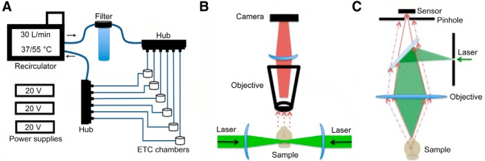
Figure 1
Schematic of electrophoresis and imaging components. Schematic of the electrophoresis system used for active clearing of tissue. A , A recirculating water bath filled with clearing solution is connect...
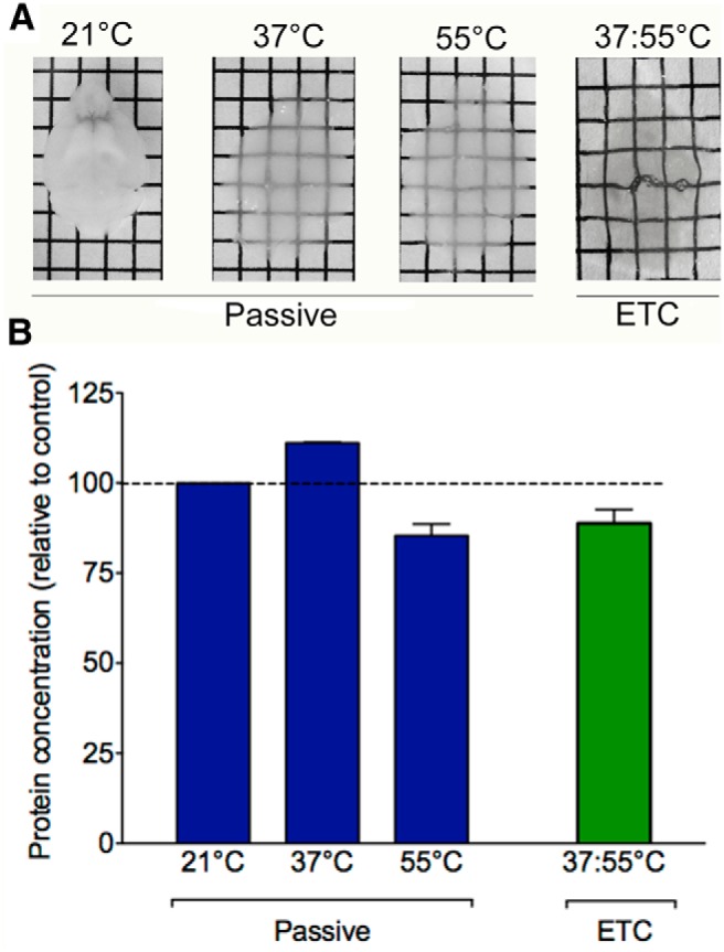
Figure 2
Active clearing of brains with ETC is more efficient than passive clearing. A , Passive clearing is temperature dependent with very little clearing at 21u00b0C occurring after 1 month in SDS. At 37 an...
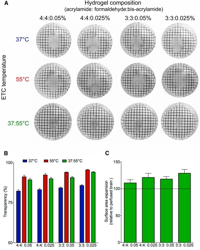
Figure 3
Clearing conditions for optimal transparency. A , Comparison of ETC temperature and hydrogel composition on tissue transparency and expansion. There were significant main effects of clearing temperatu...
Figure 4
The effect of ETC duration and temperature on imaging depth. A 2mm thick coronal section before (A) and after (B) clearing with out optimized protocol. C, Coronal mouse brain section (2 mm thick) clea...
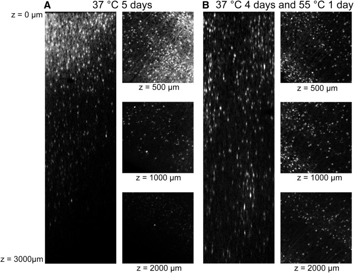
Figure 5
Imaging depth optimization. Using ArcTRAP-tdTomato mice we visualized the endogenous fluorescence signal in mice brains that underwent 5 d of ETC at 37u00b0C ( A ) or were cleared for 4 d at 37u00b0C ...
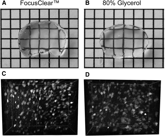
Figure 6
Examining effects of different imaging media (FocusClear vs glycerol). Refractive matching of clarified tissue with either FocusClear or glycerol produces equally transparent tissue, and high-quality ...
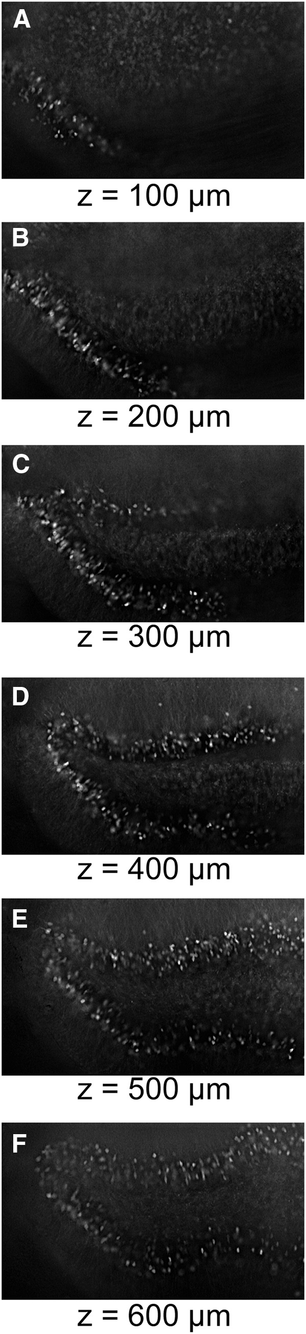
Figure 7
Examining effects of different microscopes to image clear brains. Although a light sheet or confocal microscope may be ideal, when sufficiently clear, tissue may be imaged using even a very basic epif...
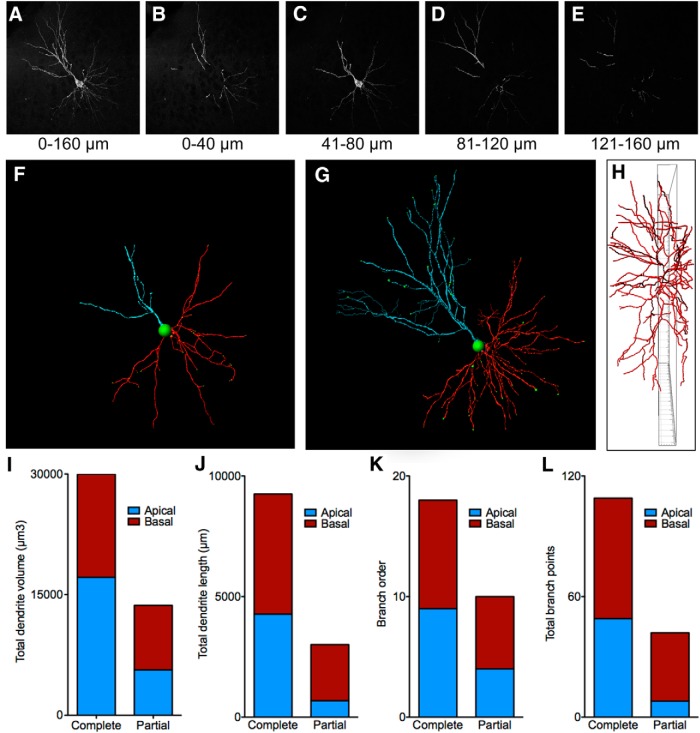
Figure 8
Using CLARITY to analyze neuron morphology. A , GFP-labeled cortical pyramidal neuron visualized in a CLARITY-processed tissue section (HSV-GFP viral vector). Bu2013E , The same neuron shown with the ...
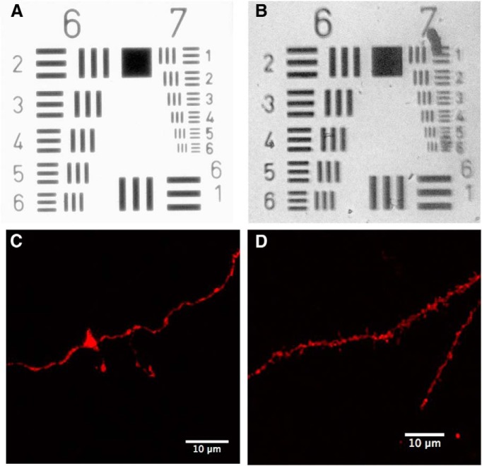
Figure 9
Image resolution in cleared tissue. A , The USAF test target pattern imaged with a 10u00d7 air objective on a Nikon light microscope. The lines in element 6-6 and 7-6 are u223c4.4 and 2.2 u00b5m thick...
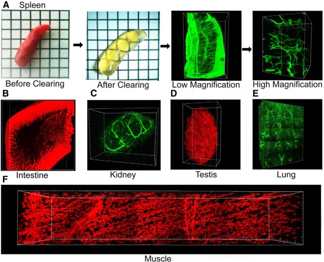
Figure 10
Using CLARITY to clear and image other organs. A , Mouse spleen before and after CLARITY and low- and high-magnification images of CD34 immunohistochemistry on cleared tissue. B , Cleared mouse intest...
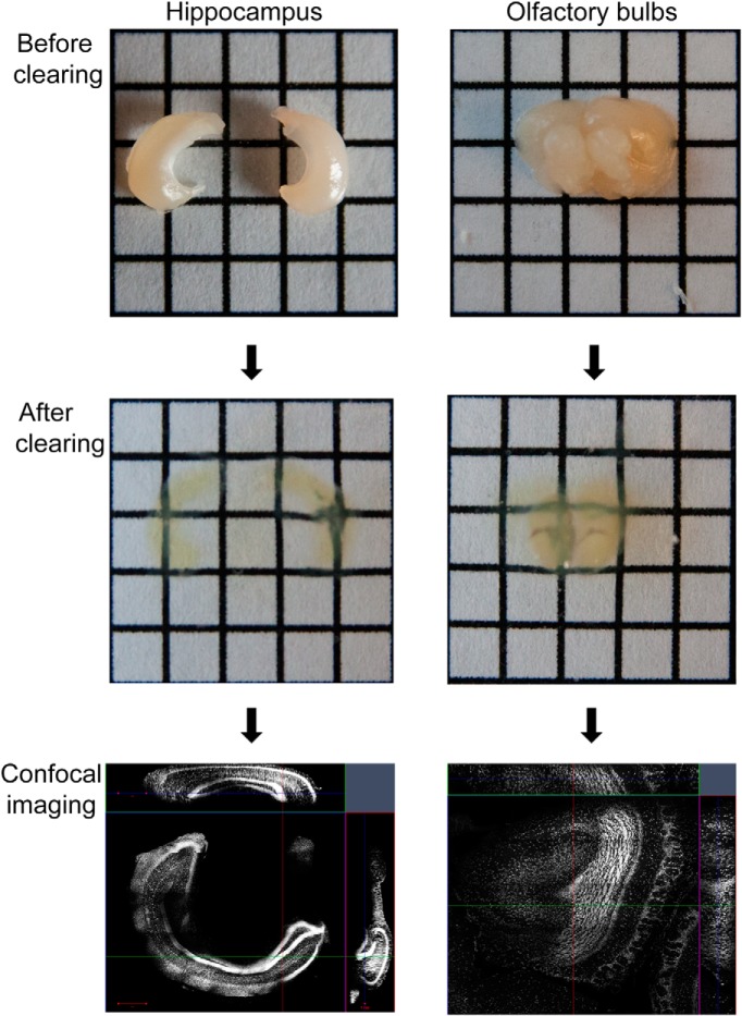
Figure 11
Dissection and clearing of brain subregions. As it may not be necessary to clear the entire brain, it is more efficient to dissect the region of interest before clearing and imaging. Examples of disse...
Figure images are served from the NIH/NLM PubMed Central Open Access Subset or Europe PMC; copyright remains with the publishers and authors.
💬 Discussion
0 commentsNo comments yet. Be the first to start a discussion!
Leave a Comment