Abstract
AbstractMost of the excitatory synapses on principal neurons of the forebrain are located on specialized structures called dendritic spines. Their morphology, comprising a spine head connected to the dendritic branch via a thin neck, provides biochemical and electrical compartmentalization during signal transmission. Spine shape is defined and tightly controlled by the organization of the actin cytoskeleton. Alterations in synaptic strength correlate with changes in the morphological appearance of the spine head and neck. Therefore, it is important to get a better understanding of the nanoscale organization of the actin cytoskeleton in dendritic spines. A periodic organization of the actin/spectrin lattice was recently discovered in axons and a small fraction of dendrites using super-resolution microscopy. Here we use a small probe phalloidin-Atto647N, to label F-actin in mature hippocampal primary neurons and in living hippocampal slices. STED nanoscopy reveals that in contrast to β-II spectrin antibody labelling, phalloidin-Atto647N stains periodic actin structures in all dendrites and the neck of nearly all dendritic spines, including filopodia-like spines. These findings extend the current view on F-actin organization in dendritic spines and may provide new avenues for understanding the structural changes in the spine neck during induction of synaptic plasticity, active organelle transport or tethering.
🔬 Techniques
🔭 Microscopes
💻 Software
✨ Fluorophores
🧪 Sample Preparation
🔬 Cell Lines
🏭 Microscope Brands
🧪 Reagent Suppliers
💻 Software Details
🏛️ Research Organizations (ROR)
Affiliated research institutions:
📋 Methods
Hippocampal neuronal primary cultures, transfections and immunocytochemistry Sacrificing of animals was done in accordance with the Animal Welfare Law of the Federal Republic of Germany (Tierschutzgesetz der Bundesrepublik Deutschland, TierSchG) and with the approval of local authorities of the city-state Hamburg (Behörde für Gesundheit und Verbraucherschutz, Fachbereich Veterinärwesen, from 21.04.2015) and the animal care committee of the University Medical Center Hamburg-Eppendorf. For the procedure of sacrificing rats for subsequent isolation of hippocampal neurons or preparation of hippocampal slices, all regulations and guidelines given in §4 TierSchG are followed. According to §7 Abs. 2 Satz 3 TierSchG sacrificing of animals is not an experiment on animals, therefore no specific authorization or notification is required. Primary hippocampal cultures were prepared with slight modification as described previously 30 . Shortly, brains of E18 Wistar rat embryos were dissected, hippocampi removed, washed in ice cold Hanks’ balanced salt solution (HBSS, Sigma Aldrich), and trypsinized for 15 min by addition of 100 μl per hippocampus of 0.25% trypsin/0.02% ethylenediaminetetraacetic acid (EDTA, Thermo Fisher Scientific) at 37 °C. After washing with warm HBSS, cells were triturated by repeated pipetting through glass needles (20 G, 26 G), filtered via a 100 μm strainer (Greiner), counted and plated in 1 ml DMEM (Thermo Fisher Scientific supplemented with 10% foetal calf serum, 2 mM glutamine, and 1x penicillin/streptomycin) on poly-L-lysin coated glass coverslips at a density of 30000 cells per coverslip in 12-well-plates. The medium was exchanged to neurobasal medium (Thermo Fisher Scientific, supplemented with 2% B27, 0.25% glutamax, 1x penicillin/streptomycin) after cells adhered. Cells were incubated in an incubator at 37 °C, 5% CO 2 and 95% humidity. At DIV 15 neurons were transfected with pEGFP-N1 (Clontech) as a cell fill using Lipofectamine2000 (Invitrogen) as described earlier 30 and fixed with 4% PFA (Roti-Histofix, Carl-Roth)/4% sucrose in PBS for 10–15 min at room temperature (RT) at DIV16. Non-transfected cells were fixed at DIV21 in a same way as DIV16, washed with PBS and transferred into a humidified chamber for immunostaining. Cells were permeabilized for 10 min with 0.2% Triton X-100 in PBS, washed and blocked in blocking buffer (BB, 0.1% Triton X-100, 10% horse serum in PBS). Incubation with primary antibodies (mouse anti-β-II spectrin (BD Biosciences #612562, 1:150), mouse anti-bassoon (clone SAP7F407 Stressgen, 1:500) or mouse anti-cortactin (Millipore, 1:500), and rabbit anti-MAP2 (Abcam #ab32454, 1:400)) diluted in BB was done over night at 4 °C. Secondary antibodies (anti-rabbit-Alexa488 1:500, anti-mouse Abberior Star 580, Abberior GmbH Göttingen, Germany 1:200) were applied in BB for 1.5 h after 3 washing steps at RT. Incubation with phalloidin-Atto647N (0.165 nM in PBS) was performed at RT for 2.5 h and subsequently over night at 4 °C. Cells were washed with PBS and mounted onto objective slides with Mowiol (Sigma Aldrich). Preparation of organotypic and acute hippocampal slices and phalloidin uptake assay Phalloidin staining in slices was optimized in organotypic hippocampal slice cultures prepared from P4-6 Wistar female rat pups. The hippocampi were dissected in ice-cooled dissection medium (in mM: 1 CaCl 2 , 5 MgCl 2 , 10 glucose, 4 KCl, 26 NaHCO 3 , 248 sucrose, 2 kynurenic acid, 0.001% phenol red, Sigma-Aldrich) and cut into 400 μm slices using a tissue chopper (McIlwain Mickle Laboratory Engineering, Surrey, UK). Slices were cultured on millicell membranes (Merck Millipore) in culturing medium (MEM (Invitrogen), containing 20% horse serum, 1 mM L-glutamine, 0.00125% ascorbic acid, 0.01 mg/ml insulin, 1 mM CaCl 2 , 2 mM MgSO 4 , 13 mM D-glucose, 1 M HEPES pH ~7,28, Sigma-Aldrich) and cultured in the incubator (5% CO 2 , 37 °C, and 95% humidity). Slices were fed twice per week by replacing 700 μl of the medium. Two to three weeks old organotypic slices cultures were stained with phalloidin-A647N by pipetting a small droplet (2–4 μl, 6.6 μM) on top of the slice. After >1 hour of incubation, the organotypic slices were imaged at an Olympus Fv1000 scanning Confocal/multi-photon upright microscope and the Abberior STED system. Acute hippocampal slices were prepared from a 9–weeks old male Wistar rat as described above. Within 24 hours slices were live stained with phalloidin-Atto647N. A small droplet of Phalloidin-Atto647N (2–4 μl, 6.6 μM) was pipetted on top of the slice and imaged >1 hour after incubation at a Leica TCS SP8-3X gated STED microscope.
Show full methods section
Hippocampal neuronal primary cultures, transfections and immunocytochemistry Sacrificing of animals was done in accordance with the Animal Welfare Law of the Federal Republic of Germany (Tierschutzgesetz der Bundesrepublik Deutschland, TierSchG) and with the approval of local authorities of the city-state Hamburg (Behörde für Gesundheit und Verbraucherschutz, Fachbereich Veterinärwesen, from 21.04.2015) and the animal care committee of the University Medical Center Hamburg-Eppendorf. For the procedure of sacrificing rats for subsequent isolation of hippocampal neurons or preparation of hippocampal slices, all regulations and guidelines given in §4 TierSchG are followed. According to §7 Abs. 2 Satz 3 TierSchG sacrificing of animals is not an experiment on animals, therefore no specific authorization or notification is required. Primary hippocampal cultures were prepared with slight modification as described previously 30 . Shortly, brains of E18 Wistar rat embryos were dissected, hippocampi removed, washed in ice cold Hanks’ balanced salt solution (HBSS, Sigma Aldrich), and trypsinized for 15 min by addition of 100 μl per hippocampus of 0.25% trypsin/0.02% ethylenediaminetetraacetic acid (EDTA, Thermo Fisher Scientific) at 37 °C. After washing with warm HBSS, cells were triturated by repeated pipetting through glass needles (20 G, 26 G), filtered via a 100 μm strainer (Greiner), counted and plated in 1 ml DMEM (Thermo Fisher Scientific supplemented with 10% foetal calf serum, 2 mM glutamine, and 1x penicillin/streptomycin) on poly-L-lysin coated glass coverslips at a density of 30000 cells per coverslip in 12-well-plates. The medium was exchanged to neurobasal medium (Thermo Fisher Scientific, supplemented with 2% B27, 0.25% glutamax, 1x penicillin/streptomycin) after cells adhered. Cells were incubated in an incubator at 37 °C, 5% CO 2 and 95% humidity. At DIV 15 neurons were transfected with pEGFP-N1 (Clontech) as a cell fill using Lipofectamine2000 (Invitrogen) as described earlier 30 and fixed with 4% PFA (Roti-Histofix, Carl-Roth)/4% sucrose in PBS for 10–15 min at room temperature (RT) at DIV16. Non-transfected cells were fixed at DIV21 in a same way as DIV16, washed with PBS and transferred into a humidified chamber for immunostaining. Cells were permeabilized for 10 min with 0.2% Triton X-100 in PBS, washed and blocked in blocking buffer (BB, 0.1% Triton X-100, 10% horse serum in PBS). Incubation with primary antibodies (mouse anti-β-II spectrin (BD Biosciences #612562, 1:150), mouse anti-bassoon (clone SAP7F407 Stressgen, 1:500) or mouse anti-cortactin (Millipore, 1:500), and rabbit anti-MAP2 (Abcam #ab32454, 1:400)) diluted in BB was done over night at 4 °C. Secondary antibodies (anti-rabbit-Alexa488 1:500, anti-mouse Abberior Star 580, Abberior GmbH Göttingen, Germany 1:200) were applied in BB for 1.5 h after 3 washing steps at RT. Incubation with phalloidin-Atto647N (0.165 nM in PBS) was performed at RT for 2.5 h and subsequently over night at 4 °C. Cells were washed with PBS and mounted onto objective slides with Mowiol (Sigma Aldrich). Preparation of organotypic and acute hippocampal slices and phalloidin uptake assay Phalloidin staining in slices was optimized in organotypic hippocampal slice cultures prepared from P4-6 Wistar female rat pups. The hippocampi were dissected in ice-cooled dissection medium (in mM: 1 CaCl 2 , 5 MgCl 2 , 10 glucose, 4 KCl, 26 NaHCO 3 , 248 sucrose, 2 kynurenic acid, 0.001% phenol red, Sigma-Aldrich) and cut into 400 μm slices using a tissue chopper (McIlwain Mickle Laboratory Engineering, Surrey, UK). Slices were cultured on millicell membranes (Merck Millipore) in culturing medium (MEM (Invitrogen), containing 20% horse serum, 1 mM L-glutamine, 0.00125% ascorbic acid, 0.01 mg/ml insulin, 1 mM CaCl 2 , 2 mM MgSO 4 , 13 mM D-glucose, 1 M HEPES pH ~7,28, Sigma-Aldrich) and cultured in the incubator (5% CO 2 , 37 °C, and 95% humidity). Slices were fed twice per week by replacing 700 μl of the medium. Two to three weeks old organotypic slices cultures were stained with phalloidin-A647N by pipetting a small droplet (2–4 μl, 6.6 μM) on top of the slice. After >1 hour of incubation, the organotypic slices were imaged at an Olympus Fv1000 scanning Confocal/multi-photon upright microscope and the Abberior STED system. Acute hippocampal slices were prepared from a 9–weeks old male Wistar rat as described above. Within 24 hours slices were live stained with phalloidin-Atto647N. A small droplet of Phalloidin-Atto647N (2–4 μl, 6.6 μM) was pipetted on top of the slice and imaged >1 hour after incubation at a Leica TCS SP8-3X gated STED microscope.
Confocal and STED imaging
Overview confocal images of organotypic hippocampal slice cultures were acquired at an Olympus Fv1000 scanning confocal/multi-photon upright microscope controlled by Fluoview software (version 4.2). Slices were submerged in HEPES-buffered artificial cerebrospinal fluid (ACSF) containing (in mM): 145 NaCl, 10 HEPES, 12.5 D-glucose, 1.25 NaH 2 PO 4 , 2.5 KCl, 1 MgCl 2 , 2 CaCl 2 , (Sigma-Aldrich), pH ~7.4 at RT. Images were taken with an 25x water immersion objective (Olympus XLPlan N, 1.05 NA) with 3.5x zoom. Phalloidin-Atto647 was excited by a 635 nm laser and detected by an alkali-photodetector. Optimal labelling of dendritic structures and best signal-to-noise ratio was found ~10 μm below the slice surface. Single plane STED images were acquired on a Leica TCS SP8-3X gated STED microscope equipped with a pulsed 775 nm depletion laser and a pulsed white light laser (WLL) for excitation. For acquiring images the Leica objective HC APO CS2 100x/1.40 oil was used. Primary neuron samples were excited by the WLL at 650 nm (phalloidin), 488 nm (MAP2) and 561 nm (bassoon/β-II spectrin), respectively. MAP2 staining was only imaged in confocal mode. For STED imaging emission was acquired between 660–730 nm for Atto647N and 580–620 nm for Abberior Star 580. The detector time gates for both STED channels were set from 0.5–1 ns to 6 ns. Both dyes were depleted with 775 nm. Respective confocal channels uses the same settings as STED channels, except the excitation power was reduced and the detection time gates were set to 300 ps to 6 ns for both channels. The format for all images were set to 1024 × 1024 pixels. With an optical zoom of 5, the resulting voxel size is 23 nm for xy. Images were taken with 600 lines per second and line averaging of 16. STED images from adult acute hippocampal slices were acquired by placing the slices up-side-down in a live imaging chamber contain HEPES-buffered ACSF (in mM: 145 NaCl, 10 HEPES, 12.5 D-glucose, 1.25 NaH 2 PO 4 , 2.5 KCl, 1 MgCl 2 , 2 CaCl 2 , (Sigma-Aldrich), pH ~7.4 at RT). The format for all images was set to 1024 × 1024 pixels. With an optical zoom of 6, the resulting resolution is 18.9 nm per pixel for x-y. Images where taken with 600 lines per second and line averaging of 8. Time gates were set to 0.5 to 6 ns. STED images from organotypic slice cultures were acquired with Abberior Instruments STED microscope using 60x P-Apo oil objective 1.4 NA. The imaging conditions were the same as for acute slices. Phalloidin-A647N was excited by a 640 nm laser and depleted by a 775 nm pulsed laser. Pinhole size was set to 0.83 airy units, pixel size 20 nm.
Data analysis
Spine length was measured manually in raw STED images in Fiji 31 by drawing a segmented line from the spine base along the neck to the tip of the protrusion. Spines were defined as dendritic protrusions showing a clear presynaptic contact as visualized by bassoon or an enrichment of cortactin at the postsynaptic site, independent of spine length. Differentiation between mushroom and thin spines was purely defined by appearance of a clear spine head, which is at least 1.5x thicker than the neck. Mushroom-like spines were called ‘long mushroom’ when the total spine length was exceeding 2 μm. Protrusions without bassoon staining are classified as filopodia-like (same with over entire length) or lamelliopodia-like (amorphic protrusions). Data from at least three independent neuronal preparations for DIV21 and one for DIV16 were used for the analysis. Intensity profiles of segmented lines (width1–3pixels) along axons, dendrites and spine necks were created in Fiji using the plot profile option, values exported and graphs generated in Prism 6 (GraphPad, La Jolla, CA, USA). Local maxima of intensities profiles of raw STED images were used for quantification of actin ring spacing. They were defined manually by a researcher blind to the identity of the measured profile. Inter-peak distances between at least 3 subsequent local intensity maxima were calculated. To improve image quality for representation, raw data of STED images were deconvolved using the Huygens Professional (SVI, v 15.10) STED package as follows: to calculate the theoretical point spread function (PSF) the optical microscopic parameters provided by the lif-file itself were used. Within the Deconvolution wizard, images were subjected to a manual background correction. For deconvolution, the signal to noise ratio was set to 15. The Optimized iteration mode of the CMLE was applied until it reached a Quality threshold of 0.05. For representation of confocal and STED images, contrast was enhanced by linear methods using Fiji for the complete image for each channel separately.
Statistics
Differences in F-actin periodicity in spines, dendrites and axons was analysed by 1-way analysis of variances (ANOVA) after testing for equal variances between the groups (Brown-Forsythe test) using Prism 6. Correlation between spine length and actin spacing was analysed using linear regression mode in Prism 6. Prism 6 was also used to create all graphs. Origin 2015 (OriginLab Corporation) was used to perform fast Fourier transformation (FFT) of a single selected spine profile.
📊 Figures
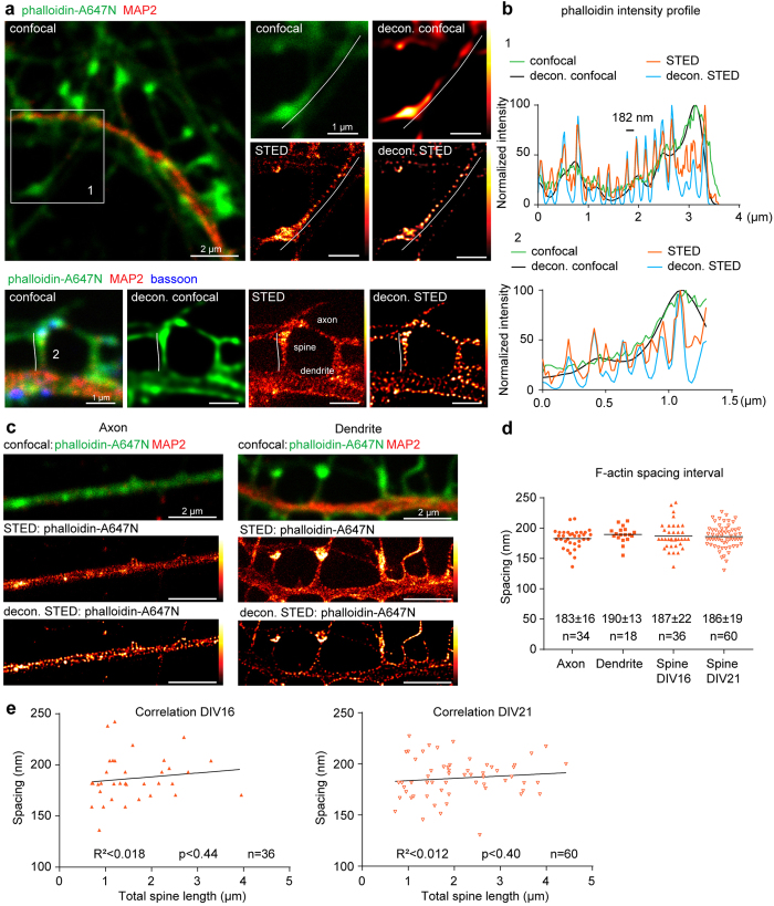
Figure 1
Periodic F-actin lattice is present in nearly all necks of dendritic spines.
(a) Top: Confocal image of a primary hippocampal neuron (DIV21) stained with anti-MAP2 antibodies and phalloidin-A647N with corresponding raw/deconvolved confocal and STED images of F-actin in higher ...
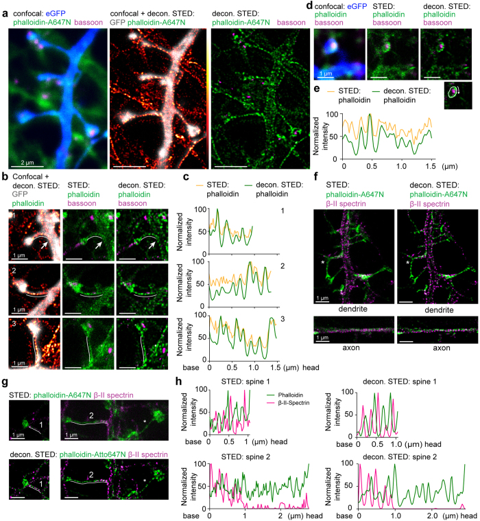
Figure 2
Periodic structures are found in mushroom-like spines of primary hippocampal neurons and can be better identified by phalloidin-A647N than u03b2-II spectrin staining.
( a ) Confocal image of a DIV16 hippocampal neuron filled with eGFP (blue) and stained for phalloidin (green) and bassoon (magenta) with corresponding deconvolved STED images. Note that phalloidin fil...
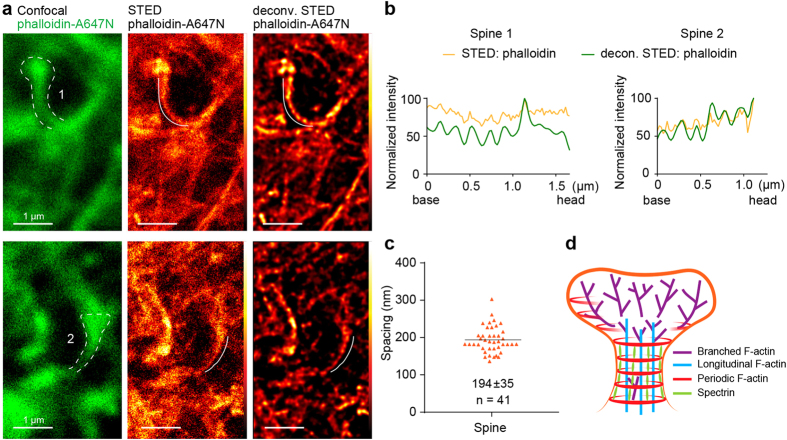
Figure 3
Phalloidin-A647N labels the periodic actin lattice in dendritic spines in acute hippocampal slices.
( a ) Representative confocal images of 2 spines from hippocampal slices with phalloidin-Atto647N (green, left panel) and corresponding raw and deconvolved STED image (middle/right). ( b ) Normalized ...
Figure images are served from the NIH/NLM PubMed Central Open Access Subset or Europe PMC; copyright remains with the publishers and authors.
💬 Discussion
0 commentsNo comments yet. Be the first to start a discussion!
Leave a Comment