Abstract
The opportunistic pathogen Pseudomonas aeruginosa is a major cause of antibiotic-tolerant infections in humans. P. aeruginosa evades antibiotics in bacterial biofilms by up-regulating expression of a symbiotic filamentous inoviral prophage, Pf4. We investigated the mechanism of phage-mediated antibiotic tolerance using biochemical reconstitution combined with structural biology and high-resolution cellular imaging. We resolved electron cryomicroscopy atomic structures of Pf4 with and without its linear single-stranded DNA genome, and studied Pf4 assembly into liquid crystalline droplets using optical microscopy and electron cryotomography. By biochemically replicating conditions necessary for antibiotic protection, we found that phage liquid crystalline droplets form phase-separated occlusive compartments around rod-shaped bacteria leading to increased bacterial survival. Encapsulation by these compartments was observed even when inanimate colloidal rods were used to mimic rod-shaped bacteria, suggesting that shape and size complementarity profoundly influences the process. Filamentous inoviruses are pervasive across prokaryotes, and in particular, several Gram-negative bacterial pathogens including Neisseria meningitidis , Vibrio cholerae, and Salmonella enterica harbor these prophages. We propose that biophysical occlusion mediated by secreted filamentous molecules such as Pf4 may be a general strategy of bacterial survival in harsh environments.
🔬 Techniques
🧬 Organisms
💻 Software
🏭 Microscope Brands
🧪 Reagent Suppliers
💻 Software Details
💾 Data Repositories
🏛️ Research Organizations (ROR)
Affiliated research institutions:
📋 Methods
Pf4 Phage Preparation. Native Pf4 phage was initially generated from a static biofilm of a P. aeruginosa PAO1 strain, further amplified by infection of PAO1 on Luria-Bertani plates and isolated by polyethylene glycol precipitation as described previously ( 20 ) and in further detail in SI Appendix , Materials and Methods . Cryo-EM and Cryo-ET Data Collection. Cryo-EM data of the Pf4 phage were collected as movie frame stacks using the EPU software on a Titan Krios microscope (ThermoFisher) operating at 300 kV fitted with a Quantum energy filter and a K2 Summit direct electron detector (Gatan) operating in counting mode. Tilt series data for cryo-ET were collected on the same microscope using the SerialEM software ( 29 ) in two directions starting from 0° between ±60° with 1° tilt increments. Further details on grid preparation and data collection conditions are provided in SI Appendix , Materials and Methods and Table S1 . Cryo-EM Image Processing and Data Analysis. Cryo-EM refinement was performed using the real-space helical reconstruction procedure implemented in Relion 3.0 ( 30 ). Fourier transforms of class averages produced using the Spring software ( 31 ), which showed high-resolution features, were indexed to determine initial helical symmetry parameters used for 3D refinement in Relion. Pf4 compositional variation was accounted for by splitting the dataset into segments from classes showing density at the core of the filament and classes where this density was absent. Model building was performed iteratively using Coot ( 32 ) along with real-space refinement against the density map in PHENIX ( 33 ). Further details are provided in SI Appendix , Materials and Methods . Tomogram Reconstruction from Cryo-ET Data. Tilt series alignment using gold fiducials and tomogram reconstruction were both carried out using a set of image processing, modeling and display programs (IMOD) ( 34 ) as described previously ( 35 ). Visualization of data was performed in IMOD and UCSF Chimera ( 36 ). Fluorescence Microscopy and FRAP. Samples for fluorescence microscopy were either immobilized onto agar pads constructed using Gene Frames (ThermoFisher), or applied directly to glass slides and imaged using a Zeiss AxioImager M2 widefield or Zeiss LSM Exciter confocal microscope, respectively. For FRAP experiments, samples were placed in a capillary on a glass slide and regions of interest were bleached using the Zen software bleaching mode on the Zeiss LSM exciter confocal microscope. Further details on sample preparation, imaging conditions, and image analysis are provided in SI Appendix , Materials and Methods . Materials and Data Availability. Cryo-EM maps reported in this study have been deposited at the Electron Microscopy Data Bank with accession codes EMD-10593 and EMD-10594. Atomic models have been deposited in the Protein Data Bank with PDB ID codes 6TUP and 6TUQ. Reagents generated in this study will be made available on request, but we may require a payment and/or a completed Materials Transfer Agreement if there is potential for commercial application.
Show full methods section
Pf4 Phage Preparation. Native Pf4 phage was initially generated from a static biofilm of a P. aeruginosa PAO1 strain, further amplified by infection of PAO1 on Luria-Bertani plates and isolated by polyethylene glycol precipitation as described previously ( 20 ) and in further detail in SI Appendix , Materials and Methods . Cryo-EM and Cryo-ET Data Collection. Cryo-EM data of the Pf4 phage were collected as movie frame stacks using the EPU software on a Titan Krios microscope (ThermoFisher) operating at 300 kV fitted with a Quantum energy filter and a K2 Summit direct electron detector (Gatan) operating in counting mode. Tilt series data for cryo-ET were collected on the same microscope using the SerialEM software ( 29 ) in two directions starting from 0° between ±60° with 1° tilt increments. Further details on grid preparation and data collection conditions are provided in SI Appendix , Materials and Methods and Table S1 . Cryo-EM Image Processing and Data Analysis. Cryo-EM refinement was performed using the real-space helical reconstruction procedure implemented in Relion 3.0 ( 30 ). Fourier transforms of class averages produced using the Spring software ( 31 ), which showed high-resolution features, were indexed to determine initial helical symmetry parameters used for 3D refinement in Relion. Pf4 compositional variation was accounted for by splitting the dataset into segments from classes showing density at the core of the filament and classes where this density was absent. Model building was performed iteratively using Coot ( 32 ) along with real-space refinement against the density map in PHENIX ( 33 ). Further details are provided in SI Appendix , Materials and Methods . Tomogram Reconstruction from Cryo-ET Data. Tilt series alignment using gold fiducials and tomogram reconstruction were both carried out using a set of image processing, modeling and display programs (IMOD) ( 34 ) as described previously ( 35 ). Visualization of data was performed in IMOD and UCSF Chimera ( 36 ). Fluorescence Microscopy and FRAP. Samples for fluorescence microscopy were either immobilized onto agar pads constructed using Gene Frames (ThermoFisher), or applied directly to glass slides and imaged using a Zeiss AxioImager M2 widefield or Zeiss LSM Exciter confocal microscope, respectively. For FRAP experiments, samples were placed in a capillary on a glass slide and regions of interest were bleached using the Zen software bleaching mode on the Zeiss LSM exciter confocal microscope. Further details on sample preparation, imaging conditions, and image analysis are provided in SI Appendix , Materials and Methods . Materials and Data Availability. Cryo-EM maps reported in this study have been deposited at the Electron Microscopy Data Bank with accession codes EMD-10593 and EMD-10594. Atomic models have been deposited in the Protein Data Bank with PDB ID codes 6TUP and 6TUQ. Reagents generated in this study will be made available on request, but we may require a payment and/or a completed Materials Transfer Agreement if there is potential for commercial application.
Materials and Data Availability. Cryo-EM maps reported in this study have been deposited at the Electron Microscopy Data Bank with accession codes EMD-10593 and EMD-10594. Atomic models have been deposited in the Protein Data Bank with PDB ID codes 6TUP and 6TUQ. Reagents generated in this study will be made available on request, but we may require a payment and/or a completed Materials Transfer Agreement if there is potential for commercial application.
Supplementary Material Supplementary File Supplementary File Supplementary File Supplementary File Supplementary File Supplementary File Supplementary File
📊 Figures
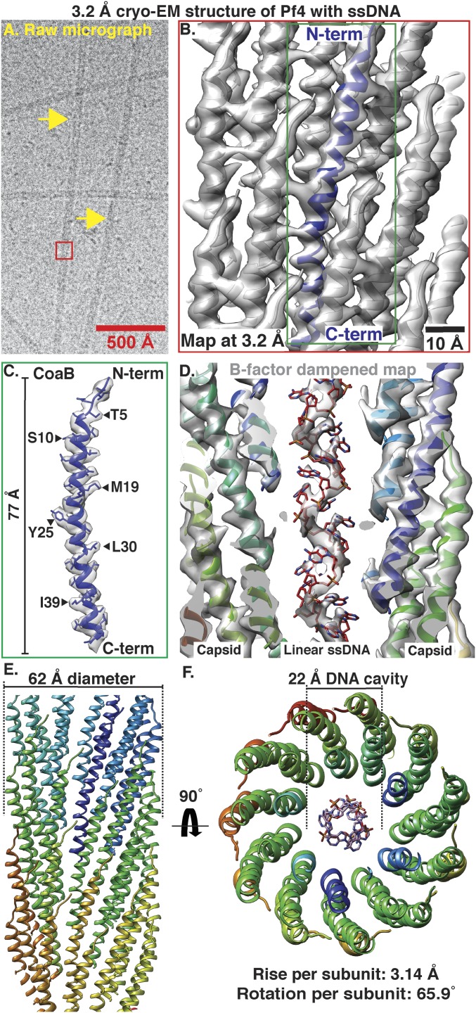
Fig. 1.
Cryo-EM structure of Pf4 phage at 3.2-u00c5 resolution. ( A ) Cryo-EM image of native Pf4 phage (yellow arrows) purified from biofilms (red box represents reconstructed segment). ( B ) Single-particle...
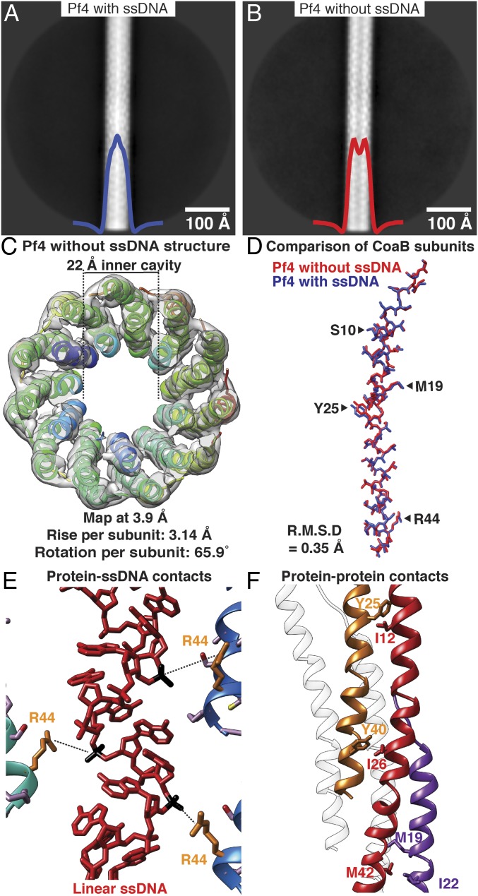
Fig. 2.
Cryo-EM structure of Pf4 filament without ssDNA at 3.9-u00c5 resolution. ( A ) Two-dimensional class average of Pf4 with ssDNA, showing density in the core of the phage, indicated by peak in the horiz...
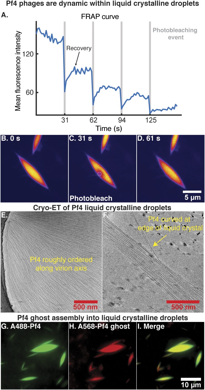
Fig. 3.
Pf4 assembly into liquid crystalline droplets is dynamic with strong orientational ordering of filaments. ( A ) A region within a liquid crystalline droplet was photobleached multiple times and the FR...
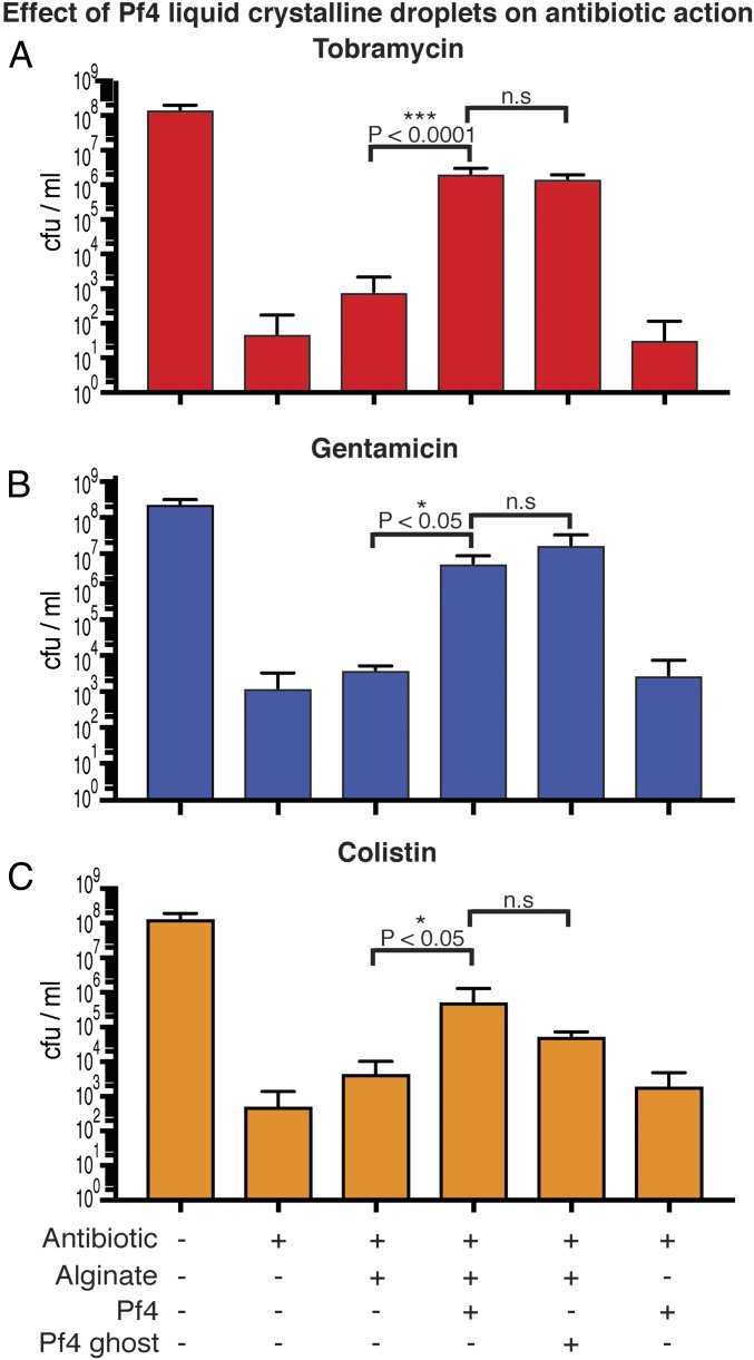
Fig. 4.
Pf4 liquid crystalline droplets with and without ssDNA protect P. aeruginosa cells against antibiotics. ( A ) Bar graph shows colony-forming units (cfu) per ml, a measure of P. aeruginosa culture cell...
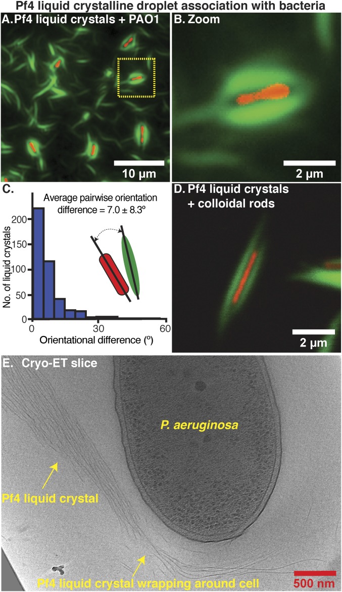
Fig. 5.
Pf4 liquid crystalline droplets form protective sheaths around P. aeruginosa cells. ( A ) Optical microscopy of the condition from the antibiotic protection assay presented in Fig. 4 , containing P. a...
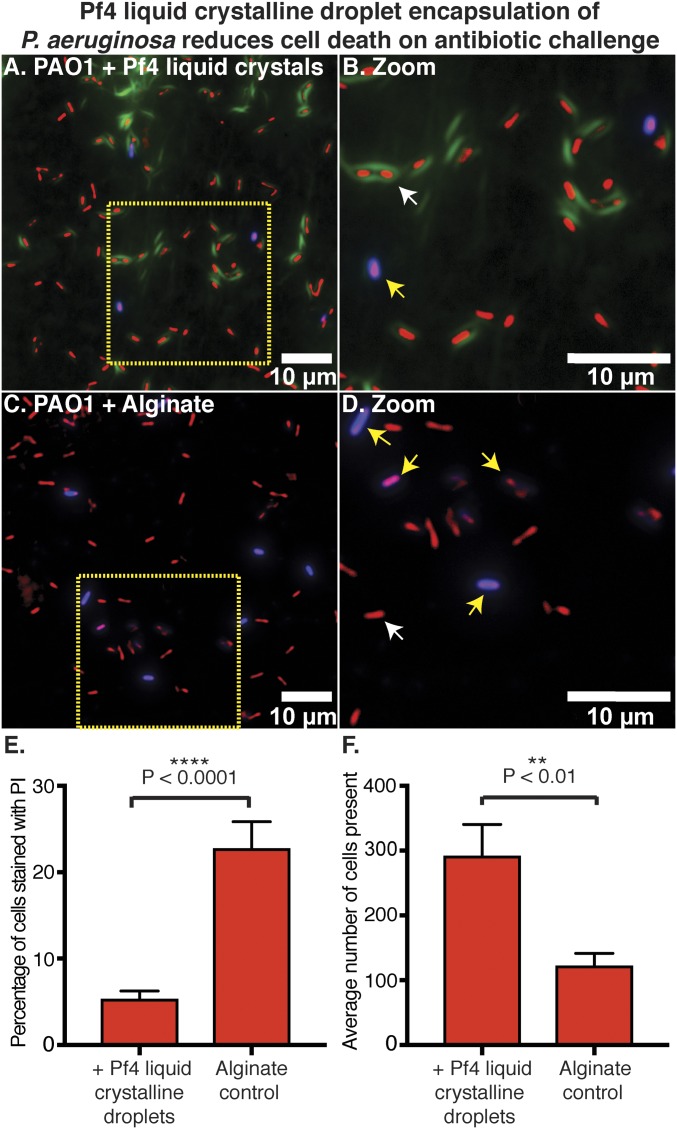
Fig. 6.
Pf4 liquid crystalline droplet encapsulation of P. aeruginosa prevents cell death on antibiotic treatment. ( A ) Optical microscopy of the antibiotic protection assay presented in Fig. 4 , containing ...
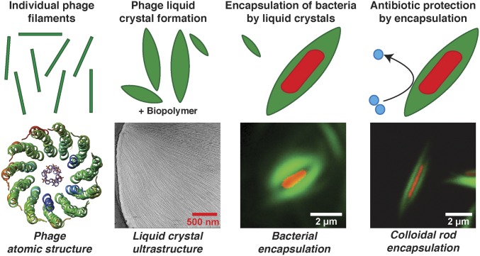
Fig. 7.
Schematic model of the mechanism of Pf4 phage-mediated antibiotic tolerance revealed in this study. Individual Pf4 phage filaments self-assemble into higher-order dynamic spindle-shaped liquid crystal...
Figure images are served from the NIH/NLM PubMed Central Open Access Subset or Europe PMC; copyright remains with the publishers and authors.
💬 Discussion
0 commentsNo comments yet. Be the first to start a discussion!
Leave a Comment