Abstract
Stress granules (SGs) are membrane-less dynamic structures consisting of mRNA and protein aggregates that form rapidly in response to a wide range of environmental cellular stresses and viral infections. They act as storage sites for translationally silenced mRNAs under stress conditions. During viral infection, SG formation results in the modulation of innate antiviral immune responses, and several viruses have the ability to either promote or prevent SG assembly. Here, we show that rabies virus (RABV) induces SG formation in infected cells, as revealed by the detection of SG-marker proteins Ras GTPase-activating protein-binding protein 1 (G3BP1), T-cell intracellular antigen 1 (TIA-1) and poly(A)-binding protein (PABP) in the RNA granules formed during viral infection. As shown by live cell imaging, RABV-induced SGs are highly dynamic structures that increase in number, grow in size by fusion events, and undergo assembly/disassembly cycles. Some SGs localize in close proximity to cytoplasmic viral factories, known as Negri bodies (NBs). Three dimensional reconstructions reveal that both structures remain distinct even when they are in close contact. In addition, viral mRNAs synthesized in NBs accumulate in the SGs during viral infection, revealing material exchange between both compartments. Although RABV-induced SG formation is not affected in MEFs lacking TIA-1, TIA-1 depletion promotes viral translation which results in an increase of viral replication indicating that TIA-1 has an antiviral effect. Inhibition of PKR expression significantly prevents RABV-SG formation and favors viral replication by increasing viral translation. This is correlated with a drastic inhibition of IFN-B gene expression indicating that SGs likely mediate an antiviral response which is however not sufficient to fully counteract RABV infection.
🔬 Techniques
🔭 Microscopes
✨ Fluorophores
🧪 Sample Preparation
🔬 Cell Lines
🏭 Microscope Brands
🧪 Reagent Suppliers
💻 Software Details
🏛️ Research Organizations (ROR)
Affiliated research institutions:
📋 Methods
Cells and viruses BSR cells, cloned from BHK 21 (baby hamster kidney) were obtained from A. Flamand (I2BC, Département de Virologie, former Laboratoire de Génétique des Virus, Gif, France), N2A cells (mouse neuroblastoma) and U373-MG cells (human gliobastoma astrocytoma) were purchased from the ATTC organization ( http://www.lgcstandards-atcc.org ). All the cells were grown in Dulbeco’s modified eagle medium (DMEM) supplemented with 10% FCS (fetal calf serum). Immortalized murine embryonic fibroblasts (MEFs) from wild-type (WT) and TIA1-knockout mice [ 59 ] were obtained from P. Anderson (Harvard University). Mouse primary neurons were obtained from JM. Peyrin (Université Pierre-et-Marie Curie, CNRS UMR 7102, Paris) as described [ 60 ]. Briefly, cortical cells were isolated from mouse embryos E16 embryos of Swiss mice (Janvier Labs), and cultured for 5 days in complete neuronal culture medium DMEM glutamax (Life Technologies, Inc., Gaithersburg, MD, USA) supplemented with serum-free Neurobasal (Gibco) and 2% B-27 supplement (Gibco). The SADB19, PV (Pasteur Virus), and CVS (Chalenge virus standard) strains of rabies virus were grown in BSR cells.
Plasmids
The plasmid encoding G3BP-eGFP, described by [ 18 ] was kindly provided by R. Lloyd (Department of Molecular Virology and Microbiology, Baylor College of Medicine, Houston, USA).
Construction and recovery of the recombinant CVS
N2C virus expressing a P-mCherry fusion protein The full-length recombinant N2C (prCVSN2C) infectious clone was described previously [ 61 ]. The authentic P coding sequence was replaced with the P-mCherry fusion encoding sequence. The original full-length genomic plasmid was digested with AvrII and NruI restriction enzymes. Three overlapping fragments were amplified by PCR. The first one going from the AvrII site in the N gene to the end of the P coding sequence, the second one corresponding to the mCherry coding sequence and the third one going from the end of the P coding sequence to the NruI site in the G gene. The PCR products and the digested plasmid were assembled using Gibson Assembly kit (New England Biolabs) to obtain the resulting plasmid, prN2C-P-mCherry. Recombinant viruses were recovered as described previously [ 62 , 63 ]. Briefly, N2A cells (10 6 cells) were transfected using lipofectamine 2000 (Invitrogen) with 0.85 μg of full-length prCVSN2C-P-mCherry, in addition to 0.4 μg pTIT-N, 0.2 μg pTIT-P, 0.2 μg pTIT-L and 0.15 μg pTIT-G, which encode respectively the N, P, L and G proteins of SAD-L16 rabies virus strain. These plasmids were cotransfected with 0.25 μg of a plasmid encoding the T7 RNA polymerase. Six days posttransfection, the supernatant was passaged on fresh N2A cells, and infectious recombinant viruses were detected three days later by the fluorescence of the P-mCherry protein.
Show full methods section
Cells and viruses BSR cells, cloned from BHK 21 (baby hamster kidney) were obtained from A. Flamand (I2BC, Département de Virologie, former Laboratoire de Génétique des Virus, Gif, France), N2A cells (mouse neuroblastoma) and U373-MG cells (human gliobastoma astrocytoma) were purchased from the ATTC organization ( http://www.lgcstandards-atcc.org ). All the cells were grown in Dulbeco’s modified eagle medium (DMEM) supplemented with 10% FCS (fetal calf serum). Immortalized murine embryonic fibroblasts (MEFs) from wild-type (WT) and TIA1-knockout mice [ 59 ] were obtained from P. Anderson (Harvard University). Mouse primary neurons were obtained from JM. Peyrin (Université Pierre-et-Marie Curie, CNRS UMR 7102, Paris) as described [ 60 ]. Briefly, cortical cells were isolated from mouse embryos E16 embryos of Swiss mice (Janvier Labs), and cultured for 5 days in complete neuronal culture medium DMEM glutamax (Life Technologies, Inc., Gaithersburg, MD, USA) supplemented with serum-free Neurobasal (Gibco) and 2% B-27 supplement (Gibco). The SADB19, PV (Pasteur Virus), and CVS (Chalenge virus standard) strains of rabies virus were grown in BSR cells.
Plasmids
The plasmid encoding G3BP-eGFP, described by [ 18 ] was kindly provided by R. Lloyd (Department of Molecular Virology and Microbiology, Baylor College of Medicine, Houston, USA).
Construction and recovery of the recombinant CVS
N2C virus expressing a P-mCherry fusion protein The full-length recombinant N2C (prCVSN2C) infectious clone was described previously [ 61 ]. The authentic P coding sequence was replaced with the P-mCherry fusion encoding sequence. The original full-length genomic plasmid was digested with AvrII and NruI restriction enzymes. Three overlapping fragments were amplified by PCR. The first one going from the AvrII site in the N gene to the end of the P coding sequence, the second one corresponding to the mCherry coding sequence and the third one going from the end of the P coding sequence to the NruI site in the G gene. The PCR products and the digested plasmid were assembled using Gibson Assembly kit (New England Biolabs) to obtain the resulting plasmid, prN2C-P-mCherry. Recombinant viruses were recovered as described previously [ 62 , 63 ]. Briefly, N2A cells (10 6 cells) were transfected using lipofectamine 2000 (Invitrogen) with 0.85 μg of full-length prCVSN2C-P-mCherry, in addition to 0.4 μg pTIT-N, 0.2 μg pTIT-P, 0.2 μg pTIT-L and 0.15 μg pTIT-G, which encode respectively the N, P, L and G proteins of SAD-L16 rabies virus strain. These plasmids were cotransfected with 0.25 μg of a plasmid encoding the T7 RNA polymerase. Six days posttransfection, the supernatant was passaged on fresh N2A cells, and infectious recombinant viruses were detected three days later by the fluorescence of the P-mCherry protein.
Antibodies and drugs
The rabbit polyclonal anti-P antibody was previously described [ 23 ]. The mouse monoclonal anti-G3BP-1 (2 F3) antibody was obtained from Sigma. The rabbit polyclonal anti-P antibody was previously described [ 23 ]. The rabbit anti-phospho eIF2α (04342) was obtained from Millipore. The Rabbit anti-MDA5 (33H12L34) was from Invitrogen, the rabbit anti-RIG-I (AT111) was from Enzo life; the goat monoclonal anti-TIA-1 (C20) and mouse anti-PABP (10E10) antibodies were from Santa-Cruz Biotechnology. Secondary fluorescent antibodies were purchased from Molecular Probes (Alexa fluor 488-, 568- or 647- conjugated) and Cell Signaling (Fluor 800 or Fluor 680 conjugated). Nocodazole (M1404), actinomycin D (A9415), sodiumarsenite (S7400) and cycloheximide (C7698) were obtained from Sigma.
Immunofluorescence staining and confocal microscopy
Cells were fixed for 15 min with 4% PFA (paraformaldehyde) and permeabilized for 5 min with 0.1% Triton X-100 in PBS. Cells were incubated with the indicated primary antibodies for 1 h at RT, washed and incubated for 1h with Alexa fluor conjugated secondary antibodies. Following washing, cells were mounted with Vectashield (Vector labs) containing DAPI. Images were captured using a Leica SP8 confocal microscope (63X oil-immersion objective). For 3D reconstruction, confocal stacks were treated with Chimera Software and 3D rendering was carried out using the volume viewer tool in Chimera Software.
Quantification of stress granule positive cells
To determine the number of stress granule-positive cells three wide-field 20X images were captured per experiment. Cells displaying ponctate immunofluorescent foci of G3BP-1 were considered as stress granule positive. Counterstaining with anti-P antibody was performed to discriminate between infected and non-infected cells. Fluorescence in situ hybridization (FISH) FISH was performed as previously described [ 23 ], with some modifications and by using 5’-ATTO448 modified oligonucleotides (Eurofins Genomics). The sequences of probes used to detect viral RNAs were previously described [ 23 ]. Messenger RNAs (mRNAs) were detected using 5’-ATTO488-oligo (dT). Cells infected with RABV were fixed in 4% PFA for 15 min at room temperature, permeabilized 5 min with 0.1% TX-100 in PBS, and incubated with primary and secondary antibodies as described above. After dehydration in 70% RNase-free ethanol overnight, coverslips were rehydrated in 2X SSC (1X SSC: 0.15M NaCl, 0.015 M sodium citrate) buffer, before prehybridization with 10 ng probe per coverslips in hybridization buffer (50% formamide (Sigma), 10% dextran sulfate sodium salt (Sigma), 20 μg/ml salmon sperm DNA (Invitrogen), 2X SSC) for 1 h at 60°C. Hybridization was carried out in hybridization buffer plus 10 ng probe per coverslips for 5 min at 60°C and 4 h at 37°C in the dark. Cells were washed in 2X SSC pre-warmed at 42°C and then fixed in 3.7% formaldehyde in PBS and mounted as described above.
EU labelling
Infected cells (16 h p.i.) were treated with 20 μM actinomycin D (Act D) for 1h, to inhibit cellular transcription, and then fed with 1 mM 5-ethynyl uridine (EU; Invitrogen) for 45 min. For pulse-chase analyses, the medium containing EU was replaced by new culture medium supplemented with Act D throughout the chase period. Cells were fixed using 4% PFA in PBS. EU labeling of cells was detected according to the manufacturer’s instructions (Invitrogen, Click-it RNA imaging kits). After this step, cells were washed with PBS and incubated with antibodies as described above. Quantitative real-time RT-PCR (RT-qPCR) U373 cells (2–4 10 5 ) transfected by siRNA and infected with RABV-CVS for 24 h were flash frozen in liquid nitrogen and stored at -80°C. Total RNA was extracted with Nucleospin RNA II kit (Macherey Nagel, France) and 1 μg of RNA was used for first strand cDNA synthesis by reverse transcription with AffinityScript QPCR cDNA synthesis kit (Agilent Technologies, USA) and oligo(dT) primers (200 ng). The quantitative real-time PCR was performed on the Mx3000P apparatus (Stratagene, USA) in a total volume of 20 μl containing the first strand cDNA template, 200 nM of each primer and 1x Mesa green QPCR master mix plus solution (Eurogentec, France). Standard curves for Interferon-B gene (IFN-B) and the housekeeping gene glyceraldehyde-3-phosphate dehydrogenase (GAPDH) were obtained by using serial dilutions of human genomic DNA. The forward and reverse primers for mouse GADPH were: 5’-TCAACTACATGGTCTACATGTT-3’and 5’GGTCTCGCTCCTGGAAGAT-3’, respectively. Forward and reverse primers for human IFN-B and GAPDH genes were 5’- GTC TCC TCC AAA TTG CTC TC (f), 5’- ACA GGA GCT TCT GAC ACT GA (r), 5’- ACA GCC TCA AGA TCA TCA GC (f) and 5’- TCT TCT GGG TGG CAG TGA T (r), respectively. IFN-B mRNA levels were normalized to GAPDH expression levels that remained unaffected during viral infection. The plasmid pCW278 containing the cDNA of RABV-CVS-N2C genome was used to establish standard curves for quantification of viral N and P mRNAs and to calculate the amplification efficiency for each pair of primers used for RABV-N gene 5’- GCA GCA ATG CAG TTC TTT GA (f), 5’- GTC AAT TCC ATG CCT CCT GT (r), and RABV-P gene 5’- CTT GAG ATG GCC GAA GAG AC (f), 5’- ACG ATT GGA ACA GGA GGT TG (r), respectively. Each amplification reaction was carried out in triplicate with the following conditions: an initial denaturation at 95°C for 10 min, 40 cycles of 95°C for 10 s, 60°C for 30 s and 72°C for 10 s. The uniqueness and sizes of PCR products were checked by agarose gel electrophoresis. U373 cells mock-infected in parallel were used as controls. Small interfering RNA (siRNA) transfection A pool of 4 siRNAs targeting PKR (EIF2AK2) or TIA-1 were purchased from GE Healthcare (ON-TARGET plus human EIF2AK2 siRNA SMART pool, ON-TARGET plus human TIA-1 siRNA SMART pool and ON-TARGET plus non-targeting pool). Cells were seeded at 5x10 4 per well in 24 well plates the day before. Transient transfections were performed using Dharmafect reagent according to the manufacturer’s instructions. A final siRNA concentration of 25 nM was used. A second transfection was performed 24 h after the first one with the same siRNA concentration. Cells were infected 24 h after the second transfection. Western-blot and immunofluorescence analysis were performed 24 h post-infection. The treatment with sodium arsenite (0;5mM) was performed on non-infected cells 48 h post transfection.
Western blot analysis
Cells were washed and re-suspended in PBS, lysed in hot Laemmli sample buffer and boiled for 5min. Proteins were separated by electrophoresis on 12% SDS-PAGE and transferred onto a nitrocellulose membrane. The membrane was blocked with 10% skimmed milk in TBS for 2 h and incubated overnight at 4°C with the corresponding antibodies. The blots were then washed extensively in TBS-0.5% Tween 20 and incubated for 1 h with Fluor 800-conjugated IgG or Fluor 680-conjugated IgG secondary antibody (Cell Signaling) at room temperature. After washing, the membranes were scanned with the Odyssey infrared imaging system (LI-COR, Lincoln, NE) at a wavelength of 700 or 800 nm. Protein spot levels were determined by using Image Studio software (LI-COR).
Live cell microscopy
For live-cell imaging, U373 cells were seeded onto 35-mm micro-dishes (Ibidi) 24 h before transfection. Cells were transfected using Lipofectamine 2000 (Invitrogen) with a plasmid encoding G3BP-GFP. One hour after transfection cells were infected with CVS-N2C-P-mCherry rabies virus in DMEM FluoroBrite medium (Invitrogen) supplemented with 5% FCS. Live-cell time-lapse experiments were recovered with a Zeiss AxioObserver epifluorescence microscope (63X oil-immersion objective). Cells are maintained at 37°C and 5% CO 2 during imaging.
Supporting Information S1 Movie Dynamics of RABV-induced SGs. U373-MG cells transiently expressing G3BP-GFP were infected with rCVSN2C-PmCherry. One cell, exhibiting a persistent pattern of SGs formation, was analyzed by time-lapse microscopy at 14 h p.i. One frame was taken every 1 min. G3BP-GFP signals (white) and P-mCherry signals (red) are merged, the time post-infection is displayed and the scale bar corresponds to 15 μm. (AVI) Click here for additional data file. S2 Movie Dynamics of RABV-induced SGs. U373-MG cells transiently expressing G3BP-GFP were infected with rCVSN2C-PmCherry. One cell, exhibiting a transient pattern of SGs formation, was analyzed by time-lapse microscopy at 14 h p.i. as indicated for the S1 Movie . (AVI) Click here for additional data file. S1 Fig Distribution of G3BP-GFP in uninfected and infected cells. U373-MG cells transiently expressing G3BP-GFP were uninfected or infected with rCVSN2C-PmCherry. G3BP is diffuse in the cytoplasm of non-infected cells and located in RABV-induced SGs. (TIF) Click here for additional data file. S2 Fig TIA-1 depletion has no effect on the localization of viral mRNAs during viral infection. (A) WT MEF (upper panel) and TIA-1 -/- MEF (lower panel) were infected with RABV (MOI of 3) for 20 h. Cells were stained for G3BP (red) and P (purple) as in Fig 8 . FISH was performed by using 5’-ATTO448 modified oligonucleotides (Eurofins Genomics) to detect cellular mRNA (polyA) (Green). (B) WT MEF (upper panel) and TIA-1 -/- MEF (lower panel) were treated with Act D (20μM) for 1 h and then fed with 1 mM 5-ethynyl uridine for 45 min. Cells were stained for G3BP (red) and P (purple) and newly synthesized viral RNA (green) was detected as in Fig 6A . (TIF) Click here for additional data file. S3 Fig TIA-1 depletion has no effect on arsenite-induced SG formation. WT MEF (upper panel) and TIA-1 -/- MEF (lower panel) were untreated (NT) or treated with sodium arsenite (0.5 mM) for 30 min. Cells were then stained for TIA-1 and G3BP1 as above. DAPI (blue) was used to stain the nuclei (merge). Colocalization is apparent as yellow coloration in the merged panel. The scale bars correspond to 15 μm. (TIF) Click here for additional data file. S4 Fig PKR depletion does not affect arsenite-induced SG formation. U373-MG cells were transfected with non-targeting (siScr) or PKR-targeting (siPKR) siRNA and treated with sodium arsenite (0.5mM). Cells were then stained for TIA-1 and G3BP1. DAPI (blue) was used to stain the nuclei (merge). Colocalization is apparent as yellow coloration in the merged panel. The scale bars correspond to 15 μm. (TIF) Click here for additional data file. S5 Fig Localization of RIG-I in RABV-infected cells. U373-MG cells were infected with CVS (MOI of 3) for 20 h. Cells were then stained with a goat anti-TIA-1 (green), mouse anti-P (red) and the rabbit anti-RIG-I (purple). DAPI (blue) was used to stain the nuclei (merge). The scale bars correspond to 15 μm. (TIF) Click here for additional data file.
📊 Figures
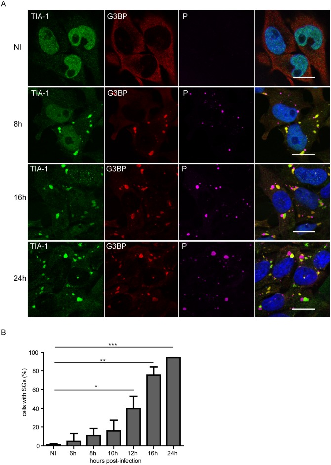
Fig 1
RABV infection induces the formation of SG-like structures.
(A) U373-MG cells grown on glass coverslips were either non-infected (NI) or infected with RABV (CVS strain) at a MOI of 3. At different times post-infection (8 h, 16 h, 24 h), cells were analyzed by ...
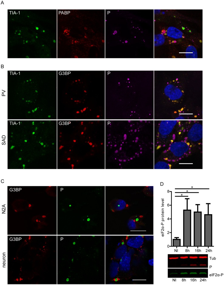
Fig 2
RABV induced-SGs contain PABP and SGs formation is independent of virus strain and cell-type.
(A) U373-MG cells were infected with RABV (CVS strain) at a MOI of 3. At 24 h p.i, cells were analyzed by confocal microscopy after co-staining with the goat anti-TIA-1, mouse anti-PABP and rabbit ant...
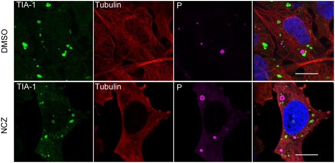
Fig 3
Effect of nocodazole on RABV-induced SGs.
U373-MG cells were infected and treated with nocodazole (NCZ) (2 u03bcM) or mock treated (DMSO) for 1 h before virus inoculation and during infection. At 20 h p.i as indicated, cells were co-stained w...
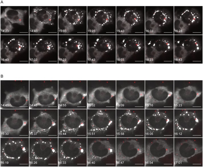
Fig 4
Live cell imaging of RABV infected cells.
U373-MG cells were transiently transfected with pG3BP-eGFP (to visualize SGs) and infected for 14 h with the recombinant virus rCVSN2C-P-mCherry. Live-cell time-lapse experiments were performed on ten...
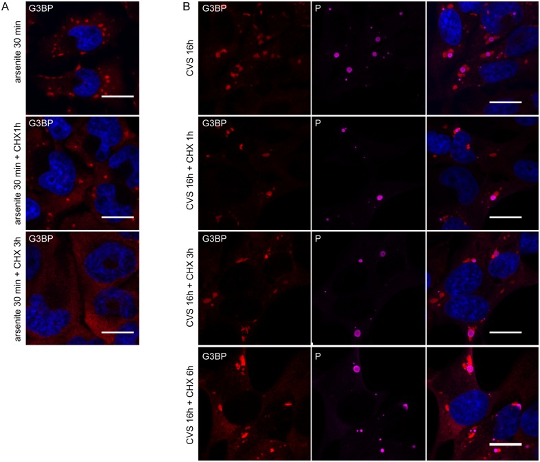
Fig 5
SGs induced by arsenite are disrupted by the protein synthesis inhibitor CHX whereas RABV-induced SGs are not.
(A) U373-MG cells were treated with arsenite (0.5mM) for 30 min and subsequently were either left untreated or treated with CHX for another 1 h or 3 h (as indicated) before processing for IF. Cells we...
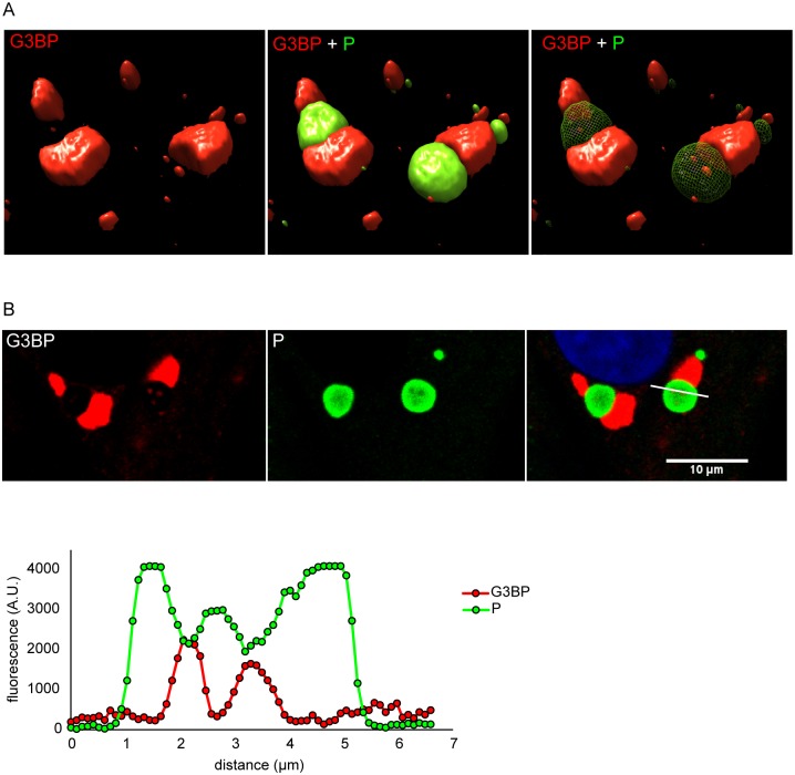
Fig 6
SGs are found adjacent to Negri bodies.
Infected U373-MG cells were analyzed by confocal microscopy after staining with rabbit anti-P antibody (green) and mouse anti-G3BP1 antibody (red) followed by incubation with secondary fluorescent IgG...
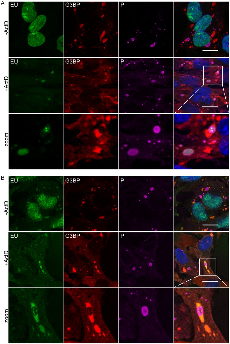
Fig 7
Localization of viral RNA and cellular RNA in SGs.
(A) U373-MG were infected for 16 h then untreated (- Act D) or treated with 20 u03bcM actinomycin D (+ Act D) for 1 h, to inhibit cellular transcription, and then fed with 1mM 5-ethynyl uridine (EU; I...
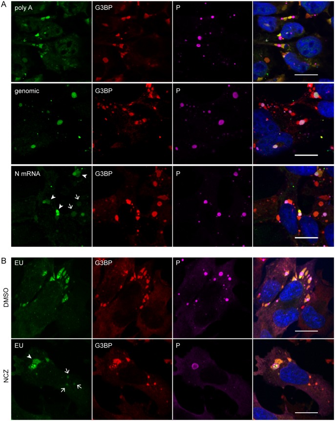
Fig 8
Viral mRNAs are specifically recruited to SGs and their transport from NBs to SGs does not require microtubules.
(A) Cells were infected (MOI = 3) for 24 h. FISH experiments were performed by using 5u2019-ATTO448 modified oligonucleotides (Eurofins Genomics) to detect cellular mRNA (polyA), viral genomic RNA and...
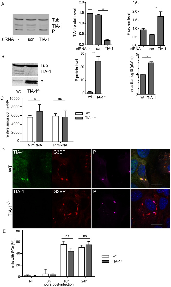
Fig 9
Effect of TIA-1 depletion on RABV infection.
(A) U373-MG cells were non-transfected (-) or transfected with non-targeting (siScr) or TIA-1 targeting (siTIA) siRNA and 48 h p.i, cells were infected with RABV (MOI of 3) for 20 h. Cells extracts we...
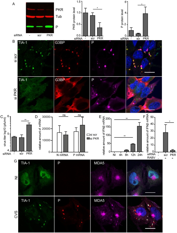
Fig 10
Effect of depletion of PKR on SGs and on RABV infection.
(A) U373-MG cells were non-transfected (-) or transfected with non-targeting (siScr) or PKR-targeting (siPKR) siRNA and 48 h post-transfection, cells were infected with RABV (MOI of 3) for 20 h. Cell ...
Figure images are served from the NIH/NLM PubMed Central Open Access Subset or Europe PMC; copyright remains with the publishers and authors.
💬 Discussion
0 commentsNo comments yet. Be the first to start a discussion!
Leave a Comment