Abstract
MicroRNA (miRNA) and RNA interference (RNAi) pathways rely on small RNAs produced by Dicer endonucleases. Mammalian Dicer primarily supports the essential gene-regulating miRNA pathway, but how it is specifically adapted to miRNA biogenesis is unknown. We show that the adaptation entails a unique structural role of Dicer's DExD/H helicase domain. Although mice tolerate loss of its putative ATPase function, the complete absence of the domain is lethal because it assures high-fidelity miRNA biogenesis. Structures of murine Dicer•-miRNA precursor complexes revealed that the DExD/H domain has a helicase-unrelated structural function. It locks Dicer in a closed state, which facilitates miRNA precursor selection. Transition to a cleavage-competent open state is stimulated by Dicer-binding protein TARBP2. Absence of the DExD/H domain or its mutations unlocks the closed state, reduces substrate selectivity, and activates RNAi. Thus, the DExD/H domain structurally contributes to mammalian miRNA biogenesis and underlies mechanistical partitioning of miRNA and RNAi pathways.
🔬 Techniques
🧬 Organisms
✨ Fluorophores
🧪 Sample Preparation
🔬 Cell Lines
🏭 Microscope Brands
🧪 Reagent Suppliers
💻 Software Details
💾 Data Repositories
🏷️ Research Resource Identifiers (RRIDs)
Verified research resources used in this paper:
🏛️ Research Organizations (ROR)
Affiliated research institutions:
📋 Methods
Key resources table REAGENT or RESOURCE SOURCE IDENTIFIER Antibodies α-HA
Rat monoclonal antibody (clone 3F10) Roche Cat# 11867431001; RRID: AB_390919 α-HA Cell Signaling Cat# 3724 Anti-HA Magnetic Beads ThermoFisher Scientific Cat# 88836; RRID: AB_2749815 α-TUBA4A Sigma-Aldrich Cat# T6074; RRID: AB_477582 α-TARBP2 ThermoFisher Scientific Cat# LF-MA0209; RRID: AB_1875916 α-FLAG Sigma-Aldrich Cat# F3165; RRID: AB_259529 mouse anti-rabbit IgG-HRP Santa-Cruz Cat# sc-2357; RRID: AB_628497 HRP-conjugated anti-mouse IgG binding protein Santa-Cruz Cat# sc-525409 goat anti-Rat IgG-HPR ThermoFisher Scientific Cat# 31470; RRID: AB_228356 Bacterial and virus strains One Shot™ TOP10 Chemically Competent cells ThermoFisher Scientific Cat# C404006 NEB 5-alpha Competent E. coli New England Biolabs part of Cat# E0554S MAX Efficiency™ DH10Bac Competent Cells ThermoFisher Scientific Cat# 10361012 P1 virus ThermoFisher Scientific N/A Sf9 cells in Sf-900™ II SFM ThermoFisher Scientific Cat# 11496015 High Five™ Cells in Express Five™ Medium ThermoFisher Scientific Cat# B85502 Chemicals, peptides, and recombinant proteins [γ- 32 P]-ATP HARTMANN ANALYTIC tCat# FP-501 2-Mercaptoethanol (50 mM) ThermoFisher Scientific Cat# 31350010 30% Acrylamide/Bis Solution, 29:1 Bio-Rad Cat# 161-0156 ALLin™ HiFi DNA Polymerase HighQu Cat# HLE0201 Benzonase® Nuclease Sigma-Merck Cat# E1014-25KU CHIR-99021 (CT99021) HCl Selleck Chemicals Cat# S2924 DAPI Sigma-Aldrich Cat# 10236276001 Decade™ Markers System ThermoFisher Scientific Cat# AM7778 DMEM Sigma-Aldrich Cat# D6429 dNTP Mix (10 mM each) ThermoFisher Scientific Cat# R0192 Fetal Bovine Serum (FCS) Sigma-Aldrich Cat# F7524 FuGENE HD Transfection Reagent Promega Cat# E2311 Gelatin from cold water fish skin Sigma-Aldrich Cat# G7765 Immobilon-P PVDF Membrane Sigma-Aldrich Cat# IPVH00010 KnockOut DMEM ThermoFisher Scientific Cat# 10829018 L-Glutamin solution Sigma-Aldrich Cat# G7513 LIF Isokine Cat# 01-A1140-100 Lipofectamine 3000 Transfection Reagent ThermoFisher Scientific Cat# L3000015 Lugol’s Iodine: Potassium iodide; Iodine Penta Cat# 7681-11-0 Cat# 17570-30500 MEM Non-Essential Amino Acids Solution (100X) ThermoFisher Scientific Cat# 11140068 Mirdametinib (PD0325901) Selleck Chemicals Cat# S1036 mouse Dicer SOM this paper N/A mouse Dicer ΔHEL1 this paper N/A NiNTA-agarose Qiagen Cat# 30210 Penicillin-Streptomycin Sigma-Aldrich Cat# P0781 PFA Penta Cat# 23700-31000 Pierce Anti-HA Magnetic Beads ThermoFisher Scientific Cat# 88836 Protease Inhibitor Cocktail Set III, Animal-Free Sigma-Aldrich Cat# 535140 Qiazol lysis reagent Qiagen Cat# 79306 RevertAid Reverse Transcriptase (200 U/μL) ThermoFisher Scientific Cat# EP0441 RNase Inhibitor, Murine New England BioLabs Cat# M0314L SuperSignal™ West Femto Maximum Sensitivity Substrate ThermoFisher Scientific Cat# 34096 SYBR™ Green PCR Master Mix ThermoFisher Scientific Cat# 4309155 T4 Polynucleotide Kinase New England BioLabs Cat# M0201L V-53D Diluent Mindray Cat# 105-000146-00 Vivaspin Turbo15 Sartorius Cat# VS15T41 Critical commercial assays Click-it EdU Imaging Kit ThermoFisher Scientific Cat# C10337 Fuji imaging plate BAS-IP MS 2025 VWR Cat# 28-9564-75 In Situ Cell Death Detection Kit, TMR red Roche Cat# 12156792910 Monarch® Genomic DNA Purification Kit New England Biolabs Cat# T1030S NEBNext® Multiplex Small RNA Library Prep Set for Illumina® NEB Cat# E7300S NEBNext® Ultra™ II RNA Library Prep Kit for Illumina® NEB Cat# E7760S NEXTflex Small RNA-Seq Kit v3 BiooScientific Cat# NOVA-5132-06 PCR Genotyping Kit Top-Bio Cat# D227 Protein Assay Kit I (Bradford assay) Bio-Rad Cat# 500-0006 Q5 Site-Directed Mutagenesis kit New England Biolabs Cat# E0554S Ribo-Zero® plus rRNA depletion Kit Illumina Cat# 20040526 RNeasy Mini Kit Qiagen Cat# 74104 Strep-Tactin™XT Superflow™ High Capacity Resin IBA Lifesciences Cat# 2-4030-010 Superdex® 75 Increase 10/300 GL Cytiva Cat# 29-1487-21 Superose® 6 Increase 10/300 GL Cytiva Cat# 29-0915-96 X-ray film Blue Cole-Parmer Cat# 21700-03 Deposited data Coordinates of Arabidopsis DCL1 in complex with pre-miRNA 166f Wei et al., 2021 PDB: 7ELE Coordinates of Arabidopsis DCL3 in complex with a 40-bp RNA Wang et al., 2021 PDB: 7VG2 Coordinates of human Dicer•TARBP2 complex Liu et al., 2018 PDB: 5ZAK Coordinates of human Dicer•TARBP2•pre-let-7 complex Liu et al., 2018 PDB: 5ZAL Coordinates of mouse Dicer this paper, Table S4 PDB: 7YZ4 Coordinates of mouse Dicer in complex with pre-miR-15a this paper, Table S4 PDB: 7YYM Coordinates of mouse Dicer O in complex with pre-miR-15a this paper, Table S4 PDB: 7YYN Coordinates of mouse Dicer in complex with pre-miR-15a and TARBP2 (pre-cleavage) this paper, Table S4 PDB: 7ZPK Coordinates of mouse Dicer in complex with pre-miR-15a and TARBP2 (cleavage) this paper, Table S4 PDB: 7ZPI RNA-seq data this paper, Table S5 GEO: GSE196310 Experimental models: Cell lines DicerX/X and Pkr-/- ESC strain this paper N/A DicerX/X ESC strain this paper N/A Hi5 cells ThermoFisher Scientific Cat# B85502 Human osteosarcoma U-2 OS ATCC Cat# HTB-96 Human osteosarcoma U-2 OS (PKR knock-out exons 3-8) this paper N/A NIH 3T3 cells ATCC Cat# CRL-1658 NIH 3T3 cells (PKR knock-out exons 3-8) this paper N/A RS7 parental ESC strain this paper Czech Centre for Phenogenomics Sf9 cells ThermoFisher Scientific Cat# 11496015 Experimental models: Organisms/strains DicerGNT mouse strain this paper N/A DicerDQCH mouse strain this paper N/A DicerX mouse strain this paper N/A DicerSOM mouse strain Taborska et al., 2019 N/A Oligonucleotides GUCCAGUUUUCCCAGGAAUCCC UUGGAUGCUAAGAUGGGGAUUC CUGGAAAUACUGUUCUUG this paper; RNA oligonucleotide Sigma-Aldrich, pre-miR-145a UAGCAGCACAUAAUGGUUUGUGG AUGUUGAAAAGGUGCAGGCCAUA CUGUGCUGCCUCA this paper; RNA oligonucleotide Sigma-Aldrich, pre-miR-15a GUGAGGCUCAGUAUGGGGUGGG GGUGUCGUCGCCUGCCCGACUG ACCACCCACUCACCCUGGACUG ACUCUCAG this paper; RNA oligonucleotide Sigma-Aldrich, pre-miR-7068 AGAGGAGAGGGACAAUCAUAAA GGCCACUCGCAAGAGUGGCCU UUAUGAUUGUCCCUCUCCUCUUU this paper; RNA oligonucleotide Sigma-Aldrich, 30bp stem-loop , AGAGGAGAGGGACAAUAGAG GAGAGGGACAAUCAUAAAG GCCGCAAGGCCUUUAUGAUU GUCCCUCUCCUCUAUUGUC CCUCUCCUCUUU this paper; RNA oligonucleotide Sigma-Aldrich, 42bp stem-loop GTACCCAAATGGATAGAA this paper; sgRNA target site in intron 2 mDcr_i2a GTTGGGATGGAGGTTGTT this paper; sgRNA target site in intron 2 mDcr_i2b GAGATGAGTCCTATAAAGGGG this paper; sgRNA target site in intron 2, No.1 mDcr_i2-1 CCCCTCTGTCTCCTAAACTGC this paper; sgRNA targeting intron 2, No.2: mDcr_i2-2 ACGGGAAGAAGAAATGGCTGG this paper; sgRNA target site in intron 2, No.3 mDcr_i2-3 ACTACGCTAGGTGTAAACAG this paper; sgRNA target site in intron 6 mDcr_i6a TGCAGTCCCCGGACGTTAAAT this paper; sgRNA target site in intron 6 mDcr_i6b GCCATCTAGATATACAGGAGG his paper, sgRNA target site in intron 8, No.1 mDcr_i8-1 CCTTACCCTTCCACACGTCAC this paper; sgRNA target site in intron 8, No.2 mDcr_i8-2 CCTTCTTTAACACTTGGCTTC this paper; sgRNA target site in intron 1 Pkr_i1a CCTGTGGTGGGTTGGAAACAC this paper; sgRNA target site in intron 1 Pkr_i1b GTGGAGTTGGTGGCCACGGGG this paper; sgRNA target site in intron 5 Pkr_i5a CCTGTGTACCAACAATGATCC this paper; sgRNA target site in intron 5 Pkr_i5b GCCTTGTTTTGACCATAAATGCCG this paper; PKR genotyping primer Pkr.fwd GTGACAACGCTAGAGGATGTTCCG this paper; PKR genotyping primer Pkr.rev GATATAACCAGCTCAAGTGTTTGC this paper; Dicer genotyping primer (1st round of nested PCR) mDcr_i1_Fwd GAGCAAAAAGTTCATCAGGAACC Dicer genotyping primer (1st round of nested PCR) mDcr_i7_Rev GCCTGGTTGGGTATAGACTGCTTG this paper; Dicer genotyping primer (2nd round of nested PCR) mDcr_i1_Fwd2 CAGAGGGCTAGAGCATACAAACAC this paper; Dicer genotyping primer (2nd round of nested PCR) mDcr_i7_Rev2 CAAGCCCGCCTCTTCTGATT this paper; DicerGNT genotyping primer Dicer_26720F ATGGCACGAATGACTGAACC this paper; DicerGNT genotyping primer Dicer_28976R CAGGTCTCATCTGCCAAGGT this paper; DicerDQCH genotyping primer DQCH_30860F TGGAAGCAAGGCTTAGGAAA this paper; DicerDQCH genotyping primer DQCH_33000R cacgacatcgactacaaggacg acgacgacaagTGAAGCGGC CGCTTCCCT this paper; cloning oligo 2xFLAG_F gtccttgtagtcaccgtcgtggtcc ttgtagtcGCTATTGGGAACCTG AGGTTGATTAGC this paper; cloning oligo 2xFLAG_Rev GGGCTTTATGAAAGACTGC this paper; cloning oligo dHEL1_F TTGCAAAGCAGGGCTTTT this paper; cloning oligo dDExD_R & dHEL1 AACACGGCCATTGGACAC this paper; cloning oligo dDExD_F & dHEL2_F TAAGACAACTGCTGTGTATCTTC this paper; cloning oligo dHEL2_R GAAGATGTGGAAATCAAGCCTCGCG this paper; cloning oligo dmHEL1_F GGACACCATGACCTCTGTGGGCTTG this paper; cloning oligo dmHEL1_R GTGGAAGCAGCTACCGACCA TAACACAATTGTGTGCTTGAA CACTGGCTCAGGGAAGACGT TCATCGCGGTCCTGCTCACC AAAGAGCTGGCCCAGCAGA TCAGGG this paper; cloning oligo VTLQC_F CAAGTTCTGACGGCTGACAC TTGTTGAGCAACCTGGTTTGC AGAGTTGACGAGGAACAC GGTCCTTTTTGCATGCGGG TTGAGGTCGCCCCTGATCT GCTGGGCCAGCTCTTTGGT this paper; cloning oligo VTLQC_R AAGGACCATAACACAATTGT GTGCTTGAACACTGGCTCAG GGAAGACGTTCATCGCGGTC AAGCTCACCAAAGAGCTGGC CAAGCAGATCAGGGGCGACC TCAACC this paper; cloning oligo LKKKK_F CTTAGTTCTGACGGCTGACA CTTGTTGAGCAACCTGGTTT GCAGAGTTGACGAGGAACAC GGTCCTTTTTGCATGCGGGT TGAGGTCGCCCCTGATCTG CTTGG this paper; cloning oligo LKKKK_R CCGTTCATTTCCCAGCCTGT this paper; genotyping Deletion confirmation - forward primer AAAACAGCCCAATTCCTTGCC this paper; genotyping Deletion confirmation - reverse primer ATCTACGGATCCACCATGGTATGGA GCCATCCTCAATTTGAAAAGGG TGGCGGGTCCGGCGGTGGGTC TGGCGGTAGCGCTTGGTCCCA CCCCCAGTTCG this paper; cloning oligo Twin-HA-TEV_Fwd GTAGATGTCGACCAGGCCCTGAA AATACAGGTTTTCGGTACCAGCGT AATCTGGAACATCGTATGGGTAGT CACCCTTCTCGAACTGGGGGTGG GACCAA this paper; cloning oligo Twin-HA-TEV_Rev TCTACAGCGGCCGCGGCGAGA ATCTCTACTTCCAAGGCGCTAG CGACTATAAGGACCACGACGG AGACTA this paper; cloning oligo C_TEV-FLAG-His_Fwd GTAGATAAGCTTAGTGATGGTG ATGGTGATGGTGGTGGGACCC ATCATGATCCTTGTAGTCTCCG TCGTGGTCCTT this paper; cloning oligo C_TEV-FLAG-His_Rev ATGTCGACGCAGGCCTGCAGC TCATGACCCC this paper; cloning oligo mDicer_SalI_Fwd CAGTCGACAGCCGTGATACAG AAGTATACAC this paper; cloning oligo mDicerO_SalI_Fwd ATGCGGCCGCTGTTAGGAACCT GAGGCTGGTTAGC this paper; cloning oligo mDicer-NotI_Rev GCTGACAAGAGCATAGCGGAC TGTGTTGCTGCACTGCTGGGC TGCTACTTAACCAGC this paper; cloning oligo mDicer E1560A Forward GCTGGTTAAGTAGCAGCCCAG CAGTGCAGCAACACAGTCCGC TATGCTCTTGTCAGC this paper; cloning oligo mDicer E1560A Reverse CAAGGCCATGGGGGACATTT TTGCATCTCTTGCTGGTGCC ATTTATAT this paper; cloning oligo mDicer E1807A Forward ATATAAATGGCACCAGCAAG AGATGCAAAAATGTCCCCCATGGCCTTG this paper; cloning oligo mDicer E1807A Reverse Recombinant DNA CRISPR-Cas9 plasmid Taborska et al., 2019 N/A DicerSOM expression plasmid Addgene Cat# 120540 DicerX expression plasmid Addgene Cat# 120541 Firefly luciferase reporter – FL plasmid Addgene Cat# 120522 Hairpin-expressing plasmid CAG-EGFP-Elavl2IR Addgene Cat# 120518 Hairpin-expressing plasmid CAG-EGFP-Lin28IR Addgene Cat# 120517 Hairpin-expressing plasmid CAG-EGFP-MosIR Addgene Cat# 120516 Hairpin-expressing plasmid CAG-EGFP-MosMos Addgene Cat# 120515 Hairpin-expressing plasmid CAG-EGFP-RlucIR this paper N/A MosIR plasmid Addgene Cat# 120516 pCIneo 5’-DICER1(dHEL1)-2xFLAG this paper N/A pCIneo 5’-DICER1(dHEL2)-2xFLAG this paper N/A pCIneo 5’-DICER1(dDExD)-2xFLAG this paper N/A pEF1-MH.Bl-mDcr OO Addgene Cat# 120541 pEF1-MH.Bl-mDcr SOM Addgene Cat# 120540 pFastBACT1 5’-TwinStrep-HA-TEV-Dicer O -TEV-2xFLAG-8xHis this paper N/A pFastBACT1 5’-TwinStrep-HA-TEV-Dicer-TEV-2xFLAG-8xHis this paper N/A pFastBACT1 TwinStrep-HA-TEV-Dicer(E1560A, E1807A)-TEV-2xFLAG-8xHis this paper N/A pFastBACT1 TwinStrep-HA-TEV-Dicer O (E1560A, E1807A)-TEV-2xFLAG-8xHis this paper N/A pFastBACT1 plasmid Invitrogen N/A puromycin selection plasmids Taborska et al., 2019 N/A Renilla luciferase reporter - RL-Lin28 plasmid Addgene Cat# 120520 Software and algorithms Alphafold Jumper et al., 2021 ; Varadi et al., 2022 https://alphafold.ebi.ac.uk/ Coot 0.9.6.2 Emsley et al., 2010 https://www2.mrc-lmb.cam.ac.uk/personal/pemsley/coot/ crYOLO 1.7.6 Wagner et al., 2019 https://cryolo.readthedocs.io/en/stable/ cryoSPARC Punjani et al., 2017 https://cryosparc.com/ cutadapt version 1.8.3 Martin, 2011 N/A DESeq2 Love et al., 2014 N/A EMAN2 Tang et al., 2007 https://blake.bcm.edu/emanwiki/EMAN2 fastx-toolkit version 0.0.14 http://hannonlab.cshl.edu/fastx_toolkit N/A featureCounts v.2.0.0 Liao et al., 2014 N/A GCTF Zhang, 2016 https://www2.mrc-lmb.cam.ac.uk/research/locally-developed-software/zhang-software/ GraphPad Prism 9.1.0 GraphPad Software https://www.graphpad.com/scientific-software/prism/ ISOLDE 1.1.0 Croll, 2018 https://isolde.cimr.cam.ac.uk/static/isolde/doc/isolde.html Molprobity Davis et al., 2004 , 2007 ; Williams et al., 2018 http://molprobity.biochem.duke.edu/?fbclid=IwAR23TiIo_fFJl0iW0JjnMBtSo2JRdRKoxNt2tsD4m7hPt3FzRzvJG08IDpU MotionCor2 Zheng et al., 2017 https://emcore.ucsf.edu/ucsf-software Multi Gauge v3.2 Fujifilm, Tokyo, Japan N/A pcaExplorer Marini and Binder, 2019 N/A PHENIX 1.19.2-4158-000 Liebschner et al., 2019 https://phenix-online.org/ Relion 3.1 Scheres, 2012 , 2016 https://www3.mrc-lmb.cam.ac.uk/relion/index.php/Main_Page RNAComposer Antczak et al., 2016 ; Popenda et al., 2012 https://rnacomposer.cs.put.poznan.pl/ SerialEM Mastronarde, 2005 N/A STAR 2.7.3a Dobin et al., 2013 N/A TOPAZ Bepler et al., 2019 http://cb.csail.mit.edu/cb/topaz/ UCSC tools Kent et al., 2010 N/A UCSF Chimera 1.16 Pettersen et al., 2004 https://www.rbvi.ucsf.edu/chimera/ UCSF ChimeraX 1.3 Pettersen et al., 2021 https://www.rbvi.ucsf.edu/chimerax Validation report at wwPDB (PDB Validation tool) Berman et al., 2003 https://validate-rcsb-1.wwpdb.org/ original codes this paper DOI: https://doi.org/10.5281/zenodo.7154385 Other Lacey carbon M300 SPI supplies Cat# 3830C-MB Mindray 5300 Vet Mindray N/A SkyScan 1272 high-resolution microCT Bruker N/A UltraAuFoil M300 (R1.2/1.3) Quantifoil Cat# Q350AR13A Resource availability Lead contact Further information and requests for resources and reagents should be directed to and will be fulfilled by the lead contact Petr Svoboda ( svobodap@img.cas.cz ).
Show full methods section
Key resources table REAGENT or RESOURCE SOURCE IDENTIFIER Antibodies α-HA
Rat monoclonal antibody (clone 3F10) Roche Cat# 11867431001; RRID: AB_390919 α-HA Cell Signaling Cat# 3724 Anti-HA Magnetic Beads ThermoFisher Scientific Cat# 88836; RRID: AB_2749815 α-TUBA4A Sigma-Aldrich Cat# T6074; RRID: AB_477582 α-TARBP2 ThermoFisher Scientific Cat# LF-MA0209; RRID: AB_1875916 α-FLAG Sigma-Aldrich Cat# F3165; RRID: AB_259529 mouse anti-rabbit IgG-HRP Santa-Cruz Cat# sc-2357; RRID: AB_628497 HRP-conjugated anti-mouse IgG binding protein Santa-Cruz Cat# sc-525409 goat anti-Rat IgG-HPR ThermoFisher Scientific Cat# 31470; RRID: AB_228356 Bacterial and virus strains One Shot™ TOP10 Chemically Competent cells ThermoFisher Scientific Cat# C404006 NEB 5-alpha Competent E. coli New England Biolabs part of Cat# E0554S MAX Efficiency™ DH10Bac Competent Cells ThermoFisher Scientific Cat# 10361012 P1 virus ThermoFisher Scientific N/A Sf9 cells in Sf-900™ II SFM ThermoFisher Scientific Cat# 11496015 High Five™ Cells in Express Five™ Medium ThermoFisher Scientific Cat# B85502 Chemicals, peptides, and recombinant proteins [γ- 32 P]-ATP HARTMANN ANALYTIC tCat# FP-501 2-Mercaptoethanol (50 mM) ThermoFisher Scientific Cat# 31350010 30% Acrylamide/Bis Solution, 29:1 Bio-Rad Cat# 161-0156 ALLin™ HiFi DNA Polymerase HighQu Cat# HLE0201 Benzonase® Nuclease Sigma-Merck Cat# E1014-25KU CHIR-99021 (CT99021) HCl Selleck Chemicals Cat# S2924 DAPI Sigma-Aldrich Cat# 10236276001 Decade™ Markers System ThermoFisher Scientific Cat# AM7778 DMEM Sigma-Aldrich Cat# D6429 dNTP Mix (10 mM each) ThermoFisher Scientific Cat# R0192 Fetal Bovine Serum (FCS) Sigma-Aldrich Cat# F7524 FuGENE HD Transfection Reagent Promega Cat# E2311 Gelatin from cold water fish skin Sigma-Aldrich Cat# G7765 Immobilon-P PVDF Membrane Sigma-Aldrich Cat# IPVH00010 KnockOut DMEM ThermoFisher Scientific Cat# 10829018 L-Glutamin solution Sigma-Aldrich Cat# G7513 LIF Isokine Cat# 01-A1140-100 Lipofectamine 3000 Transfection Reagent ThermoFisher Scientific Cat# L3000015 Lugol’s Iodine: Potassium iodide; Iodine Penta Cat# 7681-11-0 Cat# 17570-30500 MEM Non-Essential Amino Acids Solution (100X) ThermoFisher Scientific Cat# 11140068 Mirdametinib (PD0325901) Selleck Chemicals Cat# S1036 mouse Dicer SOM this paper N/A mouse Dicer ΔHEL1 this paper N/A NiNTA-agarose Qiagen Cat# 30210 Penicillin-Streptomycin Sigma-Aldrich Cat# P0781 PFA Penta Cat# 23700-31000 Pierce Anti-HA Magnetic Beads ThermoFisher Scientific Cat# 88836 Protease Inhibitor Cocktail Set III, Animal-Free Sigma-Aldrich Cat# 535140 Qiazol lysis reagent Qiagen Cat# 79306 RevertAid Reverse Transcriptase (200 U/μL) ThermoFisher Scientific Cat# EP0441 RNase Inhibitor, Murine New England BioLabs Cat# M0314L SuperSignal™ West Femto Maximum Sensitivity Substrate ThermoFisher Scientific Cat# 34096 SYBR™ Green PCR Master Mix ThermoFisher Scientific Cat# 4309155 T4 Polynucleotide Kinase New England BioLabs Cat# M0201L V-53D Diluent Mindray Cat# 105-000146-00 Vivaspin Turbo15 Sartorius Cat# VS15T41 Critical commercial assays Click-it EdU Imaging Kit ThermoFisher Scientific Cat# C10337 Fuji imaging plate BAS-IP MS 2025 VWR Cat# 28-9564-75 In Situ Cell Death Detection Kit, TMR red Roche Cat# 12156792910 Monarch® Genomic DNA Purification Kit New England Biolabs Cat# T1030S NEBNext® Multiplex Small RNA Library Prep Set for Illumina® NEB Cat# E7300S NEBNext® Ultra™ II RNA Library Prep Kit for Illumina® NEB Cat# E7760S NEXTflex Small RNA-Seq Kit v3 BiooScientific Cat# NOVA-5132-06 PCR Genotyping Kit Top-Bio Cat# D227 Protein Assay Kit I (Bradford assay) Bio-Rad Cat# 500-0006 Q5 Site-Directed Mutagenesis kit New England Biolabs Cat# E0554S Ribo-Zero® plus rRNA depletion Kit Illumina Cat# 20040526 RNeasy Mini Kit Qiagen Cat# 74104 Strep-Tactin™XT Superflow™ High Capacity Resin IBA Lifesciences Cat# 2-4030-010 Superdex® 75 Increase 10/300 GL Cytiva Cat# 29-1487-21 Superose® 6 Increase 10/300 GL Cytiva Cat# 29-0915-96 X-ray film Blue Cole-Parmer Cat# 21700-03 Deposited data Coordinates of Arabidopsis DCL1 in complex with pre-miRNA 166f Wei et al., 2021 PDB: 7ELE Coordinates of Arabidopsis DCL3 in complex with a 40-bp RNA Wang et al., 2021 PDB: 7VG2 Coordinates of human Dicer•TARBP2 complex Liu et al., 2018 PDB: 5ZAK Coordinates of human Dicer•TARBP2•pre-let-7 complex Liu et al., 2018 PDB: 5ZAL Coordinates of mouse Dicer this paper, Table S4 PDB: 7YZ4 Coordinates of mouse Dicer in complex with pre-miR-15a this paper, Table S4 PDB: 7YYM Coordinates of mouse Dicer O in complex with pre-miR-15a this paper, Table S4 PDB: 7YYN Coordinates of mouse Dicer in complex with pre-miR-15a and TARBP2 (pre-cleavage) this paper, Table S4 PDB: 7ZPK Coordinates of mouse Dicer in complex with pre-miR-15a and TARBP2 (cleavage) this paper, Table S4 PDB: 7ZPI RNA-seq data this paper, Table S5 GEO: GSE196310 Experimental models: Cell lines DicerX/X and Pkr-/- ESC strain this paper N/A DicerX/X ESC strain this paper N/A Hi5 cells ThermoFisher Scientific Cat# B85502 Human osteosarcoma U-2 OS ATCC Cat# HTB-96 Human osteosarcoma U-2 OS (PKR knock-out exons 3-8) this paper N/A NIH 3T3 cells ATCC Cat# CRL-1658 NIH 3T3 cells (PKR knock-out exons 3-8) this paper N/A RS7 parental ESC strain this paper Czech Centre for Phenogenomics Sf9 cells ThermoFisher Scientific Cat# 11496015 Experimental models: Organisms/strains DicerGNT mouse strain this paper N/A DicerDQCH mouse strain this paper N/A DicerX mouse strain this paper N/A DicerSOM mouse strain Taborska et al., 2019 N/A Oligonucleotides GUCCAGUUUUCCCAGGAAUCCC UUGGAUGCUAAGAUGGGGAUUC CUGGAAAUACUGUUCUUG this paper; RNA oligonucleotide Sigma-Aldrich, pre-miR-145a UAGCAGCACAUAAUGGUUUGUGG AUGUUGAAAAGGUGCAGGCCAUA CUGUGCUGCCUCA this paper; RNA oligonucleotide Sigma-Aldrich, pre-miR-15a GUGAGGCUCAGUAUGGGGUGGG GGUGUCGUCGCCUGCCCGACUG ACCACCCACUCACCCUGGACUG ACUCUCAG this paper; RNA oligonucleotide Sigma-Aldrich, pre-miR-7068 AGAGGAGAGGGACAAUCAUAAA GGCCACUCGCAAGAGUGGCCU UUAUGAUUGUCCCUCUCCUCUUU this paper; RNA oligonucleotide Sigma-Aldrich, 30bp stem-loop , AGAGGAGAGGGACAAUAGAG GAGAGGGACAAUCAUAAAG GCCGCAAGGCCUUUAUGAUU GUCCCUCUCCUCUAUUGUC CCUCUCCUCUUU this paper; RNA oligonucleotide Sigma-Aldrich, 42bp stem-loop GTACCCAAATGGATAGAA this paper; sgRNA target site in intron 2 mDcr_i2a GTTGGGATGGAGGTTGTT this paper; sgRNA target site in intron 2 mDcr_i2b GAGATGAGTCCTATAAAGGGG this paper; sgRNA target site in intron 2, No.1 mDcr_i2-1 CCCCTCTGTCTCCTAAACTGC this paper; sgRNA targeting intron 2, No.2: mDcr_i2-2 ACGGGAAGAAGAAATGGCTGG this paper; sgRNA target site in intron 2, No.3 mDcr_i2-3 ACTACGCTAGGTGTAAACAG this paper; sgRNA target site in intron 6 mDcr_i6a TGCAGTCCCCGGACGTTAAAT this paper; sgRNA target site in intron 6 mDcr_i6b GCCATCTAGATATACAGGAGG his paper, sgRNA target site in intron 8, No.1 mDcr_i8-1 CCTTACCCTTCCACACGTCAC this paper; sgRNA target site in intron 8, No.2 mDcr_i8-2 CCTTCTTTAACACTTGGCTTC this paper; sgRNA target site in intron 1 Pkr_i1a CCTGTGGTGGGTTGGAAACAC this paper; sgRNA target site in intron 1 Pkr_i1b GTGGAGTTGGTGGCCACGGGG this paper; sgRNA target site in intron 5 Pkr_i5a CCTGTGTACCAACAATGATCC this paper; sgRNA target site in intron 5 Pkr_i5b GCCTTGTTTTGACCATAAATGCCG this paper; PKR genotyping primer Pkr.fwd GTGACAACGCTAGAGGATGTTCCG this paper; PKR genotyping primer Pkr.rev GATATAACCAGCTCAAGTGTTTGC this paper; Dicer genotyping primer (1st round of nested PCR) mDcr_i1_Fwd GAGCAAAAAGTTCATCAGGAACC Dicer genotyping primer (1st round of nested PCR) mDcr_i7_Rev GCCTGGTTGGGTATAGACTGCTTG this paper; Dicer genotyping primer (2nd round of nested PCR) mDcr_i1_Fwd2 CAGAGGGCTAGAGCATACAAACAC this paper; Dicer genotyping primer (2nd round of nested PCR) mDcr_i7_Rev2 CAAGCCCGCCTCTTCTGATT this paper; DicerGNT genotyping primer Dicer_26720F ATGGCACGAATGACTGAACC this paper; DicerGNT genotyping primer Dicer_28976R CAGGTCTCATCTGCCAAGGT this paper; DicerDQCH genotyping primer DQCH_30860F TGGAAGCAAGGCTTAGGAAA this paper; DicerDQCH genotyping primer DQCH_33000R cacgacatcgactacaaggacg acgacgacaagTGAAGCGGC CGCTTCCCT this paper; cloning oligo 2xFLAG_F gtccttgtagtcaccgtcgtggtcc ttgtagtcGCTATTGGGAACCTG AGGTTGATTAGC this paper; cloning oligo 2xFLAG_Rev GGGCTTTATGAAAGACTGC this paper; cloning oligo dHEL1_F TTGCAAAGCAGGGCTTTT this paper; cloning oligo dDExD_R & dHEL1 AACACGGCCATTGGACAC this paper; cloning oligo dDExD_F & dHEL2_F TAAGACAACTGCTGTGTATCTTC this paper; cloning oligo dHEL2_R GAAGATGTGGAAATCAAGCCTCGCG this paper; cloning oligo dmHEL1_F GGACACCATGACCTCTGTGGGCTTG this paper; cloning oligo dmHEL1_R GTGGAAGCAGCTACCGACCA TAACACAATTGTGTGCTTGAA CACTGGCTCAGGGAAGACGT TCATCGCGGTCCTGCTCACC AAAGAGCTGGCCCAGCAGA TCAGGG this paper; cloning oligo VTLQC_F CAAGTTCTGACGGCTGACAC TTGTTGAGCAACCTGGTTTGC AGAGTTGACGAGGAACAC GGTCCTTTTTGCATGCGGG TTGAGGTCGCCCCTGATCT GCTGGGCCAGCTCTTTGGT this paper; cloning oligo VTLQC_R AAGGACCATAACACAATTGT GTGCTTGAACACTGGCTCAG GGAAGACGTTCATCGCGGTC AAGCTCACCAAAGAGCTGGC CAAGCAGATCAGGGGCGACC TCAACC this paper; cloning oligo LKKKK_F CTTAGTTCTGACGGCTGACA CTTGTTGAGCAACCTGGTTT GCAGAGTTGACGAGGAACAC GGTCCTTTTTGCATGCGGGT TGAGGTCGCCCCTGATCTG CTTGG this paper; cloning oligo LKKKK_R CCGTTCATTTCCCAGCCTGT this paper; genotyping Deletion confirmation - forward primer AAAACAGCCCAATTCCTTGCC this paper; genotyping Deletion confirmation - reverse primer ATCTACGGATCCACCATGGTATGGA GCCATCCTCAATTTGAAAAGGG TGGCGGGTCCGGCGGTGGGTC TGGCGGTAGCGCTTGGTCCCA CCCCCAGTTCG this paper; cloning oligo Twin-HA-TEV_Fwd GTAGATGTCGACCAGGCCCTGAA AATACAGGTTTTCGGTACCAGCGT AATCTGGAACATCGTATGGGTAGT CACCCTTCTCGAACTGGGGGTGG GACCAA this paper; cloning oligo Twin-HA-TEV_Rev TCTACAGCGGCCGCGGCGAGA ATCTCTACTTCCAAGGCGCTAG CGACTATAAGGACCACGACGG AGACTA this paper; cloning oligo C_TEV-FLAG-His_Fwd GTAGATAAGCTTAGTGATGGTG ATGGTGATGGTGGTGGGACCC ATCATGATCCTTGTAGTCTCCG TCGTGGTCCTT this paper; cloning oligo C_TEV-FLAG-His_Rev ATGTCGACGCAGGCCTGCAGC TCATGACCCC this paper; cloning oligo mDicer_SalI_Fwd CAGTCGACAGCCGTGATACAG AAGTATACAC this paper; cloning oligo mDicerO_SalI_Fwd ATGCGGCCGCTGTTAGGAACCT GAGGCTGGTTAGC this paper; cloning oligo mDicer-NotI_Rev GCTGACAAGAGCATAGCGGAC TGTGTTGCTGCACTGCTGGGC TGCTACTTAACCAGC this paper; cloning oligo mDicer E1560A Forward GCTGGTTAAGTAGCAGCCCAG CAGTGCAGCAACACAGTCCGC TATGCTCTTGTCAGC this paper; cloning oligo mDicer E1560A Reverse CAAGGCCATGGGGGACATTT TTGCATCTCTTGCTGGTGCC ATTTATAT this paper; cloning oligo mDicer E1807A Forward ATATAAATGGCACCAGCAAG AGATGCAAAAATGTCCCCCATGGCCTTG this paper; cloning oligo mDicer E1807A Reverse Recombinant DNA CRISPR-Cas9 plasmid Taborska et al., 2019 N/A DicerSOM expression plasmid Addgene Cat# 120540 DicerX expression plasmid Addgene Cat# 120541 Firefly luciferase reporter – FL plasmid Addgene Cat# 120522 Hairpin-expressing plasmid CAG-EGFP-Elavl2IR Addgene Cat# 120518 Hairpin-expressing plasmid CAG-EGFP-Lin28IR Addgene Cat# 120517 Hairpin-expressing plasmid CAG-EGFP-MosIR Addgene Cat# 120516 Hairpin-expressing plasmid CAG-EGFP-MosMos Addgene Cat# 120515 Hairpin-expressing plasmid CAG-EGFP-RlucIR this paper N/A MosIR plasmid Addgene Cat# 120516 pCIneo 5’-DICER1(dHEL1)-2xFLAG this paper N/A pCIneo 5’-DICER1(dHEL2)-2xFLAG this paper N/A pCIneo 5’-DICER1(dDExD)-2xFLAG this paper N/A pEF1-MH.Bl-mDcr OO Addgene Cat# 120541 pEF1-MH.Bl-mDcr SOM Addgene Cat# 120540 pFastBACT1 5’-TwinStrep-HA-TEV-Dicer O -TEV-2xFLAG-8xHis this paper N/A pFastBACT1 5’-TwinStrep-HA-TEV-Dicer-TEV-2xFLAG-8xHis this paper N/A pFastBACT1 TwinStrep-HA-TEV-Dicer(E1560A, E1807A)-TEV-2xFLAG-8xHis this paper N/A pFastBACT1 TwinStrep-HA-TEV-Dicer O (E1560A, E1807A)-TEV-2xFLAG-8xHis this paper N/A pFastBACT1 plasmid Invitrogen N/A puromycin selection plasmids Taborska et al., 2019 N/A Renilla luciferase reporter - RL-Lin28 plasmid Addgene Cat# 120520 Software and algorithms Alphafold Jumper et al., 2021 ; Varadi et al., 2022 https://alphafold.ebi.ac.uk/ Coot 0.9.6.2 Emsley et al., 2010 https://www2.mrc-lmb.cam.ac.uk/personal/pemsley/coot/ crYOLO 1.7.6 Wagner et al., 2019 https://cryolo.readthedocs.io/en/stable/ cryoSPARC Punjani et al., 2017 https://cryosparc.com/ cutadapt version 1.8.3 Martin, 2011 N/A DESeq2 Love et al., 2014 N/A EMAN2 Tang et al., 2007 https://blake.bcm.edu/emanwiki/EMAN2 fastx-toolkit version 0.0.14 http://hannonlab.cshl.edu/fastx_toolkit N/A featureCounts v.2.0.0 Liao et al., 2014 N/A GCTF Zhang, 2016 https://www2.mrc-lmb.cam.ac.uk/research/locally-developed-software/zhang-software/ GraphPad Prism 9.1.0 GraphPad Software https://www.graphpad.com/scientific-software/prism/ ISOLDE 1.1.0 Croll, 2018 https://isolde.cimr.cam.ac.uk/static/isolde/doc/isolde.html Molprobity Davis et al., 2004 , 2007 ; Williams et al., 2018 http://molprobity.biochem.duke.edu/?fbclid=IwAR23TiIo_fFJl0iW0JjnMBtSo2JRdRKoxNt2tsD4m7hPt3FzRzvJG08IDpU MotionCor2 Zheng et al., 2017 https://emcore.ucsf.edu/ucsf-software Multi Gauge v3.2 Fujifilm, Tokyo, Japan N/A pcaExplorer Marini and Binder, 2019 N/A PHENIX 1.19.2-4158-000 Liebschner et al., 2019 https://phenix-online.org/ Relion 3.1 Scheres, 2012 , 2016 https://www3.mrc-lmb.cam.ac.uk/relion/index.php/Main_Page RNAComposer Antczak et al., 2016 ; Popenda et al., 2012 https://rnacomposer.cs.put.poznan.pl/ SerialEM Mastronarde, 2005 N/A STAR 2.7.3a Dobin et al., 2013 N/A TOPAZ Bepler et al., 2019 http://cb.csail.mit.edu/cb/topaz/ UCSC tools Kent et al., 2010 N/A UCSF Chimera 1.16 Pettersen et al., 2004 https://www.rbvi.ucsf.edu/chimera/ UCSF ChimeraX 1.3 Pettersen et al., 2021 https://www.rbvi.ucsf.edu/chimerax Validation report at wwPDB (PDB Validation tool) Berman et al., 2003 https://validate-rcsb-1.wwpdb.org/ original codes this paper DOI: https://doi.org/10.5281/zenodo.7154385 Other Lacey carbon M300 SPI supplies Cat# 3830C-MB Mindray 5300 Vet Mindray N/A SkyScan 1272 high-resolution microCT Bruker N/A UltraAuFoil M300 (R1.2/1.3) Quantifoil Cat# Q350AR13A Resource availability Lead contact Further information and requests for resources and reagents should be directed to and will be fulfilled by the lead contact Petr Svoboda ( svobodap@img.cas.cz ).
Materials availability
Animals and plasmids are available upon request from the lead contact.
Experimental model and subject details Animals
Mus musculus genetically modified strains Dicer GNT , Dicer DQCH , Dicer ΔHEL1 , and Dicer SOM Animal experiments concerning Dicer GNT and Dicer DQCH model were carried out in accordance with the Italian law under a license from the Italian Ministry of Health.
Animal experiments concerning Dicer ΔHEL1 and Dicer
SOM models were carried out in accordance with the Czech law and were approved by the Institutional Animal Use and Care Committee (approval no. 34-2014). Dicer SOM and Dicer ΔHEL1 mutant mice Production of Dicer ΔHEL1 model was analogous to production of Dicer SOM described previously ( Taborska et al., 2019 ). We first produced ESCs with the Dicer ΔHEL1 allele and then used those for producing chimeric mice and establishing Dicer ΔHEL1 line upon germline transmission of the Dicer ΔHEL1 allele. Dicer ΔHEL1 allele in ESCs ( Nagy et al., 1993 ) was generated using CRISPR-Cas9 ( Ran et al., 2013 ) mediated modification of the endogenous Dicer locus. Pairs of sgRNAs were designed to cleave Dicer genomic sequence in intron 2 (sequence of DNA targets: mDcr_i2a 5′-GTACCCAAATGGATAGAA-3′, mDcr_i2b 5′-GTTGGGATGGAGGTTGTT-3′) and intron 6 (sequence of DNA targets: mDcr_i6a 5′-ACTACGCTAGGTGTAAACAG-3′, mDcr_i6b 5′-TGCAGTCCCCGGACGTTAAAT-3′). A template for homologous recombination was designed to contain an HA-tag at the N-terminus of Dicer coding sequence fused to exon 7 of Dicer and ∼ 1.5 kb overhangs on both ends ( Figure S1 A). Final genomic sequence of Dicer ΔHEL1 mice is provided in Document S1 . To produce Dicer ΔHEL1 mouse strain, we first produced mouse chimeras by ESC microinjection into eight-cell – stage embryos ( Poueymirou et al., 2007 ); host embryos were isolated from C57Bl/6NCrl mice ( Figure S1 ). We used two ESC lines with C57Bl/6NCrl background (commonly used JM8A3.N1 and homemade RS7) and one in 129 strain (R1). For the first three rounds of chimera production, we used homozygous and heterozygous mutant ESCs and obtained mice with varying degree mosaicism, but we failed to obtain transmission of the mutant allele into the next generation. During the fourth round, a heterozygous ESC clone D11 derived from R1 ESC line yielded a male with > 80% chimeric fur. Breeding of this male with ICR females finally lead to germline transmission of Dicer ΔHEL1 allele into the next generation and establishment of the Dicer ΔHEL1 mouse line. Sequences of the engineered Dicer locus in the mouse genome are provided in the File S1. Phenotype analysis was performed with N3 animals, small RNA seq was done with N7 and N8 embryos (all breedings to ICR background). Dicer GNT mutant mice The Dicer GNT allele was generated by replacing wild-type exon 3 with a mutant exon in which Lys60 was mutated to encode asparagine. The Dicer locus was targeted with a vector containing homology arms and a loxP -flanked neomycin cassette 5′ of exon 3 that contained the Lys60Asn mutation. Southern blotting of genomic SacI-digested DNA from individual ESC-derived clones with a 3′ probe was used to identify homologous recombinants, where the Dicer GNT-Neo allele displaying a 5.9-kb DNA fragment could be distinguished from the wild-type allele of 7.1-kb fragment size. Cre-mediated recombination resulted in the excision of the loxP -flanked neomycin cassette and the generation of the Dicer GNT allele. Mice analyzed in this study were on a C57Bl/6 genetic background. Dicer DQCH mutant mice The Dicer FH-DQCH allele was generated by retargeting the Dicer Neo allele, which contains a Flag-HA-HA sequence 5′ of exon 2 and a loxP -flanked neomycin cassette within intron 2 ( Comazzetto et al., 2014 ). This was achieved with a vector comprised of homology arms, an FRT-flanked hygromycin cassette and exon 5 in which the Glu166 codon was mutated to encode glutamine. Southern blotting of genomic SacI-digested DNA from individual ESC-derived clones with a 3′ probe was used to identify homologous recombinants with the Dicer FH-DQCH-Neo-Hyg allele displaying a 7.8-kb DNA fragment. Flp-mediated recombination removed the FRT-flanked hygromycin cassette and generated the Dicer FH-DQCH-Neo allele that was identified with the 3′ probe as a 5.9-kb SacI DNA fragment. Cre-mediated recombination led to the excision of the loxP -flanked neomycin cassette and the generation of the Dicer FH-DQCH allele. The targeting for both alleles was performed in A9 ES cells. Targeted ES cells were injected into C57BL/6 eight-cell-stage embryos. Targeted mice were crossed to deleter Cre mice ( Schwenk et al., 1995 ) or FLP- expressing transgenic mice ( Farley et al., 2000 ) to remove antibiotic resistance cassettes. The mice analyzed in this study were on a C57Bl/6 genetic background.
Cell culture and transfection Mouse
ESCs were cultured in 2i-LIF media: KnockOut-DMEM (ThermoFisher) supplemented with 15% fetal calf serum (Sigma), 1x L-Glutamine (Sigma), 1x non-essential amino acids (ThermoFisher), 50 μM β-Mercaptoethanol (ThermoFisher), 1000 U/mL LIF (Isokine), 1 μM PD0325901, 3 μM CHIR99021 (Selleck Chemicals), penicillin (100 U/mL), and streptomycin (100 μg/mL). All plastic was coated with 1% gelatin (Sigma) in PBS. NIH 3T3 fibroblasts were cultured in DMEM (Sigma) supplemented with 10% fetal calf serum, penicillin (100 U/mL), and streptomycin (100 μg/mL).
Method details Phenotype analyses Genotyping
Tail biopsies were processed by PCR genotyping kit (Top-Bio) according to the manufacturer’s protocol. 1 μl aliquots were used for genotyping PCR using 0.5 U/reaction of DNA polymerase (highQu). Genotyping primers are provided in the key resources table . Embryo harvest Mice were mated overnight, and the presence of a vaginal plug indicated embryonic day (E) 0.5. The embryos were washed in PBS and fixed in 4% PFA.
Proliferation assay - EdU staining and apoptosis TUNEL assay
Pregnant mice were injected with 60 μl of 10mM EdU 1.5 hour before embryo harvest at E10.5 and E14.5. The incorporation of EdU was visualized by Click-it EdU Imaging Kit (Invitrogen) in E10.5 whole mount samples and on 7μm paraffin sections from E14.5 embryos. Apoptosis was visualized in whole mount E10.5 embryos by TUNEL method using In Situ Cell Death Detection Kit, TMR red (Sigma-Aldrich). MicroCT E18.5 embryos were fixed for 1 week in 4% PFA and stained with Lugol’s Iodine solution for 2 weeks. Stock solution (10g KI and 5g I2 in 100ml H2O) was diluted to 25% working solution in water. Stained specimens were embedded in 2.5% low gelling temperature agarose. Scan was performed on SkyScan 1272 high-resolution microCT (Bruker, Belgium), with resolution set to 4 μm. Hematopoiesis panel 20 ul of blood from each E18.5 embryo was collected in tube containing anticoagulant EDTA and diluted with 175uL of V-53D Diluent (Mindray, 105-000146-00). The samples were measured in mode Complete blood count with Differentials (CBC + DIFF) on analyzer Mindray 5300 Vet. One-way Anova with Tukey posttest was used for statistical analysis.
RNAi activity in cultured cells assay
Effects of different Dicer isoforms on RNAi-mediated repression in Pkr –/– U-2 OS or 3T3 cells were monitored as described previously ( Demeter et al., 2019 ). Briefly, cells were co-transfected with a plasmid expressing a Dicer variant (or LacZ as a negative control), dsRNA (Lin28IR, RlucIR, or MosIR), a targeted Renilla luciferase reporter with complementary sequences to dsRNA from Lin28IR and RlucIR, and a non-targeted firefly luciferase. For transfection, cells were plated on 24-well plates, grown to 80% density and transfected using Lipofectamine 3000 (Thermo Fisher) according to the manufacturer’s protocol. The total amount of transfected DNA was kept constant (1 μg/well). Specific repression of the targeted Renilla luciferase was estimated as Renilla luciferase activity normalized to the non-targeted firefly luciferase activity, and non-specific effect of MosIR (expressing a non-targeting dsRNA). The value 1.0 corresponds to absence of RNAi, the value of LacZ negative control reflects repression mediated by endogenously-expressed Dicer.
Western blotting
Mouse tissues, U-2 OS cells transfected with Dicer variants or ES cells were homogenized mechanically in RIPA lysis buffer supplemented with 2x protease inhibitor cocktail set (Millipore) and loaded with SDS dye. Protein concentration was measured by Bradford assay (Bio-Rad) and 80 μg of total protein was used per lane. Proteins were separated on 5.5% polyacrylamide (PAA) gel and transferred on PVDF membrane (Millipore) using semi-dry blotting for 50 min, 35 V. The membrane was blocked in 5% skim milk in TBS-T, Dicer was detected using anti-HA 3F10 monoclonal primary antibody (High Affinity rat IgG1, Roche #11867431001; dilution 1:500), anti-HA rabbit primary antibody (Cell Signaling, #3724, dilution 1:1,000) or anti-Flag (M2 mouse monoclonal antibody, Sigma #F3165, dilution 1:10,000) and incubated overnight at 4°C. Secondary anti-Rat antibody (Goat anti-Rat IgG, HRP conjugate, ThermoFisher #31470, dilution 1:50,000), HRP-conjugated anti-Mouse Igg binding protein (Santa-Cruz #sc-525409, dilution 1:50,000) or anti-Rabbit-HRP antibody (Santa-Cruz #sc-2357, dilution 1:50,000) was incubated 1 h at room temperature. For TUBA4A and TARBP2 detection, samples were run on 10% PAA gel and incubated overnight at 4 °C with anti-Tubulin (Sigma, #T6074, dilution 1:10,000) or anti-TARBP2 (ThermoFisher #LF-MA0209, dilution 1:1,000) mouse primary antibodies. HRP-conjugated anti-mouse IgG binding protein (Santa-Cruz, #sc-525409, dilution 1:50,000) was used for detection. Signal was developed on films (X-ray film Blue, Cole-Parmer #21700-03) using SuperSignal West Femto Chemiluminescent Substrate (Thermo Scientific).
Immunoprecipitation NIH 3T3 cells transfected with plasmids expressing HA-tagged Dicer
ΔHEL1 or Dicer SOM variants were lysed in IP Lysis Buffer (10 mM phosphate buffer, pH 7.2, 120 mM NaCl, 1 mM EDTA, 0.5% v/v NP-40, 10% v/v glycerol). Insoluble material was pelleted by centrifugation. Cleared supernatants were diluted 4-times with IP Dilution Buffer (10 mM phosphate buffer, pH 7.2, 100 mM NaCl, 1 mM EDTA, 0.1% v/v NP-40) and incubated with anti-HA magnetic beads (anti-HA mAb, clone #2-2.2.14, ThermoFisher #88836) for 2 h on a rotator. Beads were washed 4-times with IP Dilution Buffer, finally re-suspended in 60 μl water and processed for western blotting. All buffers were supplemented with 1x Protease Inhibitor Cocktail Set (Millipore) and the whole procedure was performed at 4 °C.
RNA sequencing ESC small RNA-seq
Cells were plated on 6-well plates and grown to 80 % density. Cells were transfected with 2 μg/well of pCAG-EGFP-MosIR plasmid and cultured for 48 hours. Cells were washed with PBS, homogenized in Qiazol lysis reagent (Qiagen) and total RNA was isolated by Qiazol-chloroform extraction and ethanol precipitation method ( Toni et al., 2018 ). RNA quality was verified by Agilent 2100 Bioanalyzer. Small RNA libraries were constructed using NEBNext Multiplex Small RNA Library Prep Set for Illumina (New England Biolabs) according to the manufacturer’s protocol. Small RNA libraries were size selected on 6% PAAGE gel, a band of 140 - 150 bp was cut from the gel and RNA was extracted using Monarch® Genomic DNA Purification Kit. Quality of the libraries was assessed by Agilent 2100 bioanalyzer. Libraries were sequenced on the Illumina HiSeq2000 platform at the Genomics Core Facility at EMBL. E15.5 small RNA-seq E15.5 embryos were removed from the uterus and washed in PBS. The yolk sac was taken for genotyping and embryos were transferred into RNAlater (Thermo Fisher Scientific). Embryos were homogenized in Qiazol lysis reagent (Qiagen) and total RNA was isolated by Qiazol-chloroform extraction and ethanol precipitation method ( Toni et al., 2018 ). Small RNA libraries were constructed using Nextflex Small RNA-seq kit v3 for Illumina (Perkin Elmer) according to the manufacturer’s protocol; 3′ adapter ligation was performed overnight at 20 °C, 15 cycles were used for PCR amplification and NextFlex beads were used for size selection. Final libraries were sequenced by 75-nucleotide single-end reading using the Illumina NextSeq500/550 platform at the core genomics facility of IMG.
Bioinformatic analyses
RNA-seq data ( Table S5 ) were deposited in the Gene Expression Omnibus database under GEO: GSE196310 . Mapping of small RNA-seq data Small RNA-seq reads were trimmed in two rounds using fastx-toolkit version 0.0.14 ( http://hannonlab.cshl.edu/fastx_toolkit ) and cutadapt version 1.8.3 ( Martin, 2011 ). First, 4 random bases were trimmed from left side: fastx_trimmer -f 5 -i {INP}.fastq -o {TMP}.fastq Next, NEXTflex adapters were trimmed. Additionally, the N-nucleotides on ends of reads were trimmed and reads containing more than 10% of the N-nucleotides were discarded: cutadapt --format="fastq" --front=”GTTCAGAGTTCTACAGTCCGACGATCNNNN” --adapter=”NNNNTGGAATTCTCGGGTGCCAAGG” --error-rate=0.075 --times=2 --overlap=14 --minimum-length=12 --max-n=0.1 --output=”$ {TRIMMED}.fastq" --trim-n --match-read-wildcards $ {TMP}.fastq Trimmed reads were mapped to the mouse (mm10) genome using STAR aligner ( Dobin et al., 2013 ) with following parameters: STAR --readFilesIn $ {TRIMMED}.fastq.gz --runThreadN 4 --genomeDir $ {GENOME_INDEX} --genomeLoad LoadAndRemove --readFilesCommand unpigz -c --readStrand Unstranded --limitBAMsortRAM 20000000000 --outFileNamePrefix $ {FILENAME} --outReadsUnmapped Fastx --outSAMtype BAM SortedByCoordinate --outFilterMultimapNmax 99999 --outFilterMismatchNoverLmax 0.1 --outFilterMatchNminOverLread 0.66 --alignSJoverhangMin 999 --alignSJDBoverhangMin 999 miRNA expression analyses Mapped reads were counted using program featureCounts ( Liao et al., 2014 ). Only reads with lengths 19-25nt were selected from the small RNA-seq data: featureCounts -a $ {ANNOTATION_FILE} -F $ {FILE} -minOverlap 15 -fracOverlap 0.00 -s 1 -M -O -fraction -T 8 $ {FILE}.bam The GENCODE gene set ( Frankish et al., 2019 ) was used for the annotation of long RNA-seq data. The miRBase 22.1 ( Kozomara et al., 2019 ). set of miRNAs was used for the annotation of small RNA-seq data for main figures, mirGeneDB annotation of high-confidence miRNAs ( Fromm et al., 2022 ) was used to make sure that results were not biased by annotated low-confidence miRNAs from the miRBase. Statistical significance and fold changes in gene expression were computed in R using the DESeq2 package ( Love et al., 2014 ). Genes were considered to be significantly up- or down-regulated if their corresponding p-adjusted values were smaller than 0.05. miRNA expression plots – normalization of data, miRNA and miRNA ∗ sorting First, the relative position of each mature miRNA (“5p” and “3p” for the miRNA-5p and miRNA-3p, respectively) provided by miRBase 22.1. annotation ( Kozomara et al., 2019 ) was manually curated and completed. Second, the miRNA type of each mature miRNA (“miRNA” and ”miRNA ∗ ” for the guide strand and passenger strand miRNA, respectively ) provided by miRBase annotation was completed in this way: 1) The mature miRNAs were assigned into the pair by their hairpin names. The DESeq2 baseMean values of E15.5 and GNT experiments were added to each mature miRNA. 2) The pairs of mature miRNAs with complete miRNA type annotation (both, “miRNA” and “miRNA ∗ ” types were present) were preserved. 3) If there is only one mature miRNA annotated in the hairpin, it is assigned as “single_miRNA”. 4) If the baseMean values of both miRNAs in the pair are lower than 0.25, it is assigned as “lowExp”. 5) For the remaining pairs of mature miRNAs, if the baseMean value of one miRNA is at least double to the second one, it is assigned as “miRNA” / “miRNA ∗ ” or “miRNA ∗ ” / “miRNA”, respectively. Otherwise it is assigned as “notClear”. 6) Finally, the newly determined miRNA types are compared to each other. If the mature miRNAs were determined as “miRNA” in one experiment and as “miRNA ∗ ” in the other, it is assigned as “cellSpecific”. In all the other cases, it there is any discrepancy among the determined miRNA type, it is assigned as “notCLear”. The annotation of the mirtrons was taken from Ladewig et al. 2012 . mirGeneDB annotation of high-confidence miRNAs ( Fromm et al., 2022 ) was used to make sure that results were not biased by annotated low-confidence miRNAs from the miRBase The DESeq2 baseMean and fold changes were plotted and visualized by home-made R scripts. The MA plots related to the dominant or passenger strand miRNAs contain only the corresponding miRNAs, all the miRNAs otherwise. Small RNA clustering analysis Small RNA read clusters ( Figure S2 F) were identified following the algorithm used in previous studies ( Flemr et al., 2013 ; Demeter et al., 2019 ). Briefly: 1) Reads were weighted to fractional counts of 1/n where n represents the number of loci to which read maps 2) Reads were then collapsed into a unified set of regions and their fractional counts were summed 3) Clusters with less than 3 reads per million (RPM) were discarded 4) Clusters within 50 bp distance of each other were joined Only clusters appearing in all replicates of the same genotype (intersect) were considered in the final set. Union of coordinates of overlapping clusters were used to merge the clusters between the samples. Clusters were then annotated, and if a cluster overlapped more than one functional category, the following classification hierarchy was used: miRNA > transposable elements > mRNA (protein coding genes) > misc. RNA (other RNA annotated in ENSEMBL or RepeatMasker; Smit et al., 2013–2015 ) > other (all remaining annotated or not annotated regions).
Cleavage fidelity analysis
Only miRNAs with DESeq2 baseMean values >= 100 were selected. The cleavage points’ coordinates (CP) were extracted from their miRBase 22.1 annotation ( Kozomara et al., 2019 ). The reads of the lengths 19-25nt were selected from each replicate library. The starting and ending position of all reads were summed up in the CP and its vicinity (+/-15nt) and assigned as 3′-CP of miRNA-5p and 5′-CP of miRNA-3p, respectively. Then, the canonical miRBase CPs were re-defined based on our wild-type data: 1) Position with maximal counts (median among replicates) is assigned as the new CP. 2) If the new CP is more than 7nt outside the canonical one, keep the canonical one. 3) If there are multiple CPs with the same max counts, keep the canonical one. 4) If there are no data / no reads, keep the canonical one. The counts were extracted for each miRNA at the position of the newly defined CP with 5nt flanks on each side. The read counts were re-calculated into read densities. The final matrix was achieved as a subtraction between a mutant and its corresponding wild-type control. Top 50 miRNAs from Dicer mutants were selected based on the absolute value of the difference at the position of CP. Selected miRNAs were ordered by the change of ESC fidelity at the position of CP.
Partial processing analysis
All sequence reads were selected that overlapped the corresponding pre-miRNA locus in the sense direction. All coordinates (starting/ending position of the miRNA-5p/-3p) were extracted from the miRBase 22.1 annotation ( Kozomara et al., 2019 ). The categories shown in the Figures 5 D and S5 D were defined by pre-miRNA boundaries and the two annotated Dicer cleavage points (deviation of the boundaries +/-2nt allowed). Each read was unambiguously assigned into the appropriate category. The percentage from the total number of overlapping reads was calculated.
Luciferase assay
Dual luciferase activity was measured according to Hampf and Gossen ( Hampf and Gossen, 2006 ) with some modifications. Briefly, cells were washed with PBS and lyzed in PPTB lysis buffer (0.2% v/v Triton X-100 in 100 mM potassium phosphate buffer, pH 7.8). A 3-5 μl aliquots were used for measurement in 96-well plates using Modulus Microplate Multimode Reader (Turner Biosystems). First, firefly luciferase activity was measured by adding 50 μl substrate (20 mM Tricine, 1.07 mM (MgCO 3 ) 4 ·Mg(OH) 2 , 2.67 mM MgSO 4 , 0.1 mM EDTA, 33.3 mM DTT, 0.27 mM Coenzyme A, 0.53 mM ATP, 0.47 mM D-Luciferin, pH 7.8) and signal was integrated for 10 sec after a 2 sec delay. Signal was quenched by adding 50 μl Renilla substrate (25 mM Na 4 PP i , 10 mM Na-Acetate, 15 mM EDTA, 500 mM Na 2 SO 4 , 500 mM NaCl, 1.3 mM NaN 3 , 4 μM Coelenterazine, pH to 5.0) and Renilla luciferase activity was measured for 10 sec after a 2 sec delay. Hairpin-expressing plasmids and luciferase reporters are described and deposited in Addgene. RlucIR plasmid expressing a hairpin structure targeted to Renilla luciferase coding region was prepared similarly to MosIR using common cloning techniques. Recombinant plasmid preparation pCIneo plasmid carrying human DICER1 (GenBank: NM_1777438) was prepared by standard molecular cloning procedures. The C-terminal 2× FLAG tag and deletion (dHEL1, dHEL2 and dDExD) variants were prepared using Q5 Site-Directed Mutagenesis Kit (NEB) according to the manufacturer’s instructions. pFastBac plasmids carrying recombinant mouse full-length Dicer and short variant (Dicer O ) were prepared as follows. The N-terminal fragment containing TwinStrep and HA tags together with TEV protease cleavage site was PCR amplified and inserted into BamHI-SalI restriction sites in pFastBACT1 plasmid (Invitrogen). Subsequently, the C-terminal fragment containing 2xFLAG and 8xHis tags together with TEV protease cleavage site was PCR amplified and inserted into NotI-HindIII restriction sites.
Mouse Dicer and Dicer
O omitting start and stop codons were PCR-amplified from pEF1-MH.Bl-mDcr SOM (Addgene) and pEF1-MH.Bl-mDcr OO (Addgene) plasmids, respectively, and inserted in-frame into SalI-NotI sites of the modified pFastBACT1 plasmid using common cloning techniques. C-terminal 2× FLAG tag and deletion variants were prepared using Q5 Site-Directed Mutagenesis Kit (NEB) according to the manufacturer’s instructions (PCR primers: Twin-HA-TEV_Fwd, Twin-HA-TEV_Rev, 3C-FLAG-His_Fwd, 3C-FLAG-His_Rev, mDicer_SalI_Fwd, mDicerO_SalI_Fwd, mDicer-NotI_Rev). The catalytically inactive variants of Dicer/Dicer O were prepared by mutating the key residues E1560 and E1807 of the RNAse III domains into alanine residues ( Zhang et al., 2004 ) using Q5 Site-Directed Mutagenesis Kit (NEB) kit according to the manufacturer’s instructions (PCR primers: mDicer E1560A Forward, mDicer E1560A Reverse, mDicer E1807A Forward, mDicer E1807A reverse). List of all used oligonucleotides can be found in key resources table . All constructs were verified by sequencing. Dicer variants with mutations in HEL1 domain (VTLQC, LKKKK, Y1688A, and V1755A/F1760A) and with swapped HEL1 domain to the one from D. melanogaster Dcr-2 were prepared using Gibson Assembly Cloning kit (NEB) according to the manufacturer’s instructions.
Preparation of recombinant proteins
The coding sequence and the necessary regulatory sequences of mouse Dicer variants or TARBP2 were transposed into bacmid using E. coli strain DH10bac. The viral particles were obtained by transfection of the bacmids into the Sf9 cells using FuGENE Transfection Reagent (Eastport) and further amplification in Sf9 cells. Dicer variants were expressed in 200 ml of Hi5 cells (infected at 1.2×10 6 cells/ml) with the corresponding P1 virus at multiplicity of infection >1. The cells were harvested 48 hours post infection, washed by 1x PBS, and stored at -80°C. Subsequent operations were carried out at + 4°C. Pellets were resuspended in ice-cold lysis buffer containing 50 mM Tris (pH 8.0), 300 mM NaCl, 0.4% Triton X-100, 10% (v/v) glycerol, 10 mM imidazole, 1 mM DTT, 2 mM MgCl 2 , benzonase (250U), and protease inhibitors (0.66 μg/ml pepstatin, 5 μg/ml benzamidine, 4.75 μg/ml leupeptin, 2 μg/ml aprotinin) (Applichem). The resuspended cells were gently shaken for 10 min at 4°C. To aid the lysis, cells were briefly sonicated. The lysate was cleared by centrifugation at 21,000xg for 1 hr at 4°C. The supernatant was passed through a column containing 2.5 ml NiNTA-agarose (QIAGEN). The affinity matrix was washed 5-times with 15 ml of washing buffer (50 mM Tris (pH 8.0), 500 mM NaCl, 1 mM DTT, 2 mM MgCl 2 , and 10 mM imidazole). The protein was eluted three times with 3.5 ml of elution buffer (50 mM Tris (pH 8.0), 500 mM NaCl, 1 mM DTT, 2 mM MgCl 2 , and 300 mM imidazole). The fractions containing protein were pooled and concentrated to 1 ml using 100 kDa cut-off Vivaspin Turbo15 (Sartorius). The proteins were further purified on a size exclusion column (Superose 6 Increase 10/300 GL, GE Healthcare) equilibrated with a buffer containing 50 mM Tris (pH 8.0), 150 mM NaCl, 1 mM DTT, 2 mM MgCl 2 . Fractions containing protein were pooled, concentrated, snap-frozen in liquid nitrogen, and stored at -80°C until further use.
Purification of the wild-type
Dicer for structural studies included treatment by buffer containing 6 mM EDTA, prior to gel filtration. To preclude the RNA cleavage, the gel filtration buffer (and all buffers in subsequent procedures) contained 2 mM CaCl 2 instead of 2 mM MgCl 2 . TARBP2 was expressed in Sf9 cells (infected at 1.2×10 6 cells/ml) with the corresponding P1 virus at multiplicity of infection >1. The cells were harvested 48 hours post infection, washed by 1x PBS, and stored at -80°C. TARBP2 was purified as described for Dicer, except for size exclusion chromatography in which Superdex 75 Increase 10/300 GL (GE Healthcare) was used. In vitro cleavage assay Substrate preparation In vitro synthesized RNA oligonucleotides were diluted to 250 nM with nuclease-free water and mixed with T4 Polynucleotide Kinase buffer. The RNA was refolded by heating the mixture at 95°C for 3 min and snap-cooled on ice for 5 min. After addition of RNase inhibitors (NEB), T4 polynucleotide kinase (NEB), and [γ- 32 P]-ATP (HARTMANN ANALYTIC), the reaction was incubated at 37°C for 10 minutes. The 5′-radiolabelled RNA was purified on G-25 columns (GE Healthcare) and diluted to a final concentration of 50 nM. The radiolabelled RNA Decade Marker (ThermoFisher Scientific) was prepared according to the manual. The RNA and the marker were aliquoted and stored at -20°C.
Nuclease-activity assay
Time-course experiments were performed in 10 μl, containing 5 nM labelled RNA substrate, and 100 nM Dicer SOM and Dicer ΔHEL , respectively, in 30 mM Tris (pH 7.0), 30 mM NaCl, 1 mM DTT, and 2 mM MgCl 2 at 37°C. Increasing concentrations (12.5, 25, and 50) of Dicer SOM and Dicer ΔHEL1 , respectively, were mixed with 5 nM labelled RNA substrate in 30 mM Tris (pH 7.0), 30 mM NaCl, 1 mM DTT, and 2 mM MgCl 2 . After 60 min incubation at 37°C, the reactions were stopped with equal volume of 95% formamide, boiled for 5 min, and analyzed on a 20% polyacrylamide gel containing 8 M urea. After electrophoresis, the gels were exposed for 6-18 hours onto a phosphor imaging screen (Fujifilm). The signal was detected using FLA 9000 phosphorimager (Fujifilm) and analyzed in Multi Gauge v3.2 software. In vitro reconstitution of the Dicer–pre-miR-15a complex To refold pre-miR-15a RNA, it was heated for 3 min at 95°C and snap-cooled on ice for 5 min. The complex was formed by mixing 1.5 nmol of pre-miR-15a and 0.5 nmol of catalytically inactive Dicer or Dicer O variant in 50 μl of 50 mM Tris (pH 8.0), 100 mM NaCl, 1 mM DTT, and 2 mM MgCl 2 . After 30 min incubation on ice, the mixture was applied onto Superose 6 Increase 5/150 GL (Cytiva) column attached to an ÄKTA Purifier (Cytiva). Fractions containing the complex were collected and concentrated to 0.2 mg/ml. The complex of the wild-type Dicer with pre-miR-15a and TARBP2 was prepared by direct mixing of 150 pmol of pre-miR-15a, 50 pmol of Dicer and 55 pmol of TARBP2 in 50 μl of 50 mM Tris (pH 8.0), 100 mM NaCl, 1 mM DTT, and 2 mM CaCl 2 . The mixture was incubated on ice for 30 min and applied on CryoEM grid. The purity and homogeneity of the protein was assessed by SDS-PAGE, while RNA was verified by denaturing gel electrophoresis (20% polyacrylamide gel containing 8 M urea) and visualized using SYBR Gold dye (ThermoFisher Scientific).
Cryo-EM specimen preparation and data acquisition
The purified Dicer or Dicer–pre-miR-15a complex were diluted to a concentration of about 1 μM in a buffer containing 50 mM Tris (pH 8.0), 100 mM NaCl, 1 mM DTT, and 2 mM MgCl 2 . The Lacey carbon M300 grid (SPI supplies) was glow-discharged (15 sec, hydrogen-oxygen) immediately before preparing the cryo-EM specimen. In a Vitrobot Mark IV (ThermoFisher Scientific), 3.5 μl of the protein–RNA complex was applied on the grid from the plasma treated side. The grid was blotted for 5.0 sec, blot force -3, in 100% humidity at 4°C, and plunged in liquid ethane cooled by liquid nitrogen. For Dicer, UltraAuFoil M300 (R1.2/1.3) grid (Quantifoil) was glow-discharged (60 sec, argon-oxygen) and 3.5 μl of the protein was applied from the plasma treated side. The grid was blotted for 3.0 sec, blot force 0 in 100% humidity at 4°C. The data were collected using Titan Krios (ThermoFirsher Scientific) transmission electron microscope using SerialEM software (Mastronarde, 2005). The details about data acquisition, processing, structural refinement and validation are shown in Table S4 .
Image processing of electron micrographs
The movies were first processed by MotionCor2 ( Zheng et al., 2017 ) for generation of motion corrected, dose-weighted micrograph stacks. The CTF parameters were estimated using GCTF ( Zhang, 2016 ). The micrographs were further manually curated to select for astigmatism lower than 800 Å and CTF fit parameter lower than 4.5 Å. For each dataset, a set of 30-50 randomly selected micrographs was used for manual particle picking using e2boxer.py tool from the EMAN2 ( Tang et al., 2007 ) package. The manually picked particles were used for model generation using crYOLO ( Wagner et al., 2019 ). The particles obtained from full dataset picking were imported into cryoSPARC ( Punjani et al., 2017 ). Further analysis comprised the following steps, 2D classification, ab-initio modelling and 3D Refinement. The initial volume maps were used as a reference for re-analysis of the data using 3D Classification in Relion 3.1 ( Scheres, 2012 ) and/or training of TOPAZ ( Bepler et al., 2019 ) tool to improve the quality of particle picking procedure. The final 3D Refinement was performed in cryoSPARC. The detailed statistics are available in Table S4 .
Cryo-EM model building and refinement Initial
PDB coordinates of the Dicer structure were taken from AlphaFold database ( Jumper et al., 2021 ). Regions of low confidence prediction (pLDDT < 50) were excluded from the structure and the remaining blocks of the coordinates were fitted into the density map using UCSF Chimera’s tool ‘Fit in Map ( Pettersen et al., 2004 )’. The PDB coordinates and the density map were then imported into program Coot ( Emsley et al., 2010 ) and the tool ‘Real Space Refine Zone’ was used to achieve optimal fit of the PDB coordinates within the map. Low resolution regions and regions where the map was lacking density were excluded from the structure. The dsRBD of Dicer was docked into map with rigid body approach and fit was optimized using Phenix ‘rigid_body’ strategy ( Liebschner et al., 2019 ). The coordinates were validated using Coot’s tools ‘Ramachandran Plot’, ‘Rotamer Analysis’, and ‘Density Analysis’. The same procedure was applied to Dicer–pre-miR-15a complex. The initial coordinates of pre-miR-15a were obtained from a modeling server RNAComposer ( Antczak et al., 2016 ; Popenda et al., 2012 ). The model was fitted and refined into the density map using ProSMART Self Restraints implemented in Coot software. The model of Dicer–pre-miR-15a was fitted and refined into Dicer–pre-miR-15a–TARBP2 pre-cleavage complex density map. The TARBP2 dsRBDs were fitted into the map according to the predicted structure obtained from AlphaFold. TARBP2 dsRBD1 and dsRBD2 were fitted into the non-sharpened map. The coordinates of the Dicer structure and the Dicer–RNA complexes in the pre-cleavage states were subjected to further structural refinement in the Dicer core region using Phenix software and ISOLDE ( Croll, 2018 ). For the cleavage states of Dicer and Dicer O , initial PDB coordinates of the Dicer/Dicer O structure were predicted by AlphaFold software. After excluding low confidence prediction regions (pLDDT < 50), the structured were fitted into density maps obtained from CryoSparc as described above. Protein domains that were not resolved within the density map (residues 1–500) were excluded from the models. Modelled pre-miR-15a was manually fitted into the density map. MolProbity and PDB Validation tool was used to obtain the overall refinement and structural statistics.
Data visualization
Molecular graphics images were produced using the UCSF Chimera ( Pettersen et al., 2004 ) and ChimeraX ( Pettersen et al., 2021 ) package from the Resource for Biocomputing, Visualization, and Informatics at the University of California, San Francisco (supported by NIH P41 RR-01081) and/or Coot ( Emsley et al., 2010 ).
Quantification and statistical analysis
In general, all of the experiments were performed with at least duplicate independent biological samples. The number of replicates was influenced by limited availability of the biological material. Differential expression analysis of miRNAs and mRNAs relied on statistics integrated into the DESeq2 tool. One-way Anova with Tukey posttest was used for statistical analysis of blood data. Two sided t-test was used for analysis of RNAi effects in transfection assays. Sample sizes or number of replicates are provided in the text and in figures. No statistical method was used to predetermine sample sizes. For the quantification of the EMSA assays, the analyses were carried out using the Multi Gauge v3.2 software (Fujifilm). GraphPad Prism was used to plot the obtained values (Specific binding with Hill slope) and perform the statistical analysis. The bound fraction was determined as the disappearance of the signal corresponding to the unbound substrate (Lane 0). Each data point represents an average of at least two independent experiments. Error bars represent standard deviation (SD).
Materials availability
Animals and plasmids are available upon request from the lead contact.
Experimental model and subject details Animals
Mus musculus genetically modified strains Dicer GNT , Dicer DQCH , Dicer ΔHEL1 , and Dicer SOM Animal experiments concerning Dicer GNT and Dicer DQCH model were carried out in accordance with the Italian law under a license from the Italian Ministry of Health.
Animal experiments concerning Dicer ΔHEL1 and Dicer
SOM models were carried out in accordance with the Czech law and were approved by the Institutional Animal Use and Care Committee (approval no. 34-2014). Dicer SOM and Dicer ΔHEL1 mutant mice Production of Dicer ΔHEL1 model was analogous to production of Dicer SOM described previously ( Taborska et al., 2019 ). We first produced ESCs with the Dicer ΔHEL1 allele and then used those for producing chimeric mice and establishing Dicer ΔHEL1 line upon germline transmission of the Dicer ΔHEL1 allele. Dicer ΔHEL1 allele in ESCs ( Nagy et al., 1993 ) was generated using CRISPR-Cas9 ( Ran et al., 2013 ) mediated modification of the endogenous Dicer locus. Pairs of sgRNAs were designed to cleave Dicer genomic sequence in intron 2 (sequence of DNA targets: mDcr_i2a 5′-GTACCCAAATGGATAGAA-3′, mDcr_i2b 5′-GTTGGGATGGAGGTTGTT-3′) and intron 6 (sequence of DNA targets: mDcr_i6a 5′-ACTACGCTAGGTGTAAACAG-3′, mDcr_i6b 5′-TGCAGTCCCCGGACGTTAAAT-3′). A template for homologous recombination was designed to contain an HA-tag at the N-terminus of Dicer coding sequence fused to exon 7 of Dicer and ∼ 1.5 kb overhangs on both ends ( Figure S1 A). Final genomic sequence of Dicer ΔHEL1 mice is provided in Document S1 . To produce Dicer ΔHEL1 mouse strain, we first produced mouse chimeras by ESC microinjection into eight-cell – stage embryos ( Poueymirou et al., 2007 ); host embryos were isolated from C57Bl/6NCrl mice ( Figure S1 ). We used two ESC lines with C57Bl/6NCrl background (commonly used JM8A3.N1 and homemade RS7) and one in 129 strain (R1). For the first three rounds of chimera production, we used homozygous and heterozygous mutant ESCs and obtained mice with varying degree mosaicism, but we failed to obtain transmission of the mutant allele into the next generation. During the fourth round, a heterozygous ESC clone D11 derived from R1 ESC line yielded a male with > 80% chimeric fur. Breeding of this male with ICR females finally lead to germline transmission of Dicer ΔHEL1 allele into the next generation and establishment of the Dicer ΔHEL1 mouse line. Sequences of the engineered Dicer locus in the mouse genome are provided in the File S1. Phenotype analysis was performed with N3 animals, small RNA seq was done with N7 and N8 embryos (all breedings to ICR background). Dicer GNT mutant mice The Dicer GNT allele was generated by replacing wild-type exon 3 with a mutant exon in which Lys60 was mutated to encode asparagine. The Dicer locus was targeted with a vector containing homology arms and a loxP -flanked neomycin cassette 5′ of exon 3 that contained the Lys60Asn mutation. Southern blotting of genomic SacI-digested DNA from individual ESC-derived clones with a 3′ probe was used to identify homologous recombinants, where the Dicer GNT-Neo allele displaying a 5.9-kb DNA fragment could be distinguished from the wild-type allele of 7.1-kb fragment size. Cre-mediated recombination resulted in the excision of the loxP -flanked neomycin cassette and the generation of the Dicer GNT allele. Mice analyzed in this study were on a C57Bl/6 genetic background. Dicer DQCH mutant mice The Dicer FH-DQCH allele was generated by retargeting the Dicer Neo allele, which contains a Flag-HA-HA sequence 5′ of exon 2 and a loxP -flanked neomycin cassette within intron 2 ( Comazzetto et al., 2014 ). This was achieved with a vector comprised of homology arms, an FRT-flanked hygromycin cassette and exon 5 in which the Glu166 codon was mutated to encode glutamine. Southern blotting of genomic SacI-digested DNA from individual ESC-derived clones with a 3′ probe was used to identify homologous recombinants with the Dicer FH-DQCH-Neo-Hyg allele displaying a 7.8-kb DNA fragment. Flp-mediated recombination removed the FRT-flanked hygromycin cassette and generated the Dicer FH-DQCH-Neo allele that was identified with the 3′ probe as a 5.9-kb SacI DNA fragment. Cre-mediated recombination led to the excision of the loxP -flanked neomycin cassette and the generation of the Dicer FH-DQCH allele. The targeting for both alleles was performed in A9 ES cells. Targeted ES cells were injected into C57BL/6 eight-cell-stage embryos. Targeted mice were crossed to deleter Cre mice ( Schwenk et al., 1995 ) or FLP- expressing transgenic mice ( Farley et al., 2000 ) to remove antibiotic resistance cassettes. The mice analyzed in this study were on a C57Bl/6 genetic background.
Cell culture and transfection Mouse
ESCs were cultured in 2i-LIF media: KnockOut-DMEM (ThermoFisher) supplemented with 15% fetal calf serum (Sigma), 1x L-Glutamine (Sigma), 1x non-essential amino acids (ThermoFisher), 50 μM β-Mercaptoethanol (ThermoFisher), 1000 U/mL LIF (Isokine), 1 μM PD0325901, 3 μM CHIR99021 (Selleck Chemicals), penicillin (100 U/mL), and streptomycin (100 μg/mL). All plastic was coated with 1% gelatin (Sigma) in PBS. NIH 3T3 fibroblasts were cultured in DMEM (Sigma) supplemented with 10% fetal calf serum, penicillin (100 U/mL), and streptomycin (100 μg/mL).
Method details Phenotype analyses Genotyping
Tail biopsies were processed by PCR genotyping kit (Top-Bio) according to the manufacturer’s protocol. 1 μl aliquots were used for genotyping PCR using 0.5 U/reaction of DNA polymerase (highQu). Genotyping primers are provided in the key resources table . Embryo harvest Mice were mated overnight, and the presence of a vaginal plug indicated embryonic day (E) 0.5. The embryos were washed in PBS and fixed in 4% PFA.
Proliferation assay - EdU staining and apoptosis TUNEL assay
Pregnant mice were injected with 60 μl of 10mM EdU 1.5 hour before embryo harvest at E10.5 and E14.5. The incorporation of EdU was visualized by Click-it EdU Imaging Kit (Invitrogen) in E10.5 whole mount samples and on 7μm paraffin sections from E14.5 embryos. Apoptosis was visualized in whole mount E10.5 embryos by TUNEL method using In Situ Cell Death Detection Kit, TMR red (Sigma-Aldrich). MicroCT E18.5 embryos were fixed for 1 week in 4% PFA and stained with Lugol’s Iodine solution for 2 weeks. Stock solution (10g KI and 5g I2 in 100ml H2O) was diluted to 25% working solution in water. Stained specimens were embedded in 2.5% low gelling temperature agarose. Scan was performed on SkyScan 1272 high-resolution microCT (Bruker, Belgium), with resolution set to 4 μm. Hematopoiesis panel 20 ul of blood from each E18.5 embryo was collected in tube containing anticoagulant EDTA and diluted with 175uL of V-53D Diluent (Mindray, 105-000146-00). The samples were measured in mode Complete blood count with Differentials (CBC + DIFF) on analyzer Mindray 5300 Vet. One-way Anova with Tukey posttest was used for statistical analysis.
RNAi activity in cultured cells assay
Effects of different Dicer isoforms on RNAi-mediated repression in Pkr –/– U-2 OS or 3T3 cells were monitored as described previously ( Demeter et al., 2019 ). Briefly, cells were co-transfected with a plasmid expressing a Dicer variant (or LacZ as a negative control), dsRNA (Lin28IR, RlucIR, or MosIR), a targeted Renilla luciferase reporter with complementary sequences to dsRNA from Lin28IR and RlucIR, and a non-targeted firefly luciferase. For transfection, cells were plated on 24-well plates, grown to 80% density and transfected using Lipofectamine 3000 (Thermo Fisher) according to the manufacturer’s protocol. The total amount of transfected DNA was kept constant (1 μg/well). Specific repression of the targeted Renilla luciferase was estimated as Renilla luciferase activity normalized to the non-targeted firefly luciferase activity, and non-specific effect of MosIR (expressing a non-targeting dsRNA). The value 1.0 corresponds to absence of RNAi, the value of LacZ negative control reflects repression mediated by endogenously-expressed Dicer.
Western blotting
Mouse tissues, U-2 OS cells transfected with Dicer variants or ES cells were homogenized mechanically in RIPA lysis buffer supplemented with 2x protease inhibitor cocktail set (Millipore) and loaded with SDS dye. Protein concentration was measured by Bradford assay (Bio-Rad) and 80 μg of total protein was used per lane. Proteins were separated on 5.5% polyacrylamide (PAA) gel and transferred on PVDF membrane (Millipore) using semi-dry blotting for 50 min, 35 V. The membrane was blocked in 5% skim milk in TBS-T, Dicer was detected using anti-HA 3F10 monoclonal primary antibody (High Affinity rat IgG1, Roche #11867431001; dilution 1:500), anti-HA rabbit primary antibody (Cell Signaling, #3724, dilution 1:1,000) or anti-Flag (M2 mouse monoclonal antibody, Sigma #F3165, dilution 1:10,000) and incubated overnight at 4°C. Secondary anti-Rat antibody (Goat anti-Rat IgG, HRP conjugate, ThermoFisher #31470, dilution 1:50,000), HRP-conjugated anti-Mouse Igg binding protein (Santa-Cruz #sc-525409, dilution 1:50,000) or anti-Rabbit-HRP antibody (Santa-Cruz #sc-2357, dilution 1:50,000) was incubated 1 h at room temperature. For TUBA4A and TARBP2 detection, samples were run on 10% PAA gel and incubated overnight at 4 °C with anti-Tubulin (Sigma, #T6074, dilution 1:10,000) or anti-TARBP2 (ThermoFisher #LF-MA0209, dilution 1:1,000) mouse primary antibodies. HRP-conjugated anti-mouse IgG binding protein (Santa-Cruz, #sc-525409, dilution 1:50,000) was used for detection. Signal was developed on films (X-ray film Blue, Cole-Parmer #21700-03) using SuperSignal West Femto Chemiluminescent Substrate (Thermo Scientific).
Immunoprecipitation NIH 3T3 cells transfected with plasmids expressing HA-tagged Dicer
ΔHEL1 or Dicer SOM variants were lysed in IP Lysis Buffer (10 mM phosphate buffer, pH 7.2, 120 mM NaCl, 1 mM EDTA, 0.5% v/v NP-40, 10% v/v glycerol). Insoluble material was pelleted by centrifugation. Cleared supernatants were diluted 4-times with IP Dilution Buffer (10 mM phosphate buffer, pH 7.2, 100 mM NaCl, 1 mM EDTA, 0.1% v/v NP-40) and incubated with anti-HA magnetic beads (anti-HA mAb, clone #2-2.2.14, ThermoFisher #88836) for 2 h on a rotator. Beads were washed 4-times with IP Dilution Buffer, finally re-suspended in 60 μl water and processed for western blotting. All buffers were supplemented with 1x Protease Inhibitor Cocktail Set (Millipore) and the whole procedure was performed at 4 °C.
RNA sequencing ESC small RNA-seq
Cells were plated on 6-well plates and grown to 80 % density. Cells were transfected with 2 μg/well of pCAG-EGFP-MosIR plasmid and cultured for 48 hours. Cells were washed with PBS, homogenized in Qiazol lysis reagent (Qiagen) and total RNA was isolated by Qiazol-chloroform extraction and ethanol precipitation method ( Toni et al., 2018 ). RNA quality was verified by Agilent 2100 Bioanalyzer. Small RNA libraries were constructed using NEBNext Multiplex Small RNA Library Prep Set for Illumina (New England Biolabs) according to the manufacturer’s protocol. Small RNA libraries were size selected on 6% PAAGE gel, a band of 140 - 150 bp was cut from the gel and RNA was extracted using Monarch® Genomic DNA Purification Kit. Quality of the libraries was assessed by Agilent 2100 bioanalyzer. Libraries were sequenced on the Illumina HiSeq2000 platform at the Genomics Core Facility at EMBL. E15.5 small RNA-seq E15.5 embryos were removed from the uterus and washed in PBS. The yolk sac was taken for genotyping and embryos were transferred into RNAlater (Thermo Fisher Scientific). Embryos were homogenized in Qiazol lysis reagent (Qiagen) and total RNA was isolated by Qiazol-chloroform extraction and ethanol precipitation method ( Toni et al., 2018 ). Small RNA libraries were constructed using Nextflex Small RNA-seq kit v3 for Illumina (Perkin Elmer) according to the manufacturer’s protocol; 3′ adapter ligation was performed overnight at 20 °C, 15 cycles were used for PCR amplification and NextFlex beads were used for size selection. Final libraries were sequenced by 75-nucleotide single-end reading using the Illumina NextSeq500/550 platform at the core genomics facility of IMG.
Bioinformatic analyses
RNA-seq data ( Table S5 ) were deposited in the Gene Expression Omnibus database under GEO: GSE196310 . Mapping of small RNA-seq data Small RNA-seq reads were trimmed in two rounds using fastx-toolkit version 0.0.14 ( http://hannonlab.cshl.edu/fastx_toolkit ) and cutadapt version 1.8.3 ( Martin, 2011 ). First, 4 random bases were trimmed from left side: fastx_trimmer -f 5 -i {INP}.fastq -o {TMP}.fastq Next, NEXTflex adapters were trimmed. Additionally, the N-nucleotides on ends of reads were trimmed and reads containing more than 10% of the N-nucleotides were discarded: cutadapt --format="fastq" --front=”GTTCAGAGTTCTACAGTCCGACGATCNNNN” --adapter=”NNNNTGGAATTCTCGGGTGCCAAGG” --error-rate=0.075 --times=2 --overlap=14 --minimum-length=12 --max-n=0.1 --output=”$ {TRIMMED}.fastq" --trim-n --match-read-wildcards $ {TMP}.fastq Trimmed reads were mapped to the mouse (mm10) genome using STAR aligner ( Dobin et al., 2013 ) with following parameters: STAR --readFilesIn $ {TRIMMED}.fastq.gz --runThreadN 4 --genomeDir $ {GENOME_INDEX} --genomeLoad LoadAndRemove --readFilesCommand unpigz -c --readStrand Unstranded --limitBAMsortRAM 20000000000 --outFileNamePrefix $ {FILENAME} --outReadsUnmapped Fastx --outSAMtype BAM SortedByCoordinate --outFilterMultimapNmax 99999 --outFilterMismatchNoverLmax 0.1 --outFilterMatchNminOverLread 0.66 --alignSJoverhangMin 999 --alignSJDBoverhangMin 999 miRNA expression analyses Mapped reads were counted using program featureCounts ( Liao et al., 2014 ). Only reads with lengths 19-25nt were selected from the small RNA-seq data: featureCounts -a $ {ANNOTATION_FILE} -F $ {FILE} -minOverlap 15 -fracOverlap 0.00 -s 1 -M -O -fraction -T 8 $ {FILE}.bam The GENCODE gene set ( Frankish et al., 2019 ) was used for the annotation of long RNA-seq data. The miRBase 22.1 ( Kozomara et al., 2019 ). set of miRNAs was used for the annotation of small RNA-seq data for main figures, mirGeneDB annotation of high-confidence miRNAs ( Fromm et al., 2022 ) was used to make sure that results were not biased by annotated low-confidence miRNAs from the miRBase. Statistical significance and fold changes in gene expression were computed in R using the DESeq2 package ( Love et al., 2014 ). Genes were considered to be significantly up- or down-regulated if their corresponding p-adjusted values were smaller than 0.05. miRNA expression plots – normalization of data, miRNA and miRNA ∗ sorting First, the relative position of each mature miRNA (“5p” and “3p” for the miRNA-5p and miRNA-3p, respectively) provided by miRBase 22.1. annotation ( Kozomara et al., 2019 ) was manually curated and completed. Second, the miRNA type of each mature miRNA (“miRNA” and ”miRNA ∗ ” for the guide strand and passenger strand miRNA, respectively ) provided by miRBase annotation was completed in this way: 1) The mature miRNAs were assigned into the pair by their hairpin names. The DESeq2 baseMean values of E15.5 and GNT experiments were added to each mature miRNA. 2) The pairs of mature miRNAs with complete miRNA type annotation (both, “miRNA” and “miRNA ∗ ” types were present) were preserved. 3) If there is only one mature miRNA annotated in the hairpin, it is assigned as “single_miRNA”. 4) If the baseMean values of both miRNAs in the pair are lower than 0.25, it is assigned as “lowExp”. 5) For the remaining pairs of mature miRNAs, if the baseMean value of one miRNA is at least double to the second one, it is assigned as “miRNA” / “miRNA ∗ ” or “miRNA ∗ ” / “miRNA”, respectively. Otherwise it is assigned as “notClear”. 6) Finally, the newly determined miRNA types are compared to each other. If the mature miRNAs were determined as “miRNA” in one experiment and as “miRNA ∗ ” in the other, it is assigned as “cellSpecific”. In all the other cases, it there is any discrepancy among the determined miRNA type, it is assigned as “notCLear”. The annotation of the mirtrons was taken from Ladewig et al. 2012 . mirGeneDB annotation of high-confidence miRNAs ( Fromm et al., 2022 ) was used to make sure that results were not biased by annotated low-confidence miRNAs from the miRBase The DESeq2 baseMean and fold changes were plotted and visualized by home-made R scripts. The MA plots related to the dominant or passenger strand miRNAs contain only the corresponding miRNAs, all the miRNAs otherwise. Small RNA clustering analysis Small RNA read clusters ( Figure S2 F) were identified following the algorithm used in previous studies ( Flemr et al., 2013 ; Demeter et al., 2019 ). Briefly: 1) Reads were weighted to fractional counts of 1/n where n represents the number of loci to which read maps 2) Reads were then collapsed into a unified set of regions and their fractional counts were summed 3) Clusters with less than 3 reads per million (RPM) were discarded 4) Clusters within 50 bp distance of each other were joined Only clusters appearing in all replicates of the same genotype (intersect) were considered in the final set. Union of coordinates of overlapping clusters were used to merge the clusters between the samples. Clusters were then annotated, and if a cluster overlapped more than one functional category, the following classification hierarchy was used: miRNA > transposable elements > mRNA (protein coding genes) > misc. RNA (other RNA annotated in ENSEMBL or RepeatMasker; Smit et al., 2013–2015 ) > other (all remaining annotated or not annotated regions).
Cleavage fidelity analysis
Only miRNAs with DESeq2 baseMean values >= 100 were selected. The cleavage points’ coordinates (CP) were extracted from their miRBase 22.1 annotation ( Kozomara et al., 2019 ). The reads of the lengths 19-25nt were selected from each replicate library. The starting and ending position of all reads were summed up in the CP and its vicinity (+/-15nt) and assigned as 3′-CP of miRNA-5p and 5′-CP of miRNA-3p, respectively. Then, the canonical miRBase CPs were re-defined based on our wild-type data: 1) Position with maximal counts (median among replicates) is assigned as the new CP. 2) If the new CP is more than 7nt outside the canonical one, keep the canonical one. 3) If there are multiple CPs with the same max counts, keep the canonical one. 4) If there are no data / no reads, keep the canonical one. The counts were extracted for each miRNA at the position of the newly defined CP with 5nt flanks on each side. The read counts were re-calculated into read densities. The final matrix was achieved as a subtraction between a mutant and its corresponding wild-type control. Top 50 miRNAs from Dicer mutants were selected based on the absolute value of the difference at the position of CP. Selected miRNAs were ordered by the change of ESC fidelity at the position of CP.
Partial processing analysis
All sequence reads were selected that overlapped the corresponding pre-miRNA locus in the sense direction. All coordinates (starting/ending position of the miRNA-5p/-3p) were extracted from the miRBase 22.1 annotation ( Kozomara et al., 2019 ). The categories shown in the Figures 5 D and S5 D were defined by pre-miRNA boundaries and the two annotated Dicer cleavage points (deviation of the boundaries +/-2nt allowed). Each read was unambiguously assigned into the appropriate category. The percentage from the total number of overlapping reads was calculated.
Luciferase assay
Dual luciferase activity was measured according to Hampf and Gossen ( Hampf and Gossen, 2006 ) with some modifications. Briefly, cells were washed with PBS and lyzed in PPTB lysis buffer (0.2% v/v Triton X-100 in 100 mM potassium phosphate buffer, pH 7.8). A 3-5 μl aliquots were used for measurement in 96-well plates using Modulus Microplate Multimode Reader (Turner Biosystems). First, firefly luciferase activity was measured by adding 50 μl substrate (20 mM Tricine, 1.07 mM (MgCO 3 ) 4 ·Mg(OH) 2 , 2.67 mM MgSO 4 , 0.1 mM EDTA, 33.3 mM DTT, 0.27 mM Coenzyme A, 0.53 mM ATP, 0.47 mM D-Luciferin, pH 7.8) and signal was integrated for 10 sec after a 2 sec delay. Signal was quenched by adding 50 μl Renilla substrate (25 mM Na 4 PP i , 10 mM Na-Acetate, 15 mM EDTA, 500 mM Na 2 SO 4 , 500 mM NaCl, 1.3 mM NaN 3 , 4 μM Coelenterazine, pH to 5.0) and Renilla luciferase activity was measured for 10 sec after a 2 sec delay. Hairpin-expressing plasmids and luciferase reporters are described and deposited in Addgene. RlucIR plasmid expressing a hairpin structure targeted to Renilla luciferase coding region was prepared similarly to MosIR using common cloning techniques. Recombinant plasmid preparation pCIneo plasmid carrying human DICER1 (GenBank: NM_1777438) was prepared by standard molecular cloning procedures. The C-terminal 2× FLAG tag and deletion (dHEL1, dHEL2 and dDExD) variants were prepared using Q5 Site-Directed Mutagenesis Kit (NEB) according to the manufacturer’s instructions. pFastBac plasmids carrying recombinant mouse full-length Dicer and short variant (Dicer O ) were prepared as follows. The N-terminal fragment containing TwinStrep and HA tags together with TEV protease cleavage site was PCR amplified and inserted into BamHI-SalI restriction sites in pFastBACT1 plasmid (Invitrogen). Subsequently, the C-terminal fragment containing 2xFLAG and 8xHis tags together with TEV protease cleavage site was PCR amplified and inserted into NotI-HindIII restriction sites.
Mouse Dicer and Dicer
O omitting start and stop codons were PCR-amplified from pEF1-MH.Bl-mDcr SOM (Addgene) and pEF1-MH.Bl-mDcr OO (Addgene) plasmids, respectively, and inserted in-frame into SalI-NotI sites of the modified pFastBACT1 plasmid using common cloning techniques. C-terminal 2× FLAG tag and deletion variants were prepared using Q5 Site-Directed Mutagenesis Kit (NEB) according to the manufacturer’s instructions (PCR primers: Twin-HA-TEV_Fwd, Twin-HA-TEV_Rev, 3C-FLAG-His_Fwd, 3C-FLAG-His_Rev, mDicer_SalI_Fwd, mDicerO_SalI_Fwd, mDicer-NotI_Rev). The catalytically inactive variants of Dicer/Dicer O were prepared by mutating the key residues E1560 and E1807 of the RNAse III domains into alanine residues ( Zhang et al., 2004 ) using Q5 Site-Directed Mutagenesis Kit (NEB) kit according to the manufacturer’s instructions (PCR primers: mDicer E1560A Forward, mDicer E1560A Reverse, mDicer E1807A Forward, mDicer E1807A reverse). List of all used oligonucleotides can be found in key resources table . All constructs were verified by sequencing. Dicer variants with mutations in HEL1 domain (VTLQC, LKKKK, Y1688A, and V1755A/F1760A) and with swapped HEL1 domain to the one from D. melanogaster Dcr-2 were prepared using Gibson Assembly Cloning kit (NEB) according to the manufacturer’s instructions.
Preparation of recombinant proteins
The coding sequence and the necessary regulatory sequences of mouse Dicer variants or TARBP2 were transposed into bacmid using E. coli strain DH10bac. The viral particles were obtained by transfection of the bacmids into the Sf9 cells using FuGENE Transfection Reagent (Eastport) and further amplification in Sf9 cells. Dicer variants were expressed in 200 ml of Hi5 cells (infected at 1.2×10 6 cells/ml) with the corresponding P1 virus at multiplicity of infection >1. The cells were harvested 48 hours post infection, washed by 1x PBS, and stored at -80°C. Subsequent operations were carried out at + 4°C. Pellets were resuspended in ice-cold lysis buffer containing 50 mM Tris (pH 8.0), 300 mM NaCl, 0.4% Triton X-100, 10% (v/v) glycerol, 10 mM imidazole, 1 mM DTT, 2 mM MgCl 2 , benzonase (250U), and protease inhibitors (0.66 μg/ml pepstatin, 5 μg/ml benzamidine, 4.75 μg/ml leupeptin, 2 μg/ml aprotinin) (Applichem). The resuspended cells were gently shaken for 10 min at 4°C. To aid the lysis, cells were briefly sonicated. The lysate was cleared by centrifugation at 21,000xg for 1 hr at 4°C. The supernatant was passed through a column containing 2.5 ml NiNTA-agarose (QIAGEN). The affinity matrix was washed 5-times with 15 ml of washing buffer (50 mM Tris (pH 8.0), 500 mM NaCl, 1 mM DTT, 2 mM MgCl 2 , and 10 mM imidazole). The protein was eluted three times with 3.5 ml of elution buffer (50 mM Tris (pH 8.0), 500 mM NaCl, 1 mM DTT, 2 mM MgCl 2 , and 300 mM imidazole). The fractions containing protein were pooled and concentrated to 1 ml using 100 kDa cut-off Vivaspin Turbo15 (Sartorius). The proteins were further purified on a size exclusion column (Superose 6 Increase 10/300 GL, GE Healthcare) equilibrated with a buffer containing 50 mM Tris (pH 8.0), 150 mM NaCl, 1 mM DTT, 2 mM MgCl 2 . Fractions containing protein were pooled, concentrated, snap-frozen in liquid nitrogen, and stored at -80°C until further use.
Purification of the wild-type
Dicer for structural studies included treatment by buffer containing 6 mM EDTA, prior to gel filtration. To preclude the RNA cleavage, the gel filtration buffer (and all buffers in subsequent procedures) contained 2 mM CaCl 2 instead of 2 mM MgCl 2 . TARBP2 was expressed in Sf9 cells (infected at 1.2×10 6 cells/ml) with the corresponding P1 virus at multiplicity of infection >1. The cells were harvested 48 hours post infection, washed by 1x PBS, and stored at -80°C. TARBP2 was purified as described for Dicer, except for size exclusion chromatography in which Superdex 75 Increase 10/300 GL (GE Healthcare) was used. In vitro cleavage assay Substrate preparation In vitro synthesized RNA oligonucleotides were diluted to 250 nM with nuclease-free water and mixed with T4 Polynucleotide Kinase buffer. The RNA was refolded by heating the mixture at 95°C for 3 min and snap-cooled on ice for 5 min. After addition of RNase inhibitors (NEB), T4 polynucleotide kinase (NEB), and [γ- 32 P]-ATP (HARTMANN ANALYTIC), the reaction was incubated at 37°C for 10 minutes. The 5′-radiolabelled RNA was purified on G-25 columns (GE Healthcare) and diluted to a final concentration of 50 nM. The radiolabelled RNA Decade Marker (ThermoFisher Scientific) was prepared according to the manual. The RNA and the marker were aliquoted and stored at -20°C.
Nuclease-activity assay
Time-course experiments were performed in 10 μl, containing 5 nM labelled RNA substrate, and 100 nM Dicer SOM and Dicer ΔHEL , respectively, in 30 mM Tris (pH 7.0), 30 mM NaCl, 1 mM DTT, and 2 mM MgCl 2 at 37°C. Increasing concentrations (12.5, 25, and 50) of Dicer SOM and Dicer ΔHEL1 , respectively, were mixed with 5 nM labelled RNA substrate in 30 mM Tris (pH 7.0), 30 mM NaCl, 1 mM DTT, and 2 mM MgCl 2 . After 60 min incubation at 37°C, the reactions were stopped with equal volume of 95% formamide, boiled for 5 min, and analyzed on a 20% polyacrylamide gel containing 8 M urea. After electrophoresis, the gels were exposed for 6-18 hours onto a phosphor imaging screen (Fujifilm). The signal was detected using FLA 9000 phosphorimager (Fujifilm) and analyzed in Multi Gauge v3.2 software. In vitro reconstitution of the Dicer–pre-miR-15a complex To refold pre-miR-15a RNA, it was heated for 3 min at 95°C and snap-cooled on ice for 5 min. The complex was formed by mixing 1.5 nmol of pre-miR-15a and 0.5 nmol of catalytically inactive Dicer or Dicer O variant in 50 μl of 50 mM Tris (pH 8.0), 100 mM NaCl, 1 mM DTT, and 2 mM MgCl 2 . After 30 min incubation on ice, the mixture was applied onto Superose 6 Increase 5/150 GL (Cytiva) column attached to an ÄKTA Purifier (Cytiva). Fractions containing the complex were collected and concentrated to 0.2 mg/ml. The complex of the wild-type Dicer with pre-miR-15a and TARBP2 was prepared by direct mixing of 150 pmol of pre-miR-15a, 50 pmol of Dicer and 55 pmol of TARBP2 in 50 μl of 50 mM Tris (pH 8.0), 100 mM NaCl, 1 mM DTT, and 2 mM CaCl 2 . The mixture was incubated on ice for 30 min and applied on CryoEM grid. The purity and homogeneity of the protein was assessed by SDS-PAGE, while RNA was verified by denaturing gel electrophoresis (20% polyacrylamide gel containing 8 M urea) and visualized using SYBR Gold dye (ThermoFisher Scientific).
Cryo-EM specimen preparation and data acquisition
The purified Dicer or Dicer–pre-miR-15a complex were diluted to a concentration of about 1 μM in a buffer containing 50 mM Tris (pH 8.0), 100 mM NaCl, 1 mM DTT, and 2 mM MgCl 2 . The Lacey carbon M300 grid (SPI supplies) was glow-discharged (15 sec, hydrogen-oxygen) immediately before preparing the cryo-EM specimen. In a Vitrobot Mark IV (ThermoFisher Scientific), 3.5 μl of the protein–RNA complex was applied on the grid from the plasma treated side. The grid was blotted for 5.0 sec, blot force -3, in 100% humidity at 4°C, and plunged in liquid ethane cooled by liquid nitrogen. For Dicer, UltraAuFoil M300 (R1.2/1.3) grid (Quantifoil) was glow-discharged (60 sec, argon-oxygen) and 3.5 μl of the protein was applied from the plasma treated side. The grid was blotted for 3.0 sec, blot force 0 in 100% humidity at 4°C. The data were collected using Titan Krios (ThermoFirsher Scientific) transmission electron microscope using SerialEM software (Mastronarde, 2005). The details about data acquisition, processing, structural refinement and validation are shown in Table S4 .
Image processing of electron micrographs
The movies were first processed by MotionCor2 ( Zheng et al., 2017 ) for generation of motion corrected, dose-weighted micrograph stacks. The CTF parameters were estimated using GCTF ( Zhang, 2016 ). The micrographs were further manually curated to select for astigmatism lower than 800 Å and CTF fit parameter lower than 4.5 Å. For each dataset, a set of 30-50 randomly selected micrographs was used for manual particle picking using e2boxer.py tool from the EMAN2 ( Tang et al., 2007 ) package. The manually picked particles were used for model generation using crYOLO ( Wagner et al., 2019 ). The particles obtained from full dataset picking were imported into cryoSPARC ( Punjani et al., 2017 ). Further analysis comprised the following steps, 2D classification, ab-initio modelling and 3D Refinement. The initial volume maps were used as a reference for re-analysis of the data using 3D Classification in Relion 3.1 ( Scheres, 2012 ) and/or training of TOPAZ ( Bepler et al., 2019 ) tool to improve the quality of particle picking procedure. The final 3D Refinement was performed in cryoSPARC. The detailed statistics are available in Table S4 .
Cryo-EM model building and refinement Initial
PDB coordinates of the Dicer structure were taken from AlphaFold database ( Jumper et al., 2021 ). Regions of low confidence prediction (pLDDT < 50) were excluded from the structure and the remaining blocks of the coordinates were fitted into the density map using UCSF Chimera’s tool ‘Fit in Map ( Pettersen et al., 2004 )’. The PDB coordinates and the density map were then imported into program Coot ( Emsley et al., 2010 ) and the tool ‘Real Space Refine Zone’ was used to achieve optimal fit of the PDB coordinates within the map. Low resolution regions and regions where the map was lacking density were excluded from the structure. The dsRBD of Dicer was docked into map with rigid body approach and fit was optimized using Phenix ‘rigid_body’ strategy ( Liebschner et al., 2019 ). The coordinates were validated using Coot’s tools ‘Ramachandran Plot’, ‘Rotamer Analysis’, and ‘Density Analysis’. The same procedure was applied to Dicer–pre-miR-15a complex. The initial coordinates of pre-miR-15a were obtained from a modeling server RNAComposer ( Antczak et al., 2016 ; Popenda et al., 2012 ). The model was fitted and refined into the density map using ProSMART Self Restraints implemented in Coot software. The model of Dicer–pre-miR-15a was fitted and refined into Dicer–pre-miR-15a–TARBP2 pre-cleavage complex density map. The TARBP2 dsRBDs were fitted into the map according to the predicted structure obtained from AlphaFold. TARBP2 dsRBD1 and dsRBD2 were fitted into the non-sharpened map. The coordinates of the Dicer structure and the Dicer–RNA complexes in the pre-cleavage states were subjected to further structural refinement in the Dicer core region using Phenix software and ISOLDE ( Croll, 2018 ). For the cleavage states of Dicer and Dicer O , initial PDB coordinates of the Dicer/Dicer O structure were predicted by AlphaFold software. After excluding low confidence prediction regions (pLDDT < 50), the structured were fitted into density maps obtained from CryoSparc as described above. Protein domains that were not resolved within the density map (residues 1–500) were excluded from the models. Modelled pre-miR-15a was manually fitted into the density map. MolProbity and PDB Validation tool was used to obtain the overall refinement and structural statistics.
Data visualization
Molecular graphics images were produced using the UCSF Chimera ( Pettersen et al., 2004 ) and ChimeraX ( Pettersen et al., 2021 ) package from the Resource for Biocomputing, Visualization, and Informatics at the University of California, San Francisco (supported by NIH P41 RR-01081) and/or Coot ( Emsley et al., 2010 ).
Supplemental information Document S1. Figures S1–S7, Tables S2–S5, and supplemental references Table S1. miRNA expression in mutants, related to Figure 1 Document S2. Article plus supplemental information
📊 Figures
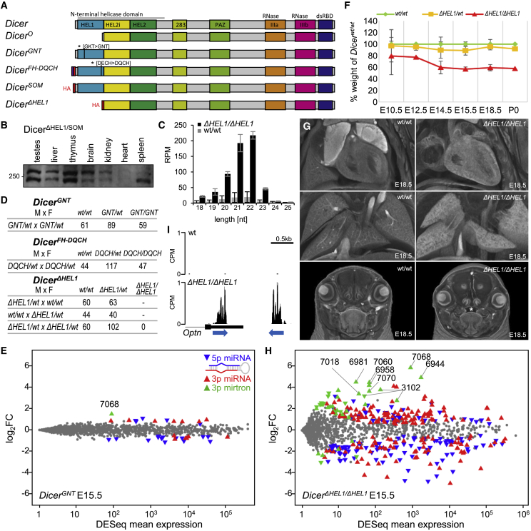
Figureu00a01
DExD/HEL1 domain of Dicer but not its ATPase activity is essential for miRNA homeostasis and normal mouse development (A) Studied mouse Dicer protein variants and mutants. Dicer (full-length) and Dice...
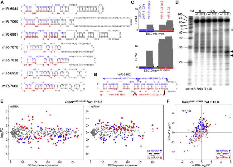
Figureu00a02
HEL1 restricts processing of mirtrons and 3p passenger strand loading (A) Strongly upregulated mirtrons have extended stems and larger loops. Secondary structures were adopted from miRBase ( Kozomara ...
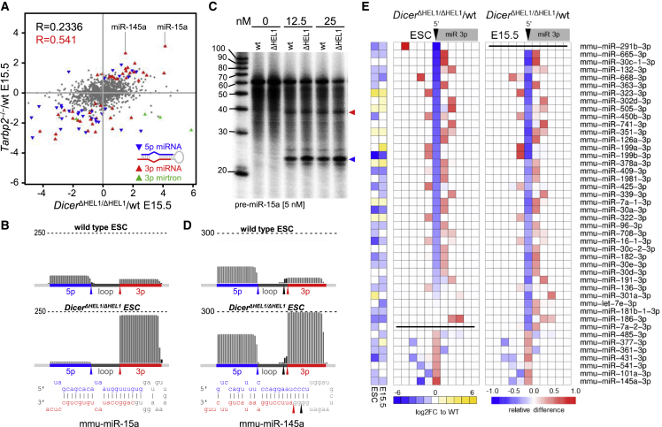
Figureu00a03
HEL1 is important for pre-miRNA cleavage fidelity (A) Comparison of relative changes of miRNAs in Tarbp2 u2212/u2212 and Dicer u0394HEL1/u0394HEL1 E15.5 embryos. Highlighted are miRNAs significantly d...
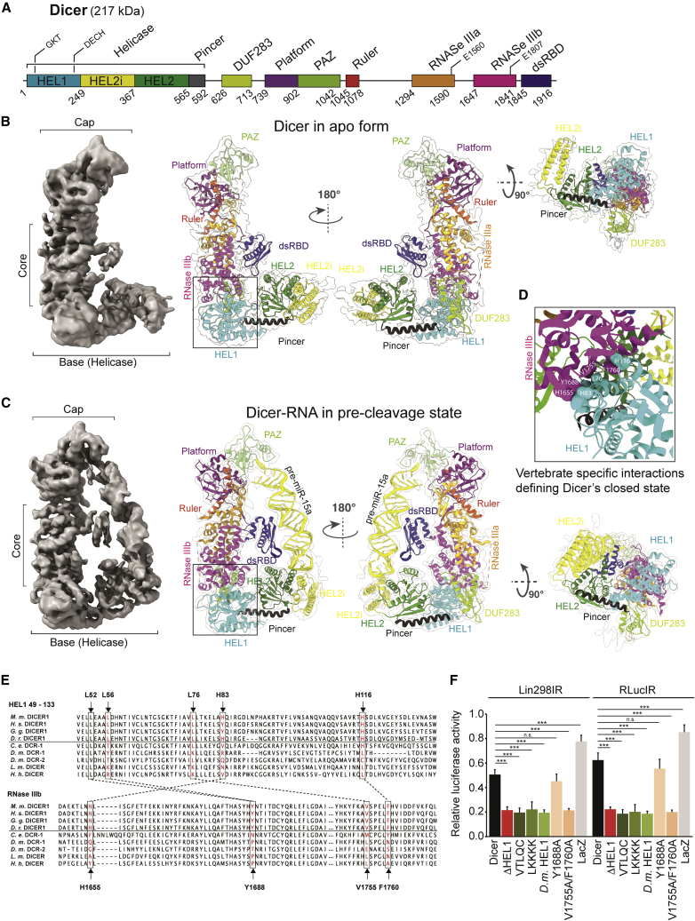
Figureu00a04
Cryo-EM structures of mouse full-length Dicer alone and in complex with Diceru2022pre-miRNA reveal the molecular basis of locking Dicer in the closed state (A) Domain architecture of full-length mouse...
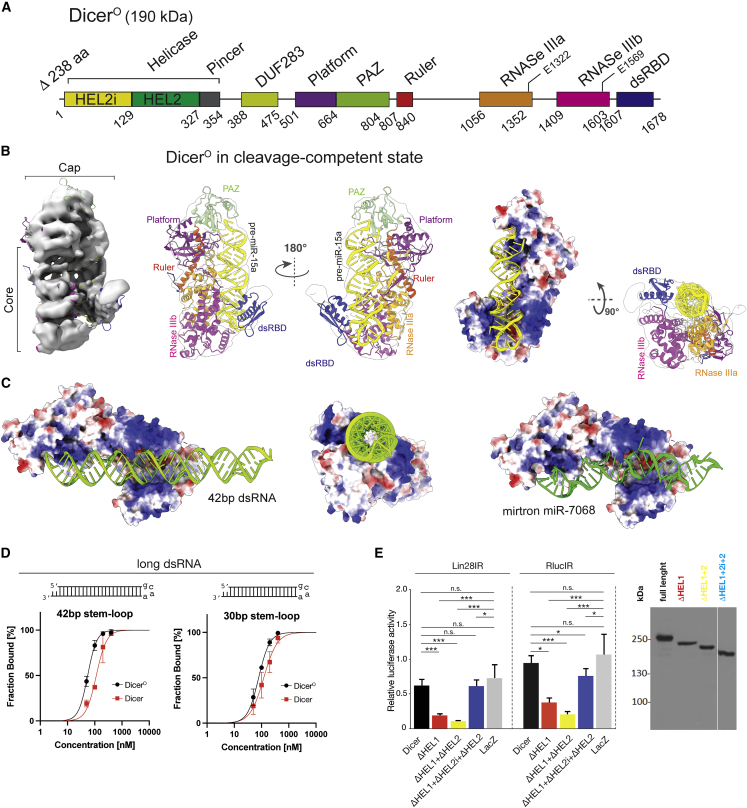
Figureu00a05
Cryo-EM structure of mouse Dicer O u2022RNA complex reveals why the absence of HEL1 makes Dicer active and promiscuous (A) Domain architecture of Dicer O numbered at boundaries. (B) Overall structure ...
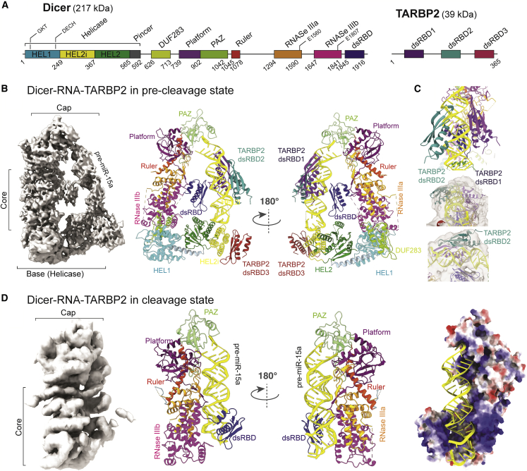
Figureu00a06
Cryo-EM structures of mouse Diceru2022RNAu2022TARBP2 complexes reveal that TARBP2 allosterically stimulates the transition from pre-cleavage to cleavage state (A) Domain architecture of Dicer (left) a...
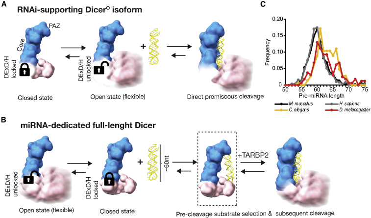
Figureu00a07
Model of Dicer function and miRNA and RNAi pathway partitioning in mammals (A) Dicer exhibits conformational dynamics of its helicase domain, which exists in two conformations: a major conformation, t...
Figure images are served from the NIH/NLM PubMed Central Open Access Subset or Europe PMC; copyright remains with the publishers and authors.
💬 Discussion
0 commentsNo comments yet. Be the first to start a discussion!
Leave a Comment