Abstract
The isolation of CCoV-HuPn-2018 from a child respiratory swab indicates that more coronaviruses are spilling over to humans than previously appreciated. We determined the structures of the CCoV-HuPn-2018 spike glycoprotein trimer in two distinct conformational states and showed that its domain 0 recognizes sialosides. We identified that the CCoV-HuPn-2018 spike binds canine, feline, and porcine aminopeptidase N (APN) orthologs, which serve as entry receptors, and determined the structure of the receptor-binding B domain in complex with canine APN. The introduction of an oligosaccharide at position N739 of human APN renders cells susceptible to CCoV-HuPn-2018 spike-mediated entry, suggesting that single-nucleotide polymorphisms might account for viral detection in some individuals. Human polyclonal plasma antibodies elicited by HCoV-229E infection and a porcine coronavirus monoclonal antibody inhibit CCoV-HuPn-2018 spike-mediated entry, underscoring the cross-neutralizing activity among ɑ-coronaviruses. These data pave the way for vaccine and therapeutic development targeting this zoonotic pathogen representing the eighth human-infecting coronavirus.
🔬 Techniques
🔭 Microscopes
🧬 Organisms
💻 Software
✨ Fluorophores
🧪 Sample Preparation
🔬 Cell Lines
🏭 Microscope Brands
🧪 Reagent Suppliers
📷 Detectors
💻 Software Details
💾 Data Repositories
🏛️ Research Organizations (ROR)
Affiliated research institutions:
📋 Methods
Key resources table REAGENT or RESOURCE SOURCE IDENTIFIER Bacterial strains
E. coli DH10B Competent Cells Invitrogen Cat# 44-0099 Deposited data
CCoV-HuPn-2018-S swung out conformation CryoEM https://www.ebi.ac.uk/pdbe/emdb/ PDB 7USA EMD-26730 CCoV-HuPn-2018-S proximal conformation CryoEM https://www.ebi.ac.uk/pdbe/emdb/ PDB 7US6 EMD-26727 CCoV-HuPn-2018-D0 swung out conformation CryoEM https://www.ebi.ac.uk/pdbe/emdb/ PDB 7USB EMD-26731 CCoV-HuPn-2018-D0 proximal conformation CryoEM map https://www.ebi.ac.uk/pdbe/emdb/ PDB 7US9 EMD-26729 CCoV-HuPn-2018_RBD + canine APN ectodomain https://www.rcsb.org PDB 7U0L Experimental models: Cell lines HEK293T cells ATCC Cat# CRL-11268 ExpiCHO cells ThermoFisher Scientific Cat# A29127 Oligonucleotides humanAPN_R741T_Fwd: aataccaacaactggacc gagatcc Integrated DNA Technologies, Inc. N/A humanAPN_R741T_Rev: atttctgaagtgaatgaagagg Integrated DNA Technologies, Inc. N/A canineAPN_T749R_Fwd: aactggcgggaccaccctc agacac Integrated DNA Technologies, Inc. N/A canineAPN_T749R_Rev: ctgtgtgatcttctcaaagtgattg Integrated DNA Technologies, Inc. N/A humanAPN_R741G_Fwd: aataccaacaactggggcgagatcc Integrated DNA Technologies, Inc. N/A Recombinant DNA pcDNA3.1(+): CCoV-HuPn-2018_S_2P_avi_his This study N/A pcDNA3.1(+): CCoV-HuPn-2018_S _Full-length This study N/A pcDNA3.1(+): HCoV-229E S_Full-length This study N/A pcDNA3.1(+): TGEV S_Full-length This study N/A pcDNA3.1(+): CCoV-HuPn-2018_RBD domain This study N/A pcDNA3.1(+): HCoV-229E RBD domain This study N/A pcDNA3.1(+): TGEV RBD domain This study N/A pcDNA3.1(+): human APN_full-length This study N/A pcDNA3.1(+): human APN_full-length_R741T This study N/A pcDNA3.1(+): canine APN_full-length This study N/A pcDNA3.1(+): canine APN_full-length_T749R This study N/A pcDNA3.1(+): feline APN_full-length This study N/A pcDNA3.1(+): porcine APN_full-length This study N/A pcDNA3.1(+): human APN_ectodomain_Fc This study N/A pcDNA3.1(+): human APN_ectodomain_R741T_Fc This study N/A pcDNA3.1(+): canine APN_ectodomain_Fc This study N/A pcDNA3.1(+): canine APN_ectodomain_T749R_Fc This study N/A pcDNA3.1(+): feline APN_ectodomain_Fc This study N/A pcDNA3.1(+): porcine APN_ectodomain_Fc This study N/A pcDNA3.1(+): human APN_full-length R741G This study N/A pcDNA3.1(+): CCoV-HuP_0_domain_I53-50A This study N/A pcDNA3.1(+): CCoV-HuP_A_domain_I53-50A This study N/A pcDNA3.1(+): CCoV-HuP_B_domain_I53-50A This study N/A pCMVR: HKU1_S_I53-50A This study N/A pCMVR: HA-ferritin ( Kanekiyo et al., 2013 ) N/A pCMVR: NC99-NA ( Kanekiyo et al., 2013 ) N/A TMPRSS2 plasmid Addgene https://www.addgene.org/53887/ Biological samples BirA biotin-protein ligase standard reaction kit Avidity Cat# 341113 Octet Kinetics Buffer (10X) Sartorius Cat# 18-1105 Anti-Human IgG Fc Capture (Biosensors) Forté Bio Cat# 18-0015 Protein A (ProA) Biosensors Forté Bio Cat# 18-0004 Alexa Fluor 680 goat anti-human IgG Jackson ImmunoResearch Cat# 109-625-098 Bio-Glo™ Luciferase Assay System Promega Cat# G7940 ExpiFectamine™ CHO Transfection Kit Life technologies Cat# A29130 Kapa HiFi HotStart Ready Mix Roche Cat# 7958935001 EndoFree Plasmid Mega kit Qiagen Cat#12381 QIAprep Spin Miniprep Kit Qiagen Cat#27106X4 Monarch PCR & DNA cleanup kit New England Biolabs Cat# T1030S T4 DNA Ligase New England Biolabs Cat# M0202 T4 Ligase buffer New England Biolabs Cat# B0202S Rat Red Blood Cells Fitzgerald 88R-R002 Turkey red blood cells Lampire 7249409 Dog red blood cells Fitzgerald 88R-D004 Pig Red Blood Cells Fitzgerald 88R-P002 Human red blood cells Rockland R407-0050 Software and algorithms cryoSPARC v3.0.1 ( Punjani et al., 2017 ) https://cryosparc.com Relion v3.0 ( Zivanov et al., 2018 ) https://www3.mrc-lmb.cam.ac.uk/relion Coot ( Emsley et al., 2010 ) https://www2.mrc-lmb.cam.ac.uk/personal/pemsley/coot/ Phenix-Refine ( Liebschner et al., 2019 ) https://www.phenix-online.org/download/ Phenix-Phaser ( McCoy et al., 2007 ) https://www.phenix-online.org/download/ XDS ( Kabsch, 2010 ) http://xds.mpimf-heidelberg.mpg.de Prism 9 GraphPad Software https://www.graphpad.com/scientific-software/prism/ Top Draw ( Bond, 2003 ) https://bondxray.org/software/topdraw/topdraw.html ChimeraX ( Goddard et al., 2018 ) https://www.cgl.ucsf.edu/chimerax/ The ConSurf server ( Armon et al., 2001 ) https://consurf.tau.ac.il/credits.php Resource availability Lead contact Further information and requests for resources and reagents should be directed to and will be fulfilled by the lead contact, David Veesler ( dveesler@uw.edu ).
Show full methods section
Key resources table REAGENT or RESOURCE SOURCE IDENTIFIER Bacterial strains
E. coli DH10B Competent Cells Invitrogen Cat# 44-0099 Deposited data
CCoV-HuPn-2018-S swung out conformation CryoEM https://www.ebi.ac.uk/pdbe/emdb/ PDB 7USA EMD-26730 CCoV-HuPn-2018-S proximal conformation CryoEM https://www.ebi.ac.uk/pdbe/emdb/ PDB 7US6 EMD-26727 CCoV-HuPn-2018-D0 swung out conformation CryoEM https://www.ebi.ac.uk/pdbe/emdb/ PDB 7USB EMD-26731 CCoV-HuPn-2018-D0 proximal conformation CryoEM map https://www.ebi.ac.uk/pdbe/emdb/ PDB 7US9 EMD-26729 CCoV-HuPn-2018_RBD + canine APN ectodomain https://www.rcsb.org PDB 7U0L Experimental models: Cell lines HEK293T cells ATCC Cat# CRL-11268 ExpiCHO cells ThermoFisher Scientific Cat# A29127 Oligonucleotides humanAPN_R741T_Fwd: aataccaacaactggacc gagatcc Integrated DNA Technologies, Inc. N/A humanAPN_R741T_Rev: atttctgaagtgaatgaagagg Integrated DNA Technologies, Inc. N/A canineAPN_T749R_Fwd: aactggcgggaccaccctc agacac Integrated DNA Technologies, Inc. N/A canineAPN_T749R_Rev: ctgtgtgatcttctcaaagtgattg Integrated DNA Technologies, Inc. N/A humanAPN_R741G_Fwd: aataccaacaactggggcgagatcc Integrated DNA Technologies, Inc. N/A Recombinant DNA pcDNA3.1(+): CCoV-HuPn-2018_S_2P_avi_his This study N/A pcDNA3.1(+): CCoV-HuPn-2018_S _Full-length This study N/A pcDNA3.1(+): HCoV-229E S_Full-length This study N/A pcDNA3.1(+): TGEV S_Full-length This study N/A pcDNA3.1(+): CCoV-HuPn-2018_RBD domain This study N/A pcDNA3.1(+): HCoV-229E RBD domain This study N/A pcDNA3.1(+): TGEV RBD domain This study N/A pcDNA3.1(+): human APN_full-length This study N/A pcDNA3.1(+): human APN_full-length_R741T This study N/A pcDNA3.1(+): canine APN_full-length This study N/A pcDNA3.1(+): canine APN_full-length_T749R This study N/A pcDNA3.1(+): feline APN_full-length This study N/A pcDNA3.1(+): porcine APN_full-length This study N/A pcDNA3.1(+): human APN_ectodomain_Fc This study N/A pcDNA3.1(+): human APN_ectodomain_R741T_Fc This study N/A pcDNA3.1(+): canine APN_ectodomain_Fc This study N/A pcDNA3.1(+): canine APN_ectodomain_T749R_Fc This study N/A pcDNA3.1(+): feline APN_ectodomain_Fc This study N/A pcDNA3.1(+): porcine APN_ectodomain_Fc This study N/A pcDNA3.1(+): human APN_full-length R741G This study N/A pcDNA3.1(+): CCoV-HuP_0_domain_I53-50A This study N/A pcDNA3.1(+): CCoV-HuP_A_domain_I53-50A This study N/A pcDNA3.1(+): CCoV-HuP_B_domain_I53-50A This study N/A pCMVR: HKU1_S_I53-50A This study N/A pCMVR: HA-ferritin ( Kanekiyo et al., 2013 ) N/A pCMVR: NC99-NA ( Kanekiyo et al., 2013 ) N/A TMPRSS2 plasmid Addgene https://www.addgene.org/53887/ Biological samples BirA biotin-protein ligase standard reaction kit Avidity Cat# 341113 Octet Kinetics Buffer (10X) Sartorius Cat# 18-1105 Anti-Human IgG Fc Capture (Biosensors) Forté Bio Cat# 18-0015 Protein A (ProA) Biosensors Forté Bio Cat# 18-0004 Alexa Fluor 680 goat anti-human IgG Jackson ImmunoResearch Cat# 109-625-098 Bio-Glo™ Luciferase Assay System Promega Cat# G7940 ExpiFectamine™ CHO Transfection Kit Life technologies Cat# A29130 Kapa HiFi HotStart Ready Mix Roche Cat# 7958935001 EndoFree Plasmid Mega kit Qiagen Cat#12381 QIAprep Spin Miniprep Kit Qiagen Cat#27106X4 Monarch PCR & DNA cleanup kit New England Biolabs Cat# T1030S T4 DNA Ligase New England Biolabs Cat# M0202 T4 Ligase buffer New England Biolabs Cat# B0202S Rat Red Blood Cells Fitzgerald 88R-R002 Turkey red blood cells Lampire 7249409 Dog red blood cells Fitzgerald 88R-D004 Pig Red Blood Cells Fitzgerald 88R-P002 Human red blood cells Rockland R407-0050 Software and algorithms cryoSPARC v3.0.1 ( Punjani et al., 2017 ) https://cryosparc.com Relion v3.0 ( Zivanov et al., 2018 ) https://www3.mrc-lmb.cam.ac.uk/relion Coot ( Emsley et al., 2010 ) https://www2.mrc-lmb.cam.ac.uk/personal/pemsley/coot/ Phenix-Refine ( Liebschner et al., 2019 ) https://www.phenix-online.org/download/ Phenix-Phaser ( McCoy et al., 2007 ) https://www.phenix-online.org/download/ XDS ( Kabsch, 2010 ) http://xds.mpimf-heidelberg.mpg.de Prism 9 GraphPad Software https://www.graphpad.com/scientific-software/prism/ Top Draw ( Bond, 2003 ) https://bondxray.org/software/topdraw/topdraw.html ChimeraX ( Goddard et al., 2018 ) https://www.cgl.ucsf.edu/chimerax/ The ConSurf server ( Armon et al., 2001 ) https://consurf.tau.ac.il/credits.php Resource availability Lead contact Further information and requests for resources and reagents should be directed to and will be fulfilled by the lead contact, David Veesler ( dveesler@uw.edu ).
Materials availability
Materials generated in this study will be made available on request and may require a material transfer agreement.
Experimental model and subject details Cell lines
Cell lines used in this study were obtained from ATCC: Human cells epithelial embryo (HEK293T, CRL-3216), Felis catus kidney cells (CRFK, CCL-94), Canis familiaris epithelial kidney cells (MDCK, CCL-34), Canis familiaris tumor fibroblast cells (A-72, CRL-1542) and Sus scrofa pig testis fibroblast cells (ST, CRL-1746) or ThermoFisher Scientific: ExpiCHO cells and Expi293F™ cells. Cells were cultivated at 37°C, in an atmosphere of 5 % CO 2 and with 130 rpm of agitation for suspension cells. None of the cell lines used were routinely tested for mycoplasma contamination.
Plasmids
Genes used in this study were synthesized by GenScript, codon optimized for expression in mammalian cells, cloned into pcDNA3.1 (+) between KpnI and XhoI, in frame with a Kozak’s sequence to direct translation, with the signal peptide derived from the μ phosphatase: MGILPSPGMPALLSLVSLLSVLLMGCVAETGT (except for the full-length genes in which the original signal peptide was used), and avi tag and a C-terminal octa-histidine tag. CCoV-HuPn-2018-S-2P corresponds to the sequence with the entry code: QVL91811.1 and includes residues 17 to 1387. To stabilize the spike in prefusion conformation, residues E (1140) and L (1141) were mutated to P, as previously described ( Pallesen et al., 2017 ). Full-length wild-type S glycoproteins from HCoV-229E ( AAK32191.1 ) residues: 1-1,155, CCoV-HuPn-2018 ( QVL91811.1 ) residues 1-1,425 and TGEV ( ABG89335.1 ) residues: 1-1,425, harboring C-terminal deletions of 18-, 23- and 23-residues, respectively, were used to pseudotype VSVΔG-luc. HCoV-229E, CCoV-HuPn-2018 and TGEV RBDs matching with the full-length sequences indicated above, include residues: 293 to 434, 539 to 671 and 522 to 665, respectively, fused to an avi-tag and his tag. Full-length APNs from human ( NP_001141.2 ), feline ( NP_001009252.2 ), canine ( NP_001139506.1 ) and pig ( AGX93258.1 ) orthologs comprise residues: 1-967, 1-967, 1-975 and 1-963, respectively. APN ectodomains from human wildtype and R741T mutant (both comprising residues 66 to 967), feline (residues 64 to 967), canine (residues 71 to 975), and pig (residues 62 to 963) from the same sequence codes shown above, were fused to a thrombin cleavage site followed by a human Fc fragment tag at the C-terminal end. The 1AF10 Fab light and heavy sequences were obtained from PDB 4F2M and cloned separately into pcDNA3.1. Only the heavy chain was fused C-terminally to a sequence encoding for a GGSGGS linker and an 8 residue-long his-tag. CCoV-HuPn-2018 domain 0 (residues 17-259), domain A (residues 260-498) and domain B (residues 523-671) were fused to the N-terminus of the trimeric I53-50A nanoparticle component ( Walls et al., 2020b ) using a 16 residue-long glycine and serine linker. The plasmid for expression of TMPRSS2 full-length was obtained from Addgene ( https://www.addgene.org/53887/ ). HKU1 S-I53-50A, NC99-NA and NC99-ferritin constructs were also synthetized by GenScript. All the protein sequences used in this study are shown in Data S1 .
Mutagenesis
The wild-type full-length human APN encoding plasmid was used as a template to introduce a glycosylation sequon at position N739 (NWR to NWT) to generate human APN R741T in a PCR reaction mixture containing: 2X Kapa HiFi (Kapa biosystems) and the following primers, each at 10 μM: forward (Fwd), 5’-aataccaacaactggaccgagatcc-3’ (underline is the codon change R to T) and reverse (Rev), 5’-atttctgaagtgaatgaagagg-3’ from Integrated DNA Technologies (IDT). The human APN R741G mutant was generated using the same Rev primer used to generate the R741T mutant and the Fwd primer 5’-aataccaacaactggggcgagatcc-3’. Full-length wild-type canine APN encoding plasmid was used as a template to knockout the glycan at position N747 (NWT to NWR) using the primers 5’-aactggcgggaccaccctcagacac-3’ (underline is the codon change T to R) and 5-ctgtgtgatcttctcaaagtgattg-3’ also using the 2X Kapa HiFi (Kapa biosystems). After treating the PCR products with DpnI (New England Biolabs) during 1 h at 37°C, amplified plasmids were purified using PCR & DNA cleanup kit (Monarch) treated with T4 polynucleotide kinase (New England Biolabs) during 1 h at 37°C and ligated using T4 DNA ligase (New England Biolabs) at room temperature for 1 hour before being used for transformation to One Shot MAX Efficiency DH10B chemically competent cells (Invitrogen). Introduction of the desired mutations was verified by sequencing purified plasmids by GENEWIZ. Plasmid harboring the desired mutation were further amplified and purified with EndoFree mega kit (Qiagen) to be suitable for transfection into mammalian cells.
Protein expression and purification
To express the prefusion-stabilized CCoV-HuPn-2018 S glycoprotein, 200 ml of ExpiCHO cells grown to a density of 6 x 10 6 /mL and 37°C, were transfected with 200 μg of CCoV-HuPn-2018-S-2P plasmid mixed with 640 l of Expifectamine CHO reagent (ThermoFisher) following manufacturer’s recommendations. The day after transfection, feed and enhancer were added to the cells. Six days post-transfection, supernatants were clarified by centrifugation at 800 g for 10 minutes. Followed addition of 20 mM imidazole, 300 mM NaCl and 20 mM Tris-HCl pH 8.0, supernatants were further centrifuged at 14,000 g for 30 min and passed through a 1 mL His trap HP column (Cytiva) previously equilibrated with binding buffer (25 mM Tris pH 7.4 and 350 mM NaCl). CCoV-HuPn-2018-S-2P was eluted using a linear gradient of 500 mM imidazole. To express B domains from CCoV-HuPn-2018, TGEV and HCoV-229E fused to an avi- and a histidine tag, 100 ml of Expi293F cells at 3 x 10 6 /mL were transiently transfected with 320 μl of Expifectamine and 100 μg of the respective plasmids, following the manufacturer’s indications. Four days post-transfection, supernatants were clarified by centrifugation at 800 g for 10 minutes, supplemented with 20 mM imidazole, 300 mM NaCl and 25 mM Tris-HCl pH 8.0, further centrifuged at 14,000 g for 30 min and passed through a 1 mL His trap HP column (Cytiva) previously equilibrated with binding buffer (25mM Tris pH 7.4 and 350mM NaCl). B domains were eluted using a linear gradient of 500 mM imidazole. A similar protocol was used to express and purify the Fab 1AF10 fused to 8 residues histidine tag except that 50 ml of Expi293F cells were transfected with a mixture containing: 50 μg of each individual plasmid encoding the Fab light and heavy chain and 160 μl of Expifectamine. To express APN ectodomains from human, feline, canine and pig orthologs fused to Fc portion of human IgG, Expi293F cells were transiently transfected with the respective plasmids following the manufacturer’s protocols. Briefly, 50 ml of Expi293F cells at 3 x 10 6 /mL were transfected using 160 μl of Expifectamine and 50 μg of APN plasmid. Four days after transfection, supernatants were clarified by centrifugation at 800 g for 10 minutes, supplemented with 300 mM NaCl and 25 mM Tris-HCl pH 8.0, further centrifuged at 14,000 g for 30 min and passed through a 1 mL HiTrap Protein A HP column (Cytiva). Proteins were eluted using 0.1 M citric acid pH 3.0 in individual tubes containing 200 μl of 1 M Tris-HCl pH 9.0 to immediately neutralize the low pH needed for elution. Fractions containing the proteins were pooled and buffer exchanged to 25 mM Tris-HCl pH 8.0, 150 mM NaCl. To produce APNs without the Fc fusion, the Fc fragment of all APN orthologs was removed using thrombin (Millipore Sigma) in a reaction mixture containing: 3 μg of thrombin/mg of APN-Fc, 20 mM Tris-HCl pH 8.0, 150 mM NaCl and 2.5 mM CaCl 2 incubated overnight at room temperature. The reaction mixture was then loaded to a Protein A column to remove uncleaved APN-Fc and the Fc tag. Monomeric APNs were further purified by size-exclusion chromatography (SEC) on a Superdex 200 column 10/300 GL (GE Life Sciences) previously equilibrated in 25 mM Tris pH 8.0 and 150 mM NaCl. HKU1 S-I53-50A protein was produced in Expi293F cells grown in suspension using Expi293F expression medium (Life Technologies) at 33°C, 70% humidity, 8% CO2 rotating at 150 rpm. The cultures were transfected using PEI-MAX (Polyscience) with cells grown to a density of 3.0 million cells per mL and cultivated for 3 days. Supernatants were clarified by centrifugation (5 min at 4000 rpm), addition of pDADMAC solution to a final concentration of 0.0375% (Sigma Aldrich, #409014), and a second spin (5 min at 4000 rpm). HKU1-S-I53-50A protein containing His tag was purified from clarified supernatants via a batch bind method where each clarified supernatant was supplemented with 1 M Tris-HCl pH 8.0 to a final concentration of 50 mM and 5 M NaCl to a final concentration of 300 mM. Talon cobalt affinity resin (Takara) was added to the treated supernatants and allowed to incubate for 15 min with gentle shaking. Resin was collected using vacuum filtration with a 0.2 mm filter and transferred to a gravity column. The resin was washed with 20 mM Tris pH 8.0, 300 mM NaCl, and the protein was eluted with 3 column volumes of 20 mM Tris pH 8.0, 300 mM NaCl, 300 mM imidazole. The batch bind process was then repeated and the first and second elutions combined. SDS-PAGE was used to assess purity. HKU1-S-I53-50A fusion protein IMAC elution was concentrated to > 1 mg/mL and subjected to three rounds of dialysis into 50 mM Tris pH 8, 150 mM NaCl, 0.25% w/v Histidine, and 5% glycerol in a hydrated 10K molecular weight cutoff dialysis cassette (Thermo Scientific). To produce HA-ferritin nanoparticles, 1 mg/L of NC99-ferritin vector was transiently transfected together with 0.05 mg/L of NC99 neuraminidase (NA) vector into Expi293F using polyethylenimine transfection reagent. Four days after transfection, culture supernatants were collected and sterile filtered. HA-ferritin NPs were purified by lectin affinity chromatography (EY labs), followed by size-exclusion chromatography with a Superose 6 Increase 10/300 GL (GE) in 25 mM Tris, pH 8.0, 150 mM NaCl, 5% glycerol. Protein biotinylation B domains from HCoV-229E, TGEV and CCoV-HuPn-2018 were biotinylated using BirA biotin-protein ligase standard reaction kit (Avidity) following manufacturer's protocol. In a typical reaction, 40 μM of B domains were incubated overnight at 4°C with 2.5 μg of BirA enzyme in reaction mixtures containing 1X BiomixB, 1X BiomixA and 40 μM BIO200. Domains B were further separated from Bir A by SEC using Superdex 75 increase 10/300 GL (GE LifeSciences) and concentrated using 10 kDa filters (Amicon). Biolayer interferometry APNs binding measurements to B domains were performed using Biolayer interferometry. Biotinylated B domains from CCoV-HuPn-2018, TGEV and HCoV-229E were immobilized at 1 μg/mL in undiluted 10X kinetics buffer (Pall) to streptavidin (SA) biosensors that were pre-hydrated in water for at least 10 minutes and then equilibrated into 10X Kinetics Buffer (Pall). The loaded tips were then dipped into a dilution series of APN orthologs from human (wild-type or glycan knockin R741T), feline, canine (wild-type or T749R, and porcine or 1AF10 Fab in 10X Kinetics Buffer (Pall) starting at various concentrations for 300 seconds prior to 300 seconds dissociation in 10X Kinetics buffer for kinetics determination. The data were baseline subtracted and the plots fitted using the Pall FortéBio/Sartorius analysis software (v.12.0). Data were plotted in Graphpad Prism (v.9.0.2). These experiments were done side-by-side with two different batches of B domains and APN orthologs preparations. For competition BLI experiments, CCoV-HuPn-2018 or TGEV biotinylated RBD were immobilized at 1 μg/mL in undiluted 10X kinetics buffer (Pall) to SA sensors that were pre-hydrated in water for at least 10 minutes and then equilibrated into 10X Kinetics Buffer (Pall). B domain-loaded SA biosensors were sequentially dipped in a solution containing the 1AF10 Fab at concentrations ten times above the respective affinity (170 nM for CoV-HuPn-2018 or 100 nM for TGEV) and then in a solution containing the same concentration of 1AF10 supplemented with orthologs APN at concentrations 10X above their respective apparent affinity. For nanoparticles experiments, canine and human R741T APN-Fc ectodomains were immobilized at the surface of protein A biosensors at 2 μg/ml and were dipped in 1 μM of CCoV-HuPn-2018-Domain0-NPs or CCoV-HuPn-2018-DomainB-NPs diluted in 10X Kinetics Buffer (Pall). Data were baseline subtracted and fit using the Pall FortéBio/Sartorius analysis software (v.12.0). Data were plotted in Graphpad Prism (v.9.0.2).
Pull-down assays
The interaction between APN orthologs and B domains was further analyzed performing a pull-down assay. Briefly, 200 μl (2 mg) of magnetic beads Dynabeads™ Strep Isolation (ThermoFisher) were washed twice with 300 μL 1X TBS-T (2.4 g Tris-Base/L, 8 g NaCl/L, 0.1 % Tween20) using a magnetic stand (ThermoFisher) before coupling them with 200 μl of B domains in TBS at 0.125 ng/ml. The mixture beads/protein were well mixed and incubated for 1h at room temperature with constant gentle rotation. Beads were then washed two times with 200 μL TBS-T before resuspending them in a solution containing an excess of 10-20X of their calculated K D,app using BLI, of the APN-Fc orthologs and allow binding for 1 hr at RT with gentle rotation after which were washed two times with TBS-T. Flow through, last wash and beads were collected and analyzed in SDS-PAGE gel. Control assays consisted of beads alone, i.e: not coupled to B domains. Bacterial protein expression and purification of nanoparticles components The I53-50A and I53-50B proteins were expressed as described before ( Walls et al., 2020b ). Briefly, transformed Lemo21(DE3) (NEB) in LB (10 g Tryptone, 5 g Yeast Extract, 10 g NaCl) were grown at 37°C to an OD600 ∼0.8 with agitation. Expression was induced with 1 mM IPTG and temperature was reduced to 18°C. Cells were harvested after ∼16 h and lysed by microfluidization using a Microfluidics M110P at 18,000 psi in 50 mM Tris, 500 mM NaCl, 30 mM imidazole, 1 mM PMSF, 0.75% CHAPS. Lysates were clarified by centrifugation at 24,000 g for 30 min and applied to a 2.6 × 10 cm Ni Sepharose 6 FF column (Cytiva) for purification by IMAC on an AKTA Avant150 FPLC system (Cytiva). Proteins were eluted with a linear gradient of 30 mM to 500 mM imidazole in 50 mM Tris pH 8, 500 mM NaCl, 0.75% CHAPS buffer. Peak fractions were pooled, concentrated in 10K MWCO centrifugal filters (Millipore), sterile filtered (0.22 μm) and applied to either a Superdex 200 Increase 10/300, or HiLoad S200 pg GL SEC column (Cytiva) previously equilibrated in 50 mM Tris pH 8, 500 mM NaCl, 0.75% CHAPS buffer. In vitro nanoparticle assembly Concentration of purified individual nanoparticle components was determined by measuring absorbance at 280 nm and the corresponding calculated extinction coefficients. Nanoparticles were prepared by incubation of domain A-I53-50A or domain B-I53-50A or HKU1-S-I53-50A trimers with pentameric I53-50B at molar ratios 1.1:1, respectively, in 50 mM Tris pH 8, 500 mM NaCl, 0.75% w/v CHAPS. Formation of Domain 0-NPs required mixing domain 0-I53-50A with I53-50A at a molar ratio of 1:6 with pentameric I53-50B. All in vitro assemblies were incubated at room temperature with gentle rocking for at least 30 min before subsequent purification by SEC on a Superose 6 column to remove residual unassembled components. Fractions were analyzed by negative stain electron microscopy and by PAGE-SDS. Assembled particles eluted in the void volume of a Superose 6 column and were pooled and stored at 4°C.
Negative stain electron microscopy
Nanoparticles diluted to 0.01 mg/mL in 50 mM Tris pH 8, 150 mM NaCl, were adsorbed to glow-discharged home-made carbon-coated copper grids for 30 seconds. The excess liquid was blotted away with filter paper (Whatman 1) and 3 μL of 2% w/v uranyl formate stain were applied to the grids. Finally, the stain was blotted away, and the grids were allowed to air dry for 1 min. Grids were imaged on a 120kV FEI Tecnai G2 Spirit with a Gatan Ultrascan 4000 4k x 4k CCD camera at 67,000 nominal magnification using a defocus ranging between 1.0 and 2.0 μm and a pixel size of 1.6 Å.
Hemagglutination assays
The hemagglutination assays was performed according to standard procedures. Briefly, 50 μl of CCoV-HuPn-2018 domain 0, domain A, domain B NPs at 400 ng/μl were incubated with 50 μl 0.5% (diluted in PBS) turkey (Lampire biological laboratories), dog (Fitzgerald), human (Rockland Immunochemicals), pig (Fitzgerald) and rat (Fitzgerald) erythrocytes in V-bottom, 96-well plates (Greiner Bio-One) for 30 min at room temperature after which plates were photographed and hemagglutination was analyzed. In another experiment, eight 2-fold dilutions starting at 200 ng/μl of CCoV-HuPn-2018 Domain 0-NPs were incubated with 50 μl 0.5% turkey, dog, human and pig erythrocytes for 30 min at room temperature after which plates were analyzed. To determine whether sialic acids were involved in the interaction, human erythrocytes were pretreated for 3 h at 37 °C with neuraminidase (NA) from A. ureafaciens (Roche; diluted to 30 mU/mL in PBS), before incubation with different concentrations of CCoV-HuPn-2018 Domain 0-NPs for 1 h at room temperature. HKU1-S-NP at 220 ng/μl and HA-ferritin at 400 ng/μl were used as positive controls when incubated with 0.5% rat and turkey erythrocytes, respectively. VSV pseudotyped virus production CCoV-HuPn, HCoV-229E ( AAK32191.1 , P100E isolate) and TGEV S pseudotyped VSV were generated as previously described ( Tortorici et al., 2020 , 2021 ). Briefly, HEK293T cells in DMEM supplemented with 10% FBS and 1% PenStrep and seeded in poly-D-lysine coated 10-cm dishes were transfected with a mixture of 24 μg of the corresponding plasmid encoding for: CCoV-HuPn S, TGEV S or HCoV-229 S, 60 μl Lipofectamine 2000 (Life Technologies) in 3 ml of Opti-MEM, following manufacturer’s instructions. After 5 h at 37°C, DMEM supplemented with 20% FBS and 1% PenStrep was added. The next day, cells were washed three times with DMEM and were transduced with VSVΔG-luc ( Kaname et al., 2010 ). After 2 h, virus inoculum was removed and cells were washed five times with DMEM prior to the addition of DMEM supplemented with anti-VSV-G antibody [Il-mouse hybridoma supernatant diluted 1 to 25 (v/v), from CRL-2700, ATCC] to minimize parental background. After 18-24 h, supernatants containing pseudotyped VSV were harvested, centrifuged at 2,000 x g for 5 minutes to remove cellular debris, filtered with a 0,45 μm membrane, concentrated 10 times using a 30 kDa cut off membrane (Amicon), aliquoted, and frozen at -80°C. Pseudotyped VSV infections and neutralizations For pseudotyped VSV infections and neutralizations, HEK293T cells were transfected with plasmids encoding for the different full-length APN orthologs (flAPN) following the protocol described by Eguia et al. (2021) . Briefly, HEK293T cells at 90% confluency and seeded in poly-D-lysine coated 10-cm dishes were transfected with a mixture in Opti-MEM containing 8 μg of the corresponding plasmid encoding flAPN ortholog, 1 μg of the plasmid encoding full-length TMPRSS2 and 30 μl of Lipofectamine 2000 (Life Technologies) according to the manufacturer’s instructions. After 5 h at 37°C, cells were trypsinized, seeded into poly-D-lysine coated clear bottom white walled 96-well plates at 40,000-50,000 cells/well and cultured overnight at 37°C. For infections, 5-10 μl of the corresponding pseudotyped VSV were mixed with 35-30 μl of DMEM and the mixture was added to the cells previously washed three times with DMEM. After 2 h at 37°C, 40 μl of DMEM were added and cells were further incubated overnight at 37°C. For neutralizations, eleven 2-fold serial dilutions of Fab 1AF10, APN ectodomains orthologs or sera, were prepared in DMEM. 20 μl of CCoV-HuPn-2018 S, HCoV-229E S or TGEV S pseudotyped VSV were added 1:1 (v/v) to each Fab1AF10, APN ectodomains or sera dilution and mixtures were incubated for 45-60 min at 37°C. After removing their media, transfected HEK293T cells were washed three times with DMEM and 40 μL of the mixture containing virus:Fab/APN ectodomains/sera were added. One hour later, 40 μL DMEM were added to the cells. After 17-20 h, 60 μL of One-Glo-EX substrate (Promega) were added to each well and incubated on a plate shaker in the dark. After 5-15 min incubation, plates were read on a Biotek plate reader. Measurements were done in duplicate with at least two biological replicates and one representative experiment is shown. Relative luciferase units were plotted and normalized in Prism (GraphPad): cells alone without pseudotyped virus was defined as 0 % infection, and cells with virus only (no sera) was defined as 100 % infection. Most of the human sera was collected from prospective bone marrow donors in Seattle with approval from the Human Subjects Institutional Review Board in the 1980s and were stored in the Infectious Disease Sciences Biospecimen Repository at the Vaccine and Infectious Disease Division of the Fred Hutch Cancer Center. A few of the sera (the ones prefixed “FH”) are residual samples from Bloodworks Northwest that were collected from adults in Seattle. Western blot 15 μl of pseudotyped VSV were mixed with 4X SDS-PAGE loading buffer, run on a 4%–15% gradient Tris-Glycine Gel (BioRad) and transferred to a PVDF membrane using the protocol mix molecular weight of the Trans-Blot Turbo System (BioRad). The membrane was blocked with 5% milk in TBS-T (20 mM Tris-HCl pH 8.0, 150 mM NaCl) supplemented with 0.05% Tween-20 at room temperature and with agitation. After 1 h, the fusion-peptide-specific S2S8 monoclonal antibody was added at 1:250 dilution and incubated overnight at 4°C with agitation. Next day, the membrane was washed three times with TBS-T and an Alexa Fluor 680-conjugated goat anti-human secondary antibody (1:50,000 dilution, Jackson ImmunoResearch, 109-625-098) was added and incubated during 1 h at room temperature. Membrane was washed three times with TBS-T after which a LI-COR processor was used to develop the western blot. Crystallization, data collection, structure determination and analysis Crystals of the CCoV-HuPn-2018-B domain-canine APN complex were obtained at 20°C by sitting drop vapor diffusion. 15 mg/ml canine APN were incubated with 3 mg/ml CCoV-HuPn-2018-B domain for 1 hour at room temperature before mixing with mother liquor solution (200 nl final volume) containing 0.1 M Na 2 HPO 4 -citric acid pH 4.2, 1.6 M Na 2 HPO 4 /K2HPO 4 . Crystals were flash frozen in liquid nitrogen using 30% glycerol as cryoprotectant. Diffraction data were collected at the Advanced Light Source synchrotron beamline 5.0.2 and processed with the XDS software package ( Kabsch, 2010 ). Initial phases were obtained by molecular replacement using Phenix-Phaser ( McCoy et al., 2007 ) and the following models: porcine APN (PDB 4f5c) and CCoV-HuPn-2018-B domain obtained by cryo-EM in this study. Several subsequent rounds of model building, and refinement were performed using Coot ( Emsley et al., 2010 ) and Phenix-Refine ( Liebschner et al., 2019 ) to arrive at a final model for the binary complex at 3.3 Å resolution in space group P21221. Model validation was done using Molprobity ( Chen et al., 2010 ) and Privateer ( Agirre et al., 2015 ). CryoEM sample preparation, data collection and data processing 3 μl of CCoV-HuPn-2018 S at approximately 0.3 mg/ml were loaded three times onto freshly glow-discharged lacey grids covered with a thin layer of home-made continuous carbon prior to plunge freezing using a vitrobot MarkIV (ThermoFisher Scientific) with a blot force of -1 and 2.5 sec blot time at 100% humidity and 21°C. Data were acquired using the Leginon software ( Suloway et al., 2005 ) to control a FEI Titan Krios transmission electron microscope operated at 300 kV equipped with a Gatan K3 direct detector and a Gatan Quantum GIF energy filter, operated with a slit width of 20eV. The dose rate was adjusted to 3.75 counts/super- resolution pixel/s, and each movie was acquired in 75 frames of 40 ms with a pixel size of 0.843 Å and a defocus range comprised between 0.8 and 2.0 μm. Movie frame alignment, estimation of the microscope contrast-transfer function parameters, particle picking and extraction were carried out using Warp ( Tegunov and Cramer, 2019 ). Two rounds of reference-free 2D classification were performed using cryoSPARC ( Punjani et al., 2017 ) with binned particles to select well-defined particle images. Subsequently, one round of 3D classification with 50 iterations, using an ab initio-generated reference model (angular sampling 7.5° for 25 iterations and 1.8° with local search for 25 iterations) was carried out using Relion ( Zivanov et al., 2018 ) without imposing symmetry. 3D refinements were carried out using non-uniform refinement in cryoSPARC ( Punjani et al., 2020 ). Particle images were subjected to Bayesian polishing ( Zivanov et al., 2019 ) using Relion during which particles were re-extracted with a box size of 512 pixels at a pixel size of 0.843 Å which was followed by another round of non-uniform refinement in cryoSPARC followed by per-particle defocus refinement and again non-uniform refinement. To improve the density of the domain 0, particles were symmetry-expanded and subjected to a Relion focus 3D classification without refining angles and shifts using a soft mask encompassing domain 0. Local refinement, local resolution estimation, filtering, and sharpening were carried out using CryoSPARC. Reported resolutions are based on the gold-standard Fourier shell correlation using 0.143 criterion ( Rosenthal and Henderson, 2003 ) and Fourier shell correlation curves were corrected for the effects of soft masking by high-resolution noise substitution ( Chen et al., 2013 ). CryoEM model building and analysis UCSF Chimera ( Pettersen et al., 2004 ) and Coot ( Emsley et al., 2010 ) were used to fit atomic models (PDB 5SZS) into the cryoEM maps and Domain 0 was manually built. Models were refined and rebuilt into the maps using Coot and Rosetta ( Frenz et al., 2019 ; Wang et al., 2016 ). Model validation was done using Molprobity ( Chen et al., 2010 ) and Privateer ( Agirre et al., 2015 ). Figures were generated using UCSF ChimeraX ( Goddard et al., 2018 ). Topology diagrams were generated using the program TopDraw ( Bond, 2003 ).
Assessment of human ANPEP diversity
We assessed human diversity at or near the APN NWT motif (NWR in human; R741) in gnomAD ( https://gnomad.broadinstitute.org/gene/ENSG00000166825 ). Observations in approximately 125,000 humans included one heterozygous N738K, one heterozygous R741G and one heterozygous I743N. No rare defects in ANPEP are described in OMIM ( https://www.omim.org/entry/151530 )
Materials availability
Materials generated in this study will be made available on request and may require a material transfer agreement.
Experimental model and subject details Cell lines
Cell lines used in this study were obtained from ATCC: Human cells epithelial embryo (HEK293T, CRL-3216), Felis catus kidney cells (CRFK, CCL-94), Canis familiaris epithelial kidney cells (MDCK, CCL-34), Canis familiaris tumor fibroblast cells (A-72, CRL-1542) and Sus scrofa pig testis fibroblast cells (ST, CRL-1746) or ThermoFisher Scientific: ExpiCHO cells and Expi293F™ cells. Cells were cultivated at 37°C, in an atmosphere of 5 % CO 2 and with 130 rpm of agitation for suspension cells. None of the cell lines used were routinely tested for mycoplasma contamination.
Plasmids
Genes used in this study were synthesized by GenScript, codon optimized for expression in mammalian cells, cloned into pcDNA3.1 (+) between KpnI and XhoI, in frame with a Kozak’s sequence to direct translation, with the signal peptide derived from the μ phosphatase: MGILPSPGMPALLSLVSLLSVLLMGCVAETGT (except for the full-length genes in which the original signal peptide was used), and avi tag and a C-terminal octa-histidine tag. CCoV-HuPn-2018-S-2P corresponds to the sequence with the entry code: QVL91811.1 and includes residues 17 to 1387. To stabilize the spike in prefusion conformation, residues E (1140) and L (1141) were mutated to P, as previously described ( Pallesen et al., 2017 ). Full-length wild-type S glycoproteins from HCoV-229E ( AAK32191.1 ) residues: 1-1,155, CCoV-HuPn-2018 ( QVL91811.1 ) residues 1-1,425 and TGEV ( ABG89335.1 ) residues: 1-1,425, harboring C-terminal deletions of 18-, 23- and 23-residues, respectively, were used to pseudotype VSVΔG-luc. HCoV-229E, CCoV-HuPn-2018 and TGEV RBDs matching with the full-length sequences indicated above, include residues: 293 to 434, 539 to 671 and 522 to 665, respectively, fused to an avi-tag and his tag. Full-length APNs from human ( NP_001141.2 ), feline ( NP_001009252.2 ), canine ( NP_001139506.1 ) and pig ( AGX93258.1 ) orthologs comprise residues: 1-967, 1-967, 1-975 and 1-963, respectively. APN ectodomains from human wildtype and R741T mutant (both comprising residues 66 to 967), feline (residues 64 to 967), canine (residues 71 to 975), and pig (residues 62 to 963) from the same sequence codes shown above, were fused to a thrombin cleavage site followed by a human Fc fragment tag at the C-terminal end. The 1AF10 Fab light and heavy sequences were obtained from PDB 4F2M and cloned separately into pcDNA3.1. Only the heavy chain was fused C-terminally to a sequence encoding for a GGSGGS linker and an 8 residue-long his-tag. CCoV-HuPn-2018 domain 0 (residues 17-259), domain A (residues 260-498) and domain B (residues 523-671) were fused to the N-terminus of the trimeric I53-50A nanoparticle component ( Walls et al., 2020b ) using a 16 residue-long glycine and serine linker. The plasmid for expression of TMPRSS2 full-length was obtained from Addgene ( https://www.addgene.org/53887/ ). HKU1 S-I53-50A, NC99-NA and NC99-ferritin constructs were also synthetized by GenScript. All the protein sequences used in this study are shown in Data S1 .
Mutagenesis
The wild-type full-length human APN encoding plasmid was used as a template to introduce a glycosylation sequon at position N739 (NWR to NWT) to generate human APN R741T in a PCR reaction mixture containing: 2X Kapa HiFi (Kapa biosystems) and the following primers, each at 10 μM: forward (Fwd), 5’-aataccaacaactggaccgagatcc-3’ (underline is the codon change R to T) and reverse (Rev), 5’-atttctgaagtgaatgaagagg-3’ from Integrated DNA Technologies (IDT). The human APN R741G mutant was generated using the same Rev primer used to generate the R741T mutant and the Fwd primer 5’-aataccaacaactggggcgagatcc-3’. Full-length wild-type canine APN encoding plasmid was used as a template to knockout the glycan at position N747 (NWT to NWR) using the primers 5’-aactggcgggaccaccctcagacac-3’ (underline is the codon change T to R) and 5-ctgtgtgatcttctcaaagtgattg-3’ also using the 2X Kapa HiFi (Kapa biosystems). After treating the PCR products with DpnI (New England Biolabs) during 1 h at 37°C, amplified plasmids were purified using PCR & DNA cleanup kit (Monarch) treated with T4 polynucleotide kinase (New England Biolabs) during 1 h at 37°C and ligated using T4 DNA ligase (New England Biolabs) at room temperature for 1 hour before being used for transformation to One Shot MAX Efficiency DH10B chemically competent cells (Invitrogen). Introduction of the desired mutations was verified by sequencing purified plasmids by GENEWIZ. Plasmid harboring the desired mutation were further amplified and purified with EndoFree mega kit (Qiagen) to be suitable for transfection into mammalian cells.
Protein expression and purification
To express the prefusion-stabilized CCoV-HuPn-2018 S glycoprotein, 200 ml of ExpiCHO cells grown to a density of 6 x 10 6 /mL and 37°C, were transfected with 200 μg of CCoV-HuPn-2018-S-2P plasmid mixed with 640 l of Expifectamine CHO reagent (ThermoFisher) following manufacturer’s recommendations. The day after transfection, feed and enhancer were added to the cells. Six days post-transfection, supernatants were clarified by centrifugation at 800 g for 10 minutes. Followed addition of 20 mM imidazole, 300 mM NaCl and 20 mM Tris-HCl pH 8.0, supernatants were further centrifuged at 14,000 g for 30 min and passed through a 1 mL His trap HP column (Cytiva) previously equilibrated with binding buffer (25 mM Tris pH 7.4 and 350 mM NaCl). CCoV-HuPn-2018-S-2P was eluted using a linear gradient of 500 mM imidazole. To express B domains from CCoV-HuPn-2018, TGEV and HCoV-229E fused to an avi- and a histidine tag, 100 ml of Expi293F cells at 3 x 10 6 /mL were transiently transfected with 320 μl of Expifectamine and 100 μg of the respective plasmids, following the manufacturer’s indications. Four days post-transfection, supernatants were clarified by centrifugation at 800 g for 10 minutes, supplemented with 20 mM imidazole, 300 mM NaCl and 25 mM Tris-HCl pH 8.0, further centrifuged at 14,000 g for 30 min and passed through a 1 mL His trap HP column (Cytiva) previously equilibrated with binding buffer (25mM Tris pH 7.4 and 350mM NaCl). B domains were eluted using a linear gradient of 500 mM imidazole. A similar protocol was used to express and purify the Fab 1AF10 fused to 8 residues histidine tag except that 50 ml of Expi293F cells were transfected with a mixture containing: 50 μg of each individual plasmid encoding the Fab light and heavy chain and 160 μl of Expifectamine. To express APN ectodomains from human, feline, canine and pig orthologs fused to Fc portion of human IgG, Expi293F cells were transiently transfected with the respective plasmids following the manufacturer’s protocols. Briefly, 50 ml of Expi293F cells at 3 x 10 6 /mL were transfected using 160 μl of Expifectamine and 50 μg of APN plasmid. Four days after transfection, supernatants were clarified by centrifugation at 800 g for 10 minutes, supplemented with 300 mM NaCl and 25 mM Tris-HCl pH 8.0, further centrifuged at 14,000 g for 30 min and passed through a 1 mL HiTrap Protein A HP column (Cytiva). Proteins were eluted using 0.1 M citric acid pH 3.0 in individual tubes containing 200 μl of 1 M Tris-HCl pH 9.0 to immediately neutralize the low pH needed for elution. Fractions containing the proteins were pooled and buffer exchanged to 25 mM Tris-HCl pH 8.0, 150 mM NaCl. To produce APNs without the Fc fusion, the Fc fragment of all APN orthologs was removed using thrombin (Millipore Sigma) in a reaction mixture containing: 3 μg of thrombin/mg of APN-Fc, 20 mM Tris-HCl pH 8.0, 150 mM NaCl and 2.5 mM CaCl 2 incubated overnight at room temperature. The reaction mixture was then loaded to a Protein A column to remove uncleaved APN-Fc and the Fc tag. Monomeric APNs were further purified by size-exclusion chromatography (SEC) on a Superdex 200 column 10/300 GL (GE Life Sciences) previously equilibrated in 25 mM Tris pH 8.0 and 150 mM NaCl. HKU1 S-I53-50A protein was produced in Expi293F cells grown in suspension using Expi293F expression medium (Life Technologies) at 33°C, 70% humidity, 8% CO2 rotating at 150 rpm. The cultures were transfected using PEI-MAX (Polyscience) with cells grown to a density of 3.0 million cells per mL and cultivated for 3 days. Supernatants were clarified by centrifugation (5 min at 4000 rpm), addition of pDADMAC solution to a final concentration of 0.0375% (Sigma Aldrich, #409014), and a second spin (5 min at 4000 rpm). HKU1-S-I53-50A protein containing His tag was purified from clarified supernatants via a batch bind method where each clarified supernatant was supplemented with 1 M Tris-HCl pH 8.0 to a final concentration of 50 mM and 5 M NaCl to a final concentration of 300 mM. Talon cobalt affinity resin (Takara) was added to the treated supernatants and allowed to incubate for 15 min with gentle shaking. Resin was collected using vacuum filtration with a 0.2 mm filter and transferred to a gravity column. The resin was washed with 20 mM Tris pH 8.0, 300 mM NaCl, and the protein was eluted with 3 column volumes of 20 mM Tris pH 8.0, 300 mM NaCl, 300 mM imidazole. The batch bind process was then repeated and the first and second elutions combined. SDS-PAGE was used to assess purity. HKU1-S-I53-50A fusion protein IMAC elution was concentrated to > 1 mg/mL and subjected to three rounds of dialysis into 50 mM Tris pH 8, 150 mM NaCl, 0.25% w/v Histidine, and 5% glycerol in a hydrated 10K molecular weight cutoff dialysis cassette (Thermo Scientific). To produce HA-ferritin nanoparticles, 1 mg/L of NC99-ferritin vector was transiently transfected together with 0.05 mg/L of NC99 neuraminidase (NA) vector into Expi293F using polyethylenimine transfection reagent. Four days after transfection, culture supernatants were collected and sterile filtered. HA-ferritin NPs were purified by lectin affinity chromatography (EY labs), followed by size-exclusion chromatography with a Superose 6 Increase 10/300 GL (GE) in 25 mM Tris, pH 8.0, 150 mM NaCl, 5% glycerol. Protein biotinylation B domains from HCoV-229E, TGEV and CCoV-HuPn-2018 were biotinylated using BirA biotin-protein ligase standard reaction kit (Avidity) following manufacturer's protocol. In a typical reaction, 40 μM of B domains were incubated overnight at 4°C with 2.5 μg of BirA enzyme in reaction mixtures containing 1X BiomixB, 1X BiomixA and 40 μM BIO200. Domains B were further separated from Bir A by SEC using Superdex 75 increase 10/300 GL (GE LifeSciences) and concentrated using 10 kDa filters (Amicon). Biolayer interferometry APNs binding measurements to B domains were performed using Biolayer interferometry. Biotinylated B domains from CCoV-HuPn-2018, TGEV and HCoV-229E were immobilized at 1 μg/mL in undiluted 10X kinetics buffer (Pall) to streptavidin (SA) biosensors that were pre-hydrated in water for at least 10 minutes and then equilibrated into 10X Kinetics Buffer (Pall). The loaded tips were then dipped into a dilution series of APN orthologs from human (wild-type or glycan knockin R741T), feline, canine (wild-type or T749R, and porcine or 1AF10 Fab in 10X Kinetics Buffer (Pall) starting at various concentrations for 300 seconds prior to 300 seconds dissociation in 10X Kinetics buffer for kinetics determination. The data were baseline subtracted and the plots fitted using the Pall FortéBio/Sartorius analysis software (v.12.0). Data were plotted in Graphpad Prism (v.9.0.2). These experiments were done side-by-side with two different batches of B domains and APN orthologs preparations. For competition BLI experiments, CCoV-HuPn-2018 or TGEV biotinylated RBD were immobilized at 1 μg/mL in undiluted 10X kinetics buffer (Pall) to SA sensors that were pre-hydrated in water for at least 10 minutes and then equilibrated into 10X Kinetics Buffer (Pall). B domain-loaded SA biosensors were sequentially dipped in a solution containing the 1AF10 Fab at concentrations ten times above the respective affinity (170 nM for CoV-HuPn-2018 or 100 nM for TGEV) and then in a solution containing the same concentration of 1AF10 supplemented with orthologs APN at concentrations 10X above their respective apparent affinity. For nanoparticles experiments, canine and human R741T APN-Fc ectodomains were immobilized at the surface of protein A biosensors at 2 μg/ml and were dipped in 1 μM of CCoV-HuPn-2018-Domain0-NPs or CCoV-HuPn-2018-DomainB-NPs diluted in 10X Kinetics Buffer (Pall). Data were baseline subtracted and fit using the Pall FortéBio/Sartorius analysis software (v.12.0). Data were plotted in Graphpad Prism (v.9.0.2).
Pull-down assays
The interaction between APN orthologs and B domains was further analyzed performing a pull-down assay. Briefly, 200 μl (2 mg) of magnetic beads Dynabeads™ Strep Isolation (ThermoFisher) were washed twice with 300 μL 1X TBS-T (2.4 g Tris-Base/L, 8 g NaCl/L, 0.1 % Tween20) using a magnetic stand (ThermoFisher) before coupling them with 200 μl of B domains in TBS at 0.125 ng/ml. The mixture beads/protein were well mixed and incubated for 1h at room temperature with constant gentle rotation. Beads were then washed two times with 200 μL TBS-T before resuspending them in a solution containing an excess of 10-20X of their calculated K D,app using BLI, of the APN-Fc orthologs and allow binding for 1 hr at RT with gentle rotation after which were washed two times with TBS-T. Flow through, last wash and beads were collected and analyzed in SDS-PAGE gel. Control assays consisted of beads alone, i.e: not coupled to B domains. Bacterial protein expression and purification of nanoparticles components The I53-50A and I53-50B proteins were expressed as described before ( Walls et al., 2020b ). Briefly, transformed Lemo21(DE3) (NEB) in LB (10 g Tryptone, 5 g Yeast Extract, 10 g NaCl) were grown at 37°C to an OD600 ∼0.8 with agitation. Expression was induced with 1 mM IPTG and temperature was reduced to 18°C. Cells were harvested after ∼16 h and lysed by microfluidization using a Microfluidics M110P at 18,000 psi in 50 mM Tris, 500 mM NaCl, 30 mM imidazole, 1 mM PMSF, 0.75% CHAPS. Lysates were clarified by centrifugation at 24,000 g for 30 min and applied to a 2.6 × 10 cm Ni Sepharose 6 FF column (Cytiva) for purification by IMAC on an AKTA Avant150 FPLC system (Cytiva). Proteins were eluted with a linear gradient of 30 mM to 500 mM imidazole in 50 mM Tris pH 8, 500 mM NaCl, 0.75% CHAPS buffer. Peak fractions were pooled, concentrated in 10K MWCO centrifugal filters (Millipore), sterile filtered (0.22 μm) and applied to either a Superdex 200 Increase 10/300, or HiLoad S200 pg GL SEC column (Cytiva) previously equilibrated in 50 mM Tris pH 8, 500 mM NaCl, 0.75% CHAPS buffer. In vitro nanoparticle assembly Concentration of purified individual nanoparticle components was determined by measuring absorbance at 280 nm and the corresponding calculated extinction coefficients. Nanoparticles were prepared by incubation of domain A-I53-50A or domain B-I53-50A or HKU1-S-I53-50A trimers with pentameric I53-50B at molar ratios 1.1:1, respectively, in 50 mM Tris pH 8, 500 mM NaCl, 0.75% w/v CHAPS. Formation of Domain 0-NPs required mixing domain 0-I53-50A with I53-50A at a molar ratio of 1:6 with pentameric I53-50B. All in vitro assemblies were incubated at room temperature with gentle rocking for at least 30 min before subsequent purification by SEC on a Superose 6 column to remove residual unassembled components. Fractions were analyzed by negative stain electron microscopy and by PAGE-SDS. Assembled particles eluted in the void volume of a Superose 6 column and were pooled and stored at 4°C.
Negative stain electron microscopy
Nanoparticles diluted to 0.01 mg/mL in 50 mM Tris pH 8, 150 mM NaCl, were adsorbed to glow-discharged home-made carbon-coated copper grids for 30 seconds. The excess liquid was blotted away with filter paper (Whatman 1) and 3 μL of 2% w/v uranyl formate stain were applied to the grids. Finally, the stain was blotted away, and the grids were allowed to air dry for 1 min. Grids were imaged on a 120kV FEI Tecnai G2 Spirit with a Gatan Ultrascan 4000 4k x 4k CCD camera at 67,000 nominal magnification using a defocus ranging between 1.0 and 2.0 μm and a pixel size of 1.6 Å.
Hemagglutination assays
The hemagglutination assays was performed according to standard procedures. Briefly, 50 μl of CCoV-HuPn-2018 domain 0, domain A, domain B NPs at 400 ng/μl were incubated with 50 μl 0.5% (diluted in PBS) turkey (Lampire biological laboratories), dog (Fitzgerald), human (Rockland Immunochemicals), pig (Fitzgerald) and rat (Fitzgerald) erythrocytes in V-bottom, 96-well plates (Greiner Bio-One) for 30 min at room temperature after which plates were photographed and hemagglutination was analyzed. In another experiment, eight 2-fold dilutions starting at 200 ng/μl of CCoV-HuPn-2018 Domain 0-NPs were incubated with 50 μl 0.5% turkey, dog, human and pig erythrocytes for 30 min at room temperature after which plates were analyzed. To determine whether sialic acids were involved in the interaction, human erythrocytes were pretreated for 3 h at 37 °C with neuraminidase (NA) from A. ureafaciens (Roche; diluted to 30 mU/mL in PBS), before incubation with different concentrations of CCoV-HuPn-2018 Domain 0-NPs for 1 h at room temperature. HKU1-S-NP at 220 ng/μl and HA-ferritin at 400 ng/μl were used as positive controls when incubated with 0.5% rat and turkey erythrocytes, respectively. VSV pseudotyped virus production CCoV-HuPn, HCoV-229E ( AAK32191.1 , P100E isolate) and TGEV S pseudotyped VSV were generated as previously described ( Tortorici et al., 2020 , 2021 ). Briefly, HEK293T cells in DMEM supplemented with 10% FBS and 1% PenStrep and seeded in poly-D-lysine coated 10-cm dishes were transfected with a mixture of 24 μg of the corresponding plasmid encoding for: CCoV-HuPn S, TGEV S or HCoV-229 S, 60 μl Lipofectamine 2000 (Life Technologies) in 3 ml of Opti-MEM, following manufacturer’s instructions. After 5 h at 37°C, DMEM supplemented with 20% FBS and 1% PenStrep was added. The next day, cells were washed three times with DMEM and were transduced with VSVΔG-luc ( Kaname et al., 2010 ). After 2 h, virus inoculum was removed and cells were washed five times with DMEM prior to the addition of DMEM supplemented with anti-VSV-G antibody [Il-mouse hybridoma supernatant diluted 1 to 25 (v/v), from CRL-2700, ATCC] to minimize parental background. After 18-24 h, supernatants containing pseudotyped VSV were harvested, centrifuged at 2,000 x g for 5 minutes to remove cellular debris, filtered with a 0,45 μm membrane, concentrated 10 times using a 30 kDa cut off membrane (Amicon), aliquoted, and frozen at -80°C. Pseudotyped VSV infections and neutralizations For pseudotyped VSV infections and neutralizations, HEK293T cells were transfected with plasmids encoding for the different full-length APN orthologs (flAPN) following the protocol described by Eguia et al. (2021) . Briefly, HEK293T cells at 90% confluency and seeded in poly-D-lysine coated 10-cm dishes were transfected with a mixture in Opti-MEM containing 8 μg of the corresponding plasmid encoding flAPN ortholog, 1 μg of the plasmid encoding full-length TMPRSS2 and 30 μl of Lipofectamine 2000 (Life Technologies) according to the manufacturer’s instructions. After 5 h at 37°C, cells were trypsinized, seeded into poly-D-lysine coated clear bottom white walled 96-well plates at 40,000-50,000 cells/well and cultured overnight at 37°C. For infections, 5-10 μl of the corresponding pseudotyped VSV were mixed with 35-30 μl of DMEM and the mixture was added to the cells previously washed three times with DMEM. After 2 h at 37°C, 40 μl of DMEM were added and cells were further incubated overnight at 37°C. For neutralizations, eleven 2-fold serial dilutions of Fab 1AF10, APN ectodomains orthologs or sera, were prepared in DMEM. 20 μl of CCoV-HuPn-2018 S, HCoV-229E S or TGEV S pseudotyped VSV were added 1:1 (v/v) to each Fab1AF10, APN ectodomains or sera dilution and mixtures were incubated for 45-60 min at 37°C. After removing their media, transfected HEK293T cells were washed three times with DMEM and 40 μL of the mixture containing virus:Fab/APN ectodomains/sera were added. One hour later, 40 μL DMEM were added to the cells. After 17-20 h, 60 μL of One-Glo-EX substrate (Promega) were added to each well and incubated on a plate shaker in the dark. After 5-15 min incubation, plates were read on a Biotek plate reader. Measurements were done in duplicate with at least two biological replicates and one representative experiment is shown. Relative luciferase units were plotted and normalized in Prism (GraphPad): cells alone without pseudotyped virus was defined as 0 % infection, and cells with virus only (no sera) was defined as 100 % infection. Most of the human sera was collected from prospective bone marrow donors in Seattle with approval from the Human Subjects Institutional Review Board in the 1980s and were stored in the Infectious Disease Sciences Biospecimen Repository at the Vaccine and Infectious Disease Division of the Fred Hutch Cancer Center. A few of the sera (the ones prefixed “FH”) are residual samples from Bloodworks Northwest that were collected from adults in Seattle. Western blot 15 μl of pseudotyped VSV were mixed with 4X SDS-PAGE loading buffer, run on a 4%–15% gradient Tris-Glycine Gel (BioRad) and transferred to a PVDF membrane using the protocol mix molecular weight of the Trans-Blot Turbo System (BioRad). The membrane was blocked with 5% milk in TBS-T (20 mM Tris-HCl pH 8.0, 150 mM NaCl) supplemented with 0.05% Tween-20 at room temperature and with agitation. After 1 h, the fusion-peptide-specific S2S8 monoclonal antibody was added at 1:250 dilution and incubated overnight at 4°C with agitation. Next day, the membrane was washed three times with TBS-T and an Alexa Fluor 680-conjugated goat anti-human secondary antibody (1:50,000 dilution, Jackson ImmunoResearch, 109-625-098) was added and incubated during 1 h at room temperature. Membrane was washed three times with TBS-T after which a LI-COR processor was used to develop the western blot. Crystallization, data collection, structure determination and analysis Crystals of the CCoV-HuPn-2018-B domain-canine APN complex were obtained at 20°C by sitting drop vapor diffusion. 15 mg/ml canine APN were incubated with 3 mg/ml CCoV-HuPn-2018-B domain for 1 hour at room temperature before mixing with mother liquor solution (200 nl final volume) containing 0.1 M Na 2 HPO 4 -citric acid pH 4.2, 1.6 M Na 2 HPO 4 /K2HPO 4 . Crystals were flash frozen in liquid nitrogen using 30% glycerol as cryoprotectant. Diffraction data were collected at the Advanced Light Source synchrotron beamline 5.0.2 and processed with the XDS software package ( Kabsch, 2010 ). Initial phases were obtained by molecular replacement using Phenix-Phaser ( McCoy et al., 2007 ) and the following models: porcine APN (PDB 4f5c) and CCoV-HuPn-2018-B domain obtained by cryo-EM in this study. Several subsequent rounds of model building, and refinement were performed using Coot ( Emsley et al., 2010 ) and Phenix-Refine ( Liebschner et al., 2019 ) to arrive at a final model for the binary complex at 3.3 Å resolution in space group P21221. Model validation was done using Molprobity ( Chen et al., 2010 ) and Privateer ( Agirre et al., 2015 ). CryoEM sample preparation, data collection and data processing 3 μl of CCoV-HuPn-2018 S at approximately 0.3 mg/ml were loaded three times onto freshly glow-discharged lacey grids covered with a thin layer of home-made continuous carbon prior to plunge freezing using a vitrobot MarkIV (ThermoFisher Scientific) with a blot force of -1 and 2.5 sec blot time at 100% humidity and 21°C. Data were acquired using the Leginon software ( Suloway et al., 2005 ) to control a FEI Titan Krios transmission electron microscope operated at 300 kV equipped with a Gatan K3 direct detector and a Gatan Quantum GIF energy filter, operated with a slit width of 20eV. The dose rate was adjusted to 3.75 counts/super- resolution pixel/s, and each movie was acquired in 75 frames of 40 ms with a pixel size of 0.843 Å and a defocus range comprised between 0.8 and 2.0 μm. Movie frame alignment, estimation of the microscope contrast-transfer function parameters, particle picking and extraction were carried out using Warp ( Tegunov and Cramer, 2019 ). Two rounds of reference-free 2D classification were performed using cryoSPARC ( Punjani et al., 2017 ) with binned particles to select well-defined particle images. Subsequently, one round of 3D classification with 50 iterations, using an ab initio-generated reference model (angular sampling 7.5° for 25 iterations and 1.8° with local search for 25 iterations) was carried out using Relion ( Zivanov et al., 2018 ) without imposing symmetry. 3D refinements were carried out using non-uniform refinement in cryoSPARC ( Punjani et al., 2020 ). Particle images were subjected to Bayesian polishing ( Zivanov et al., 2019 ) using Relion during which particles were re-extracted with a box size of 512 pixels at a pixel size of 0.843 Å which was followed by another round of non-uniform refinement in cryoSPARC followed by per-particle defocus refinement and again non-uniform refinement. To improve the density of the domain 0, particles were symmetry-expanded and subjected to a Relion focus 3D classification without refining angles and shifts using a soft mask encompassing domain 0. Local refinement, local resolution estimation, filtering, and sharpening were carried out using CryoSPARC. Reported resolutions are based on the gold-standard Fourier shell correlation using 0.143 criterion ( Rosenthal and Henderson, 2003 ) and Fourier shell correlation curves were corrected for the effects of soft masking by high-resolution noise substitution ( Chen et al., 2013 ). CryoEM model building and analysis UCSF Chimera ( Pettersen et al., 2004 ) and Coot ( Emsley et al., 2010 ) were used to fit atomic models (PDB 5SZS) into the cryoEM maps and Domain 0 was manually built. Models were refined and rebuilt into the maps using Coot and Rosetta ( Frenz et al., 2019 ; Wang et al., 2016 ). Model validation was done using Molprobity ( Chen et al., 2010 ) and Privateer ( Agirre et al., 2015 ). Figures were generated using UCSF ChimeraX ( Goddard et al., 2018 ). Topology diagrams were generated using the program TopDraw ( Bond, 2003 ).
Assessment of human ANPEP diversity
We assessed human diversity at or near the APN NWT motif (NWR in human; R741) in gnomAD ( https://gnomad.broadinstitute.org/gene/ENSG00000166825 ). Observations in approximately 125,000 humans included one heterozygous N738K, one heterozygous R741G and one heterozygous I743N. No rare defects in ANPEP are described in OMIM ( https://www.omim.org/entry/151530 )
Supplemental information Document S1. Tables S1–S4 Data S1. Sequences of protein constructs used in this study, related to Figures 1, 2, 3, 4, 5, and 6 and STAR Methods
📊 Figures
Figureu00a01
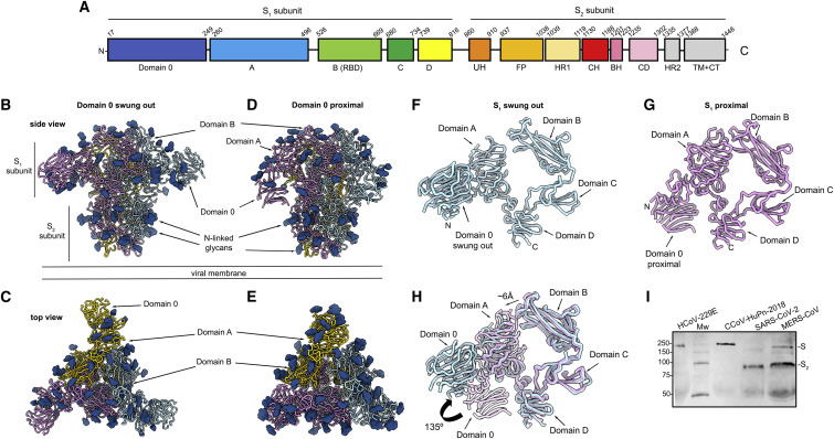
Architecture of the CCoV-HuPn-2018 infection machinery (A) Schematic diagram of the S glycoprotein organization. UH, upstream helix; FP, fusion peptide; HR1, heptad-repeat 1; CH, central helix; BH, u0...
Figureu00a0S1
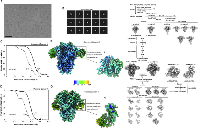
Data processing and validation of the CCoV-HuPn-2018u00a0S cryo-EM dataset, related to Figuresu00a01 and 2 (A and B) Representative electron micrograph (A)u00a0and class averages (B)u00a0of CCoV-HuPn-...
Figureu00a0S2
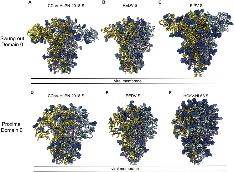
Architecture of u03b1-coronavirus S trimers harboring a domain 0, related to Figuresu00a01 and 2 (Au2013F) Side views of CCoV-HuPn-2018u00a0S with the two conformations of domain 0 (A and D), PEDV S (...
Figureu00a02
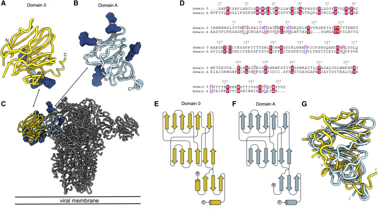
Structural conservation of CCoV-HuPn-2018u00a0S domain 0 and domain A (Au2013C) Ribbon diagrams of the CCoV-HuPn-2018u00a0S domain 0 (A)u00a0and domain A (B)u00a0oriented as shown in the prefusion S c...
Figureu00a0S3
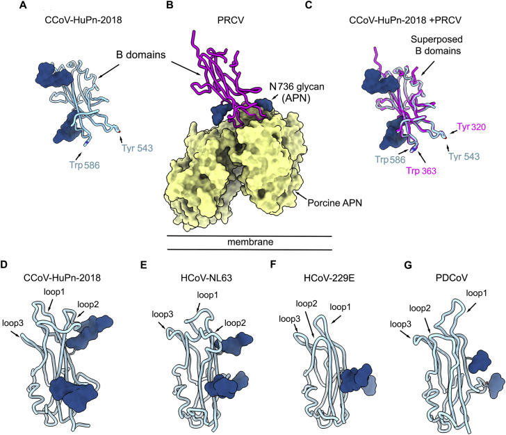
Architecture of u03b1-coronavirus B domains, related to Figuresu00a01 and 4 (Au2013C) Ribbon diagrams of the CCoV-HuPn-2018 B domain crystal structure (A), the PRCV B domain bound to porcine APN (PDB:...
Figureu00a0S4
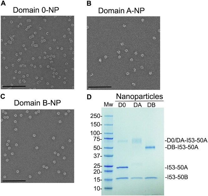
Characterization of CCoV-HuPn-2018 domain 0-, A-, and B-I53-50 nanoparticles, related to Figureu00a03 Nanoparticles (NPs) were prepared by incubation of domain 0-I53-50A, domain A-I53-50A, or domain B...
Figureu00a03
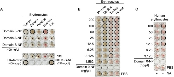
CCoV-HuPn-2018u00a0S domain 0 recognizes sialic acid and hemagglutinates erythrocytes (A) 50u00a0u03bcL of CCoV-HuPn-2018 domain 0, domain A, or domain B multivalently displayed at the surface of the ...
Figureu00a04
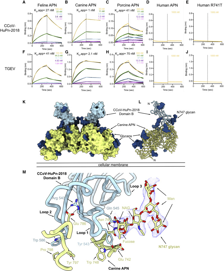
The CCoV-HuPn-2018u00a0S B domain recognizes APN (Au2013J) Biolayer interferometry kinetic analysis of feline (A and F), canine (B and G), porcine (C and H), human (D and I), and human R741T (E and J)...
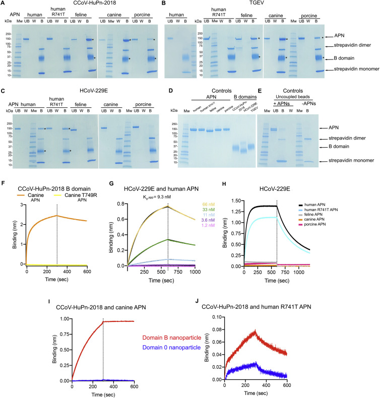
Figureu00a0S5
The CCoV-HuPn-2018 B domain recognizes APN, related to Figuresu00a04 and 5 (Au2013E) Coomassie-stained SDS-PAGE analysis of pull-downs between biotinylated B domains from CCoV-HuPn-2018 (A), TGEV (B),...
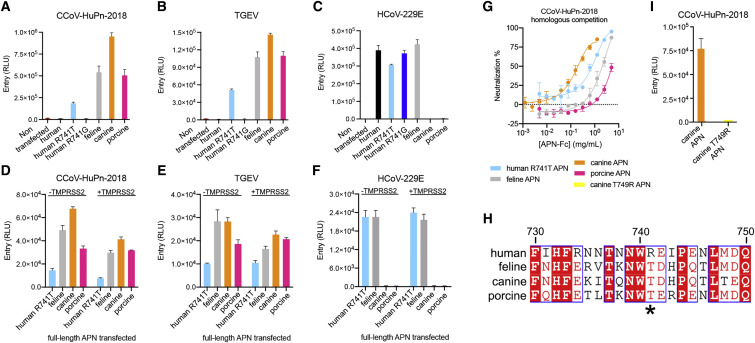
Figureu00a05
APN is a functional entry receptor for CCoV-HuPn-2018 (Au2013C) Entry of VSV particles pseudotyped with CCoV-HuPn-2018u00a0S (A), TGEV S (B), or HCoV-229E S (C)u00a0in HEK293T cells transiently transf...
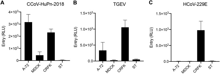
Figureu00a0S6
Cell tropism of CCoV-HuPn-2018, TGEV, and HCoV-229E S pseudoviruses, related to Figureu00a05 (Au2013C) Evaluation of pseudotyped virus mediated entry in feline, canine, and porcine cell lines. CCoV-Hu...
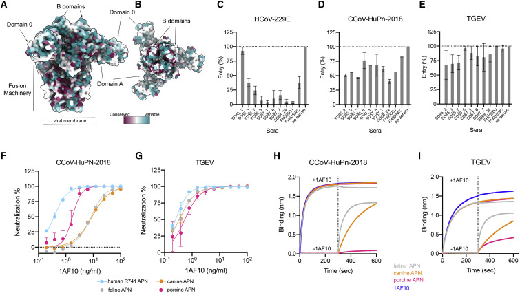
Figureu00a06
Evaluation of polyclonal and monoclonal antibody neutralization of CCoV-HuPn-2018 S-mediated entry into cells (A and B) Sequence conservation of HCoV-229E, HCoV-NL63, and CCoV-HuPn-2018u00a0S glycopro...
Figureu00a0S7
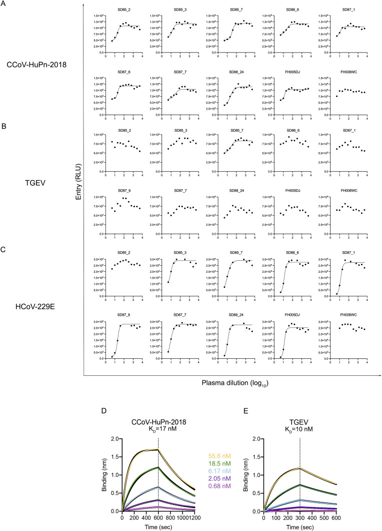
Inhibition of CCoV-HuPn-2018-S-mediated entry into cells by human polyclonal plasma antibodies and binding of CCoV-HuPn-2018 B domain to a monoclonal antibody, related to Figureu00a06 (Au2013C) CCoV-H...
Figureu00a0S8
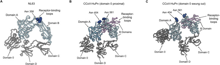
Glycan-mediated immune evasion strategy of u03b1-coronaviruses, related to Figureu00a01 (Au2013C) Ribbon diagrams of S 1 subunits from HCoV-NL63u00a0S (PDB 5SZS) (A)u00a0and from CCoV-HuPn-2018u00a0S ...
Figure images are served from the NIH/NLM PubMed Central Open Access Subset or Europe PMC; copyright remains with the publishers and authors.
💬 Discussion
0 commentsNo comments yet. Be the first to start a discussion!
Leave a Comment