Abstract
AbstractIn recent years, exploration of the brain extracellular space (ECS) has made remarkable progress, including nanoscopic characterizations. However, whether ECS precise conformation is altered during brain pathology remains unknown. Here we study the nanoscale organization of pathological ECS in adult mice under degenerative conditions. Using electron microscopy in cryofixed tissue and single nanotube tracking in live brain slices combined with super-resolution imaging analysis, we find enlarged ECS dimensions and increased nanoscale diffusion after α-synuclein-induced neurodegeneration. These animals display a degraded hyaluronan matrix in areas close to reactive microglia. Furthermore, experimental hyaluronan depletion in vivo reduces dopaminergic cell loss and α-synuclein load, induces microgliosis and increases ECS diffusivity, highlighting hyaluronan as diffusional barrier and local tissue organizer. These findings demonstrate the interplay of ECS, extracellular matrix and glia in pathology, unraveling ECS features relevant for the α-synuclein propagation hypothesis and suggesting matrix manipulation as a disease-modifying strategy.
🔬 Techniques
🔭 Microscopes
🧬 Organisms
✨ Fluorophores
🧪 Sample Preparation
🔬 Cell Lines
🏭 Microscope Brands
🧪 Reagent Suppliers
📷 Detectors
🎨 Filters
💻 Software Details
🏛️ Research Organizations (ROR)
Affiliated research institutions:
📋 Methods
Animals and surgical procedures
LB and noLB fractions were purified from fresh frozen post-mortem samples of sporadic PD-patients obtained from brains collected as part of the Brain Donation Program of the Brain Bank GIE NeuroCEB, run by a consortium of Patients Associations: ARSEP (multiple sclerosis), CSC (cerebellar ataxias), France Alzheimer and France Parkinson. The consent documents were signed by the patients themselves or their next of kin in their name, in accordance with the French Bioethical Laws. The Brain Bank GIE NeuroCEB (Bioresource Research Impact Factor number BB-0033-00011) has been declared at the Ministry of Higher Education and Research and has received approval to distribute samples (agreement AC-2013-1887). Human SNpc was dissected and homogenized before a sucrose step gradient 20 and sonicated for 5 min prior to in vivo inoculation to disrupt α-synuclein aggregates into oligomeric and fibrillary fragments. Male 10-weeks-old C57BL/6J mice (Charles River) were unilaterally injected into the substantia nigra with 2 µl of either Lewy-body (LB) fractions, containing toxic fibrillary α-synuclein, or noLB fractions, with soluble non-aggregated α-synuclein. Intranigral inoculations were achieved by stereotactic surgery (coordinates from bregma: −2.9 mm anteroposterior, 1.3 lateral, and −4.5 dorsoventral) under deep isoflurane anesthesia at a flow rate of 0.4 µl/min with a 30-gauge Hamilton syringe coupled to a microinjection pump (World Precision Instruments). The needle was left in place for 10 min to avoid leakage. Some animals were co-injected with 2 µl of LB fractions and either 1 µl (3U) of hyaluronidase (Hyase) from Streptomyces hyalurolyticus (Sigma-Aldrich), 0.025U of chondroitinase ABC (Sigma-Aldrich), low-molecular weight (200 mg/ml) or high-molecular weight hyaluronan (15 mg/ml, Sigma-Aldrich) or sterile PBS. The Institutional Animal Care and Use Committee of Bordeaux (CE50) approved experiments under license #5520-2016052514328805. 4-MU treatment 6-week-old C57BL/6J mice (Charles River) were fed with normal mouse chow or mixed with 5% (w/w) 4-methylumbelliferone (4MU, Sigma-Aldrich), a concentration reported to effectively inhibit hyaluronan production in vivo systemically 59 and in CNS 60 . The mixture was prepared fresh daily, including water and 33% of cacao to enhance the palatability of the drug. Animals were fed with 4-MU for 4 weeks prior to LB inoculation, and kept on this diet until euthanasia (4 months). Animals were weighted daily during the first month, and weekly afterwards. Despite initial weight loss in the 4MU-treated animals, mice recovered after 2 weeks. Animals showed no sign of stress or particular phenotype, other than weakness of the skin during surgery suture. The Institutional Animal Care and Use Committee of Bordeaux (CE50) approved experiments under license #10721-2017071213284522. High-pressure cryofixation and electron microscopy Bilaterally LB-inoculated mice were euthanized by cervical dislocation and brains quickly removed and sliced (180 µm thickness) in a VT1200S vibratome (Leica Microsystems) in ice-cold aCSF (see below “acute brain slice preparation” for details). Tissue discs (5 mm diameter) of substantia nigra (SN) were immediately transferred to a 200 µm-thick aluminum sample holder (6 mm diameter). Medium was cleared with a filter paper and the cavity filled with 1-hexadecene. The sample was sandwiched with a flat aluminum top plate and high-pressure frozen in a HPM100 device (Leica Microsystems). No more than 10 min elapsed from euthanasia to cryofixation. Samples were kept in liquid nitrogen until further processing. Since the pathology develops in each hemisphere independently, SNs were treated separately as individual samples. Freeze-substitution resin embedding was carried out in an AFS2 unit (Leica Microsystems). Frozen samples were incubated for 40 h using 0.1% tannic acid in anhydrous acetone at −90 °C, followed by 12 h of 2% OsO 4 in anhydrous acetone at −90 °C. The temperature was then raised to −30 °C with periodic temperature transition gradients of 1 °C/h and to −10 °C by 3 °C/h. Samples were then immersed in Epon resin/anhydrous acetone over 9 h (1/3, 1/1 and 3/1, 3 h each) whilst temperature increased to 20 °C. Finally, samples were added to 100% Epon resin at room temperature for 2 h prior to polymerization at 60 °C for 48 h. Artifact-free tissue was selected in semi-thin (0.5 mm thick) sections stained with toluidine blue. Ultra-thin sections of selected regions were post-stained with uranyl acetate (1%) and lead citrate and imaged in a transmission electron microscope (H7650, Hitachi) equipped with a SC1000 Orius camera (Gatan).
Show full methods section
Animals and surgical procedures
LB and noLB fractions were purified from fresh frozen post-mortem samples of sporadic PD-patients obtained from brains collected as part of the Brain Donation Program of the Brain Bank GIE NeuroCEB, run by a consortium of Patients Associations: ARSEP (multiple sclerosis), CSC (cerebellar ataxias), France Alzheimer and France Parkinson. The consent documents were signed by the patients themselves or their next of kin in their name, in accordance with the French Bioethical Laws. The Brain Bank GIE NeuroCEB (Bioresource Research Impact Factor number BB-0033-00011) has been declared at the Ministry of Higher Education and Research and has received approval to distribute samples (agreement AC-2013-1887). Human SNpc was dissected and homogenized before a sucrose step gradient 20 and sonicated for 5 min prior to in vivo inoculation to disrupt α-synuclein aggregates into oligomeric and fibrillary fragments. Male 10-weeks-old C57BL/6J mice (Charles River) were unilaterally injected into the substantia nigra with 2 µl of either Lewy-body (LB) fractions, containing toxic fibrillary α-synuclein, or noLB fractions, with soluble non-aggregated α-synuclein. Intranigral inoculations were achieved by stereotactic surgery (coordinates from bregma: −2.9 mm anteroposterior, 1.3 lateral, and −4.5 dorsoventral) under deep isoflurane anesthesia at a flow rate of 0.4 µl/min with a 30-gauge Hamilton syringe coupled to a microinjection pump (World Precision Instruments). The needle was left in place for 10 min to avoid leakage. Some animals were co-injected with 2 µl of LB fractions and either 1 µl (3U) of hyaluronidase (Hyase) from Streptomyces hyalurolyticus (Sigma-Aldrich), 0.025U of chondroitinase ABC (Sigma-Aldrich), low-molecular weight (200 mg/ml) or high-molecular weight hyaluronan (15 mg/ml, Sigma-Aldrich) or sterile PBS. The Institutional Animal Care and Use Committee of Bordeaux (CE50) approved experiments under license #5520-2016052514328805. 4-MU treatment 6-week-old C57BL/6J mice (Charles River) were fed with normal mouse chow or mixed with 5% (w/w) 4-methylumbelliferone (4MU, Sigma-Aldrich), a concentration reported to effectively inhibit hyaluronan production in vivo systemically 59 and in CNS 60 . The mixture was prepared fresh daily, including water and 33% of cacao to enhance the palatability of the drug. Animals were fed with 4-MU for 4 weeks prior to LB inoculation, and kept on this diet until euthanasia (4 months). Animals were weighted daily during the first month, and weekly afterwards. Despite initial weight loss in the 4MU-treated animals, mice recovered after 2 weeks. Animals showed no sign of stress or particular phenotype, other than weakness of the skin during surgery suture. The Institutional Animal Care and Use Committee of Bordeaux (CE50) approved experiments under license #10721-2017071213284522. High-pressure cryofixation and electron microscopy Bilaterally LB-inoculated mice were euthanized by cervical dislocation and brains quickly removed and sliced (180 µm thickness) in a VT1200S vibratome (Leica Microsystems) in ice-cold aCSF (see below “acute brain slice preparation” for details). Tissue discs (5 mm diameter) of substantia nigra (SN) were immediately transferred to a 200 µm-thick aluminum sample holder (6 mm diameter). Medium was cleared with a filter paper and the cavity filled with 1-hexadecene. The sample was sandwiched with a flat aluminum top plate and high-pressure frozen in a HPM100 device (Leica Microsystems). No more than 10 min elapsed from euthanasia to cryofixation. Samples were kept in liquid nitrogen until further processing. Since the pathology develops in each hemisphere independently, SNs were treated separately as individual samples. Freeze-substitution resin embedding was carried out in an AFS2 unit (Leica Microsystems). Frozen samples were incubated for 40 h using 0.1% tannic acid in anhydrous acetone at −90 °C, followed by 12 h of 2% OsO 4 in anhydrous acetone at −90 °C. The temperature was then raised to −30 °C with periodic temperature transition gradients of 1 °C/h and to −10 °C by 3 °C/h. Samples were then immersed in Epon resin/anhydrous acetone over 9 h (1/3, 1/1 and 3/1, 3 h each) whilst temperature increased to 20 °C. Finally, samples were added to 100% Epon resin at room temperature for 2 h prior to polymerization at 60 °C for 48 h. Artifact-free tissue was selected in semi-thin (0.5 mm thick) sections stained with toluidine blue. Ultra-thin sections of selected regions were post-stained with uranyl acetate (1%) and lead citrate and imaged in a transmission electron microscope (H7650, Hitachi) equipped with a SC1000 Orius camera (Gatan).
Estimation of ECS dimensions from cryofixation-EM images
ECS was manually segmented from cryofixed-EM images where membranes were easily discernible (Supplementary Fig. 2 ), using an interactive pen tablet (Wacom) and the TrakEM2 plugin in Fiji/ImageJ (NIH) 61 . Segmented images were binarized, resulting in individual ECS compartments (Fig. 1d ). The ECS volume fraction was estimated by dividing the area of ECS compartments by the total area of the image. To estimate the length of ECS compartments, binary images were skeletonized and the longest shortest path was calculated in ImageJ 62 . ECS local widths were calculated by measuring iteratively the diameter of the largest circle fitting each portion of the ECS compartment, using a 2D implementation of the Local Thickness plugin (ImageJ). The distribution of pixel values obtained from the histograms of ECS local thickness maps was converted to dimensional values using the image pixel size (1.42 nm). SWCNTs injection and acute brain slice preparation 1 mg of HiPco-synthesized carbon nanotubes (from Rice University) was suspended with 50 mg of monofunctional phospholipid-polyethylene glycol (#mPEG-DSPE-5000, Laysan Bio) in 10 ml of deuterium oxide (Sigma Aldrich). After homogenization for 15 min at 19,000 rpm with a dispersing instrument (IKA T-10 Basic), SWCNTs were further dispersed via tip sonication (20 W for 8 min) and then centrifuged at 800 × g for 60 min to remove nanotube bundles and impurities. Four months after LB or noLB inoculation, animals underwent a similar stereotactic surgery where 6 µl of SWCNT solution was injected into the cerebral lateral ventricle (coordinates from bregma: −0.5 mm anteroposterior, 1 mm mediolateral, −2.4 dorsoventral). The needle was left in place for 10 min to avoid leakage. Animals received buprenorphine (0.1 mg/kg) as analgesic and returned to their cages. 1 h after SWCNT injection, mice were euthanized by cervical dislocation. Brains were extracted and coronal sections (300 µm thick) were prepared in a Leica VT1200S vibratome. Slicing was performed in ice-cold low-Na + low-Ca 2+ high-Mg 2+ artificial cerebrospinal fluid (aCSF) solution containing 180 mM sucrose, 2.5 mM KCl, 1.2 mM NaH 2 PO 4 , 13 mM glucose, 20 mM HEPES, 10 mM MgSO 4 and 0.5 mM CaCl 2 . Slices were then transferred to oxygenated aCSF (gassed with 95% O 2 , 5% CO 2 , pH = 7.4) containing 124 mM NaCl, 2.5 mM KCl, 1.2 mM NaH 2 PO 4 , 24 mM NaHCO 3 , 13 mM glucose, 3 mM sodium pyruvate, 2 mM MgSO 4 and 2 mM CaCl 2 and kept at room temperature until imaged within 2 h.
Near-infrared microscopy
Slices were recorded at 37 °C in a 3D-printed chamber with oxygenated warm aCSF perfused by a peristaltic pump (Fig. 2b ). The overall quality of the tissue was initially checked with a standard 4× objective and a white light fiber bundle positioned at 30° above the sample (oblique illumination technique). SWCNTs with a (6,5) chirality were excited at their K-momentum dark exciton vibronic sideband 63 using a 845 nm laser (Coherent) and imaged with a long pass 900 nm filter (Semrock) using a water immersion 60× objective (NA 1.0, Nikon). Images of moving SWCNTs in ECS were collected using a 30 ms exposure time with an EM-CCD camera (Princeton Instruments). Slices were imaged for no more than 1 h. The peristaltic pump was turned off during each recording (2–4 min) to minimize sample drift.
Single-nanotube localization analysis
Image analysis was performed using custom MATLAB (MathWorks) and Python scripts. Briefly, individual diffusing SWCNTs in the ECS of live brain tissue were super-localized by fitting images with two-dimensional asymmetric Gaussian functions having arbitrary orientations. Three consecutive images were averaged for each fit to improve the localization precision (∼50 nm in water). Slow sample drift was corrected by tracking background features using a variant of the redundant cross-correlation drift estimator 64 , 65 . Briefly, a region of the image where background features were visible was initially selected for analysis. For each pair of frames t , u (1 ≤ t , u ≤ T where T is the total number of frames), the cross-correlation was computed on the region of frame t to the region on frame u . The maximum of the cross-correlation map estimates the drift between frames t and u , Δ t,u = r u − r t (where r t is the drift at time t ); the position of this maximum is estimated with subpixel accuracy by quadratic interpolation. Repeating this process over all pair of frames lead to an overdetermined system of T ( T −1)/2 equations, Δ 2,1 = r 2 − r 1 , Δ 3,1 = r 3 − r 1 , Δ 3,2 = r 3 − r 2 , etc., for the T unknowns r 1 ,…, r T . As previously described elsewhere 64 , 65 , the outlier equations (i.e. inconsistent equations) were found by comparing the residuals after solving the T ( T −1)/2-equation system in a least-squares sense (a threshold of 1 px was used for analysis). We found that a more accurate estimation of the drift could be obtained by iteratively repeating the process of solving the overdetermined system and removing outlier equations until the set of equations stabilized. Finally, the resulting drift estimate was smoothed using a local window where the drift estimate on a given frame was weighted according to the number of non-outlier equations. Local ECS dimensions and diffusivity from SWCNT images Drift-corrected SWCNT coordinates were interconnected to reconstruct individual trajectories. The SWCNT lengths were estimated using the distribution of the longest axis of the 2D asymmetric Gaussian fits when SWCNT movements were undetectable (i.e. displacements between images
📊 Figures
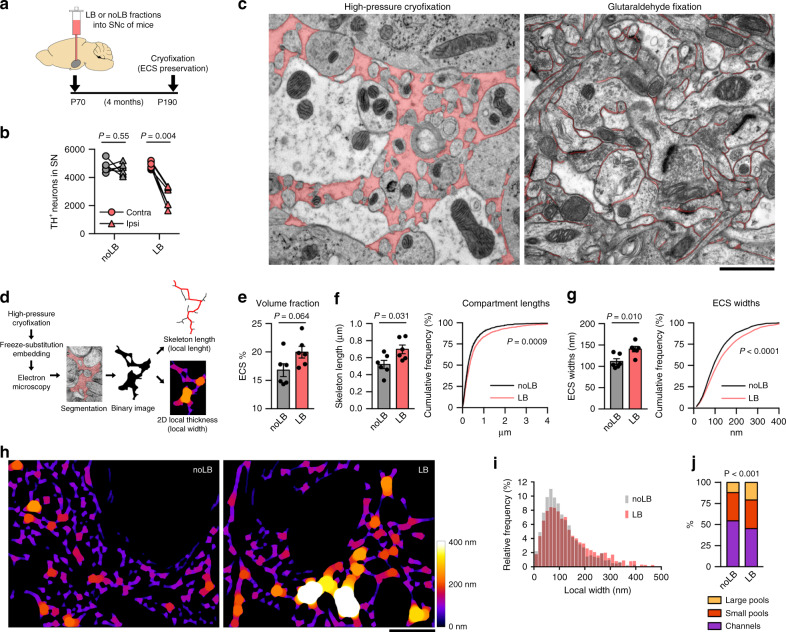
Fig. 1
u03b1-syn-induced neurodegeneration enlarges the extracellular space locally.
a Experimental timeline of the mouse model of Lewy body (LB)-induced neurodegeneration. b TH stereological cell counts confirmed 40% dopaminergic cell loss typically observed in the SN of LB-inoculate...
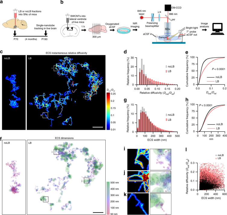
Fig. 2
Single-nanotube tracking reveals increased ECS nanoscale diffusivity and width after synucleinopathy.
a Experimental timeline. b Schematic diagram of SWCNT imaging and tracking setup. Fluorescent SWCNTs are administered in vivo into the lateral ventricle of adult mice and allowed to diffuse into the b...
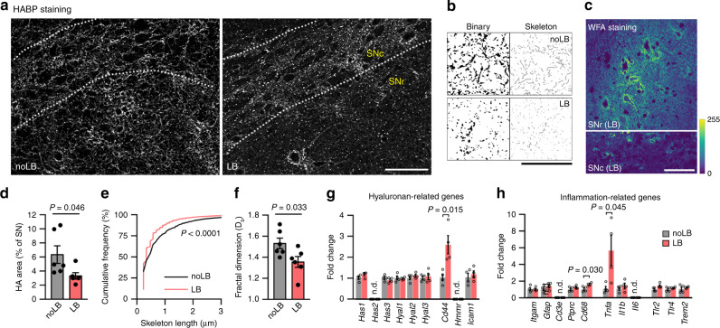
Fig. 3
The hyaluronan matrix is degraded after synucleinopathy.
a Confocal micrographs from the SN of noLB and LB-inoculated mice, showing a widespread disruption of the matrix in the degenerated SN and fewer cable-like HA structures. Scale bar, 50u2009u00b5m. b B...
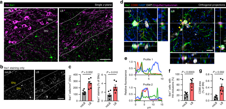
Fig. 4
Reactive microglia populate the SNc and phagocytes hyaluronan after u03b1-syn-induced neurodegeneration.
a , b Confocal images showing distribution of microglia (Iba1) in the SN of mice. In control animals, microglia populate mostly the SNr and is scarce in the SNc, packed with TH-positive neurons. This ...
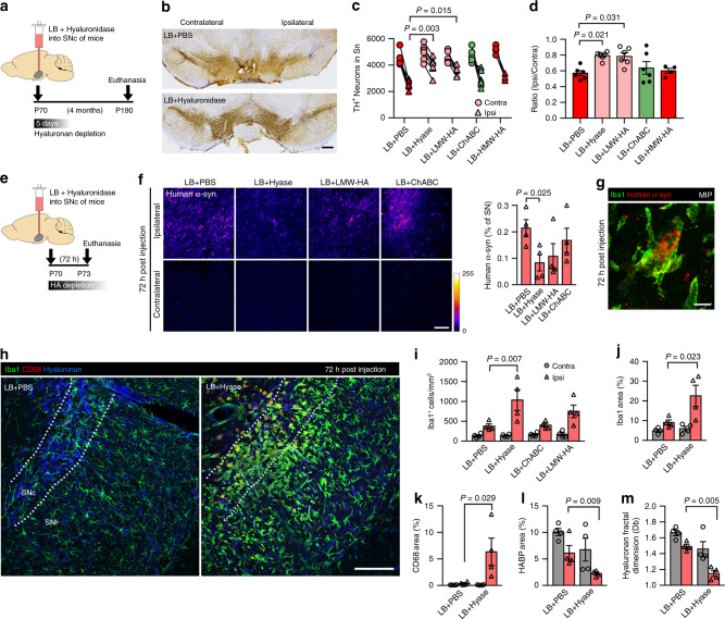
Fig. 5
Hyaluronan fragmentation ameliorates LB-induced neurodegeneration by reducing toxic-u03b1-syn load.
a Experimental timeline for ( b u2013 d ). Hyaluronidase (Hyase) effect lasts only a few days post-injection, since HA is rapidly replenished in vivo. b TH staining shows partial neuroprotection in th...
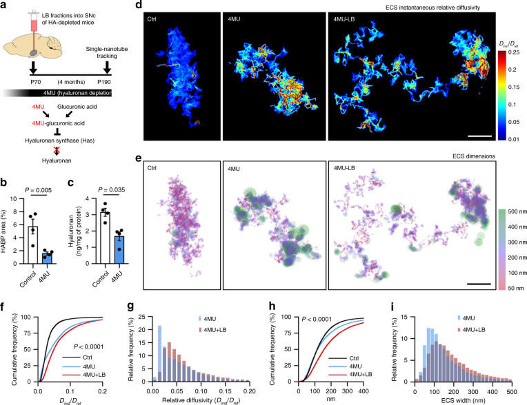
Fig. 6
Hyaluronan depletion increases ECS nanoscale diffusivity more profoundly than ECS width.
a Experimental timeline. 4-methylumbelliferone (4MU) inhibits HA synthesis in vivo. 4MU-based diet started 4 weeks prior to LB-inoculation to deplete HA chronically before the onset of pathology. b , ...
Figure images are served from the NIH/NLM PubMed Central Open Access Subset or Europe PMC; copyright remains with the publishers and authors.
💬 Discussion
0 commentsNo comments yet. Be the first to start a discussion!
Leave a Comment