Abstract
AbstractTelocytes (TCs), a particular interstitial cell type, have been recently described in a wide variety of mammalian organs (www.telocytes.com). The TCs are identified morphologically by a small cell body and extremely long (tens to hundreds of μm), thin prolongations (less than 100 nm in diameter, below the resolving power of light microscopy) called telopodes. Here, we demonstrated with electron microscopy and immunofluorescence that TCs were present in human dermis. In particular, TCs were found in the reticular dermis, around blood vessels, in the perifollicular sheath, outside the glassy membrane and surrounding sebaceous glands, arrector pili muscles and both the secretory and excretory portions of eccrine sweat glands. Immunofluorescence screening and laser scanning confocal microscopy showed two subpopulations of dermal TCs; one expressed c‐kit/CD117 and the other was positive for CD34. Both subpopulations were also positive for vimentin. The TCs were connected to each other by homocellular junctions, and they formed an interstitial 3D network. We also found TCs adjoined to stem cells in the bulge region of hair follicles. Moreover, TCs established atypical heterocellular junctions with stem cells (clusters of undifferentiated cells). Given the frequency of allergic skin pathologies, we would like to emphasize the finding that close, planar junctions were frequently observed between TCs and mast cells. In conclusion, based on TC distribution and intercellular connections, our results suggested that TCs might be involved in skin homeostasis, skin remodelling, skin regeneration and skin repair.
🔬 Techniques
🔭 Microscopes
💻 Software
✨ Fluorophores
🧪 Sample Preparation
🔬 Cell Lines
🏭 Microscope Brands
🧪 Reagent Suppliers
📷 Detectors
🔎 Objectives
💻 Software Details
🏛️ Research Organizations (ROR)
Affiliated research institutions:
📋 Methods
Biopsies of human skin were obtained from three patients (informed written consent). Normal skin samples were obtained from a re-excision procedure after removing a local melanoma. The second excisions were performed according to the Breslow index (tumoural depth), 14 days after primary excision. The samples of normal skin were taken at 1 cm distance from primary suture. Experiments were performed according to the Helsinki guidelines, in full compliance with the Bioethics Committee of the ‘Victor Babeş’ National Institute of Pathology, Bucharest regulations. In situ immunostaining and confocal analysis Paraffin embedded skin samples (7μm thick) were deparaffinised, washed for 30 min. in PBS, pH 7.4 and blocked with 2% BSA. The samples were incubated for 30 min. with 2% normal goat serum (Sigma-Aldrich Chemical, St. Louis, MO, USA). Samples were incubated overnight at 4°C in PBS with either rabbit anti c-Kit or one of the following mouse monoclonal antibodies: anti-vimentin (clone V9, 1:150), anti- CD34 (clone QBEnd-10, 1:25) (both from Dako, Glostrup, Denmark) or anti-nestin (clone 10C2, 1:100) (Millipore, Billerica, MA, USA). In addition, combinations were used in double labelling assays. After washing in PBS with 0.1% (vol/vol) Triton X- 100, the sections were incubated with Alexa Fluor-conjugated, secondary goat anti-rabbit or goat antimouse antibodies (Invitrogen, Molecular Probes, Eugene, OR, USA) for another 2 hrs, at room temperature. Following an extensive washing step, the nuclei were stained with 1 μg/ml 4′,6-diamidino-2-phenylindole (DAPI) (Sigma-Aldrich). Negative controls were performed by omitting the primary antibody from the same protocol. Epifluorescence was used to examine three to five immunolabelled sections from each biopsy on a Nikon Eclipse E600 microscope (Nikon Instruments Inc., Tokyo, Japan) with a Nikon Plan Apo 40× objective and the appropriate fluorescence filters. Digital pictures were acquired with a CCD Axiocam HRc Zeiss camera and AxioVision software (Carl Zeiss Imaging solution GmbH, Göttingen, Germany) or with confocal laser scanning microscopy, with a Nikon A1 laser microscope mounted on an ECLIPSE Ti-E inverted microscope. The confocal images were collected with a Plan Fluor 60× oil objective and 1.25-NA water ( z -axis step 0.16 μm). The following lasers and emission filters were used: Ar laser at 488 nm (used for the excitation of Alexa Fluor 488) and emission filter 500-550 nm; 561.2 nm G-HeNe laser (for Alexa Fluor 546) and emission filter 570–620 nm; and 405 nm Laser diode and 425–475 nm emission filter for DAPI. To improve the image quality, some original laser scanning microscopy data were subjected to digital deconvolution and three-dimensional reconstruction with an Imaris × 64 (version 6.3.1.) from Bitplane AG (Zürich, Switzerland). Transmission electron microscopy (TEM) The TEM was performed on small (1 mm 3 ) tissue fragments, processed according to a routine Epon-embedding procedure, as previously described [ 36 ]. Thin sections (about 60 nm) were examined with a Morgagni 286 transmission microscope (FEI Company, Eindhoven, The Netherlands) at 60 kV. Digital electron micrographs were acquired with a MegaView III CCD and iTEM-SIS software (Olympus, Soft Imaging System GmbH, Münster, Germany). To highlight the TCs and Tps, TEM images were digitally coloured in blue with Adobe © Photoshop CS3.
Show full methods section
Biopsies of human skin were obtained from three patients (informed written consent). Normal skin samples were obtained from a re-excision procedure after removing a local melanoma. The second excisions were performed according to the Breslow index (tumoural depth), 14 days after primary excision. The samples of normal skin were taken at 1 cm distance from primary suture. Experiments were performed according to the Helsinki guidelines, in full compliance with the Bioethics Committee of the ‘Victor Babeş’ National Institute of Pathology, Bucharest regulations. In situ immunostaining and confocal analysis Paraffin embedded skin samples (7μm thick) were deparaffinised, washed for 30 min. in PBS, pH 7.4 and blocked with 2% BSA. The samples were incubated for 30 min. with 2% normal goat serum (Sigma-Aldrich Chemical, St. Louis, MO, USA). Samples were incubated overnight at 4°C in PBS with either rabbit anti c-Kit or one of the following mouse monoclonal antibodies: anti-vimentin (clone V9, 1:150), anti- CD34 (clone QBEnd-10, 1:25) (both from Dako, Glostrup, Denmark) or anti-nestin (clone 10C2, 1:100) (Millipore, Billerica, MA, USA). In addition, combinations were used in double labelling assays. After washing in PBS with 0.1% (vol/vol) Triton X- 100, the sections were incubated with Alexa Fluor-conjugated, secondary goat anti-rabbit or goat antimouse antibodies (Invitrogen, Molecular Probes, Eugene, OR, USA) for another 2 hrs, at room temperature. Following an extensive washing step, the nuclei were stained with 1 μg/ml 4′,6-diamidino-2-phenylindole (DAPI) (Sigma-Aldrich). Negative controls were performed by omitting the primary antibody from the same protocol. Epifluorescence was used to examine three to five immunolabelled sections from each biopsy on a Nikon Eclipse E600 microscope (Nikon Instruments Inc., Tokyo, Japan) with a Nikon Plan Apo 40× objective and the appropriate fluorescence filters. Digital pictures were acquired with a CCD Axiocam HRc Zeiss camera and AxioVision software (Carl Zeiss Imaging solution GmbH, Göttingen, Germany) or with confocal laser scanning microscopy, with a Nikon A1 laser microscope mounted on an ECLIPSE Ti-E inverted microscope. The confocal images were collected with a Plan Fluor 60× oil objective and 1.25-NA water ( z -axis step 0.16 μm). The following lasers and emission filters were used: Ar laser at 488 nm (used for the excitation of Alexa Fluor 488) and emission filter 500-550 nm; 561.2 nm G-HeNe laser (for Alexa Fluor 546) and emission filter 570–620 nm; and 405 nm Laser diode and 425–475 nm emission filter for DAPI. To improve the image quality, some original laser scanning microscopy data were subjected to digital deconvolution and three-dimensional reconstruction with an Imaris × 64 (version 6.3.1.) from Bitplane AG (Zürich, Switzerland). Transmission electron microscopy (TEM) The TEM was performed on small (1 mm 3 ) tissue fragments, processed according to a routine Epon-embedding procedure, as previously described [ 36 ]. Thin sections (about 60 nm) were examined with a Morgagni 286 transmission microscope (FEI Company, Eindhoven, The Netherlands) at 60 kV. Digital electron micrographs were acquired with a MegaView III CCD and iTEM-SIS software (Olympus, Soft Imaging System GmbH, Münster, Germany). To highlight the TCs and Tps, TEM images were digitally coloured in blue with Adobe © Photoshop CS3.
📊 Figures
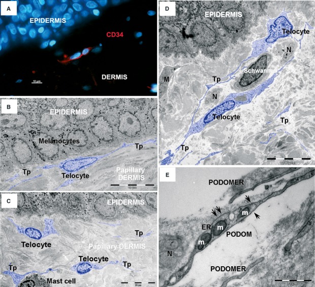
Fig 1
Telocytes in human papillary dermis. (A) Epifluorescence microscopy u2013 immunofluorescence labelling shows CD34 (red) interstitial cells. Nuclei are counterstained with DAPI (blue). Original magnifi...
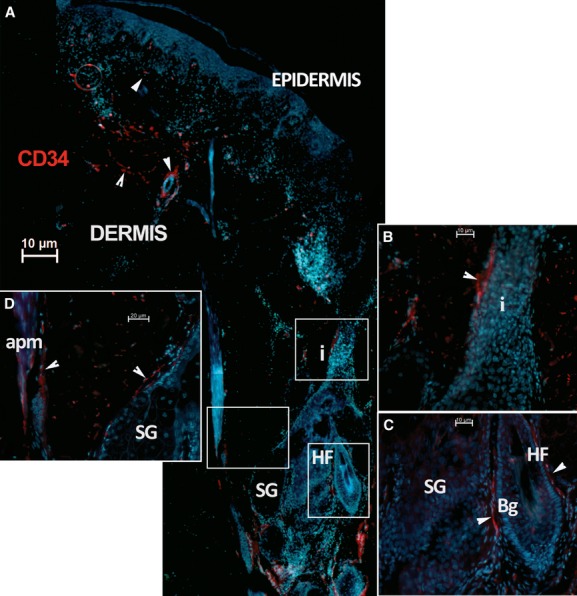
Fig 2
Telocytes in human dermis. (A) Epifluorescence microscopy: Immunofluorescence shows the distribution of CD34 positive interstitial cells (red). This image reconstruction shows scarce CD34 positive cel...
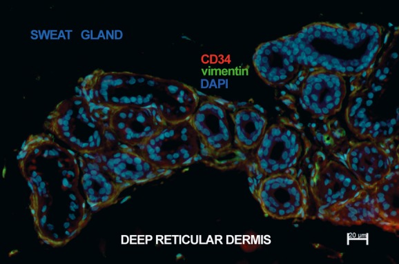
Fig 3
Telocytes in human deep reticular dermis. Epifluorescence microscopy: double immunofluorescence labelling shows co-localization (yellow) of CD34 (red) and vimentin (green) in telocytes surrounding the...
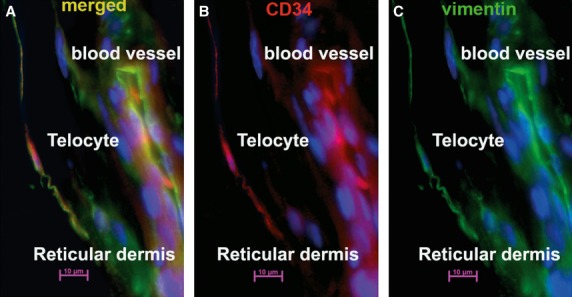
Fig 4
Telocytes in human reticular dermis vasculature. Epifluorescence microscopy: double immunofluorescence labelling shows a telocyte located in the perivascular space of a blood vessel that (A) co-expres...
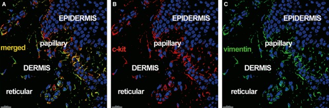
Fig 5
Telocytes in human dermis. Laser scanning confocal microscopy: three-dimensional shadow projection image. Double immunofluorescence labelling shows (A) co-localization (yellow) of (B) CD117/c-kit (red...
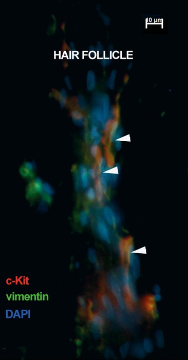
Fig 6
Telocytes surrounding human hair follicle. Epifluorescence microscopy: double immunofluorescence labelling shows co-localization (yellow) of CD117/c-kit (red) and vimentin (green) in telocytes surroun...
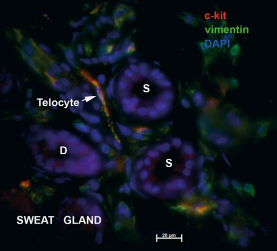
Fig 7
Telocytes associated with human sweat gland. Epifluorescence microscopy: double immunofluorescence labelling shows co-localization (yellow) of CD117/c-kit (red) and vimentin (green) in a telocyte (arr...
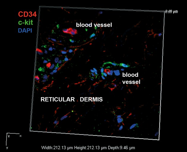
Fig 8
Distinct subsets of interstitial cells in human dermis. Laser scanning confocal microscopy: volume reconstruction. Double immunofluorescence labelling shows distinct subsets of interstitial CD34 posit...
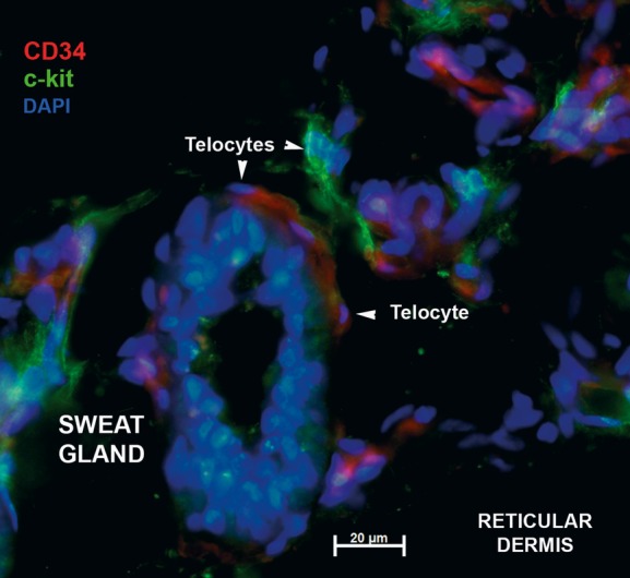
Fig 9
Distinct subsets of telocytes in connective tissue around sweat gland. Epifluorescence microscopy: double immunofluorescence labelling shows distinct CD117/c-kit (green) and CD34 (red) telocytes (arro...
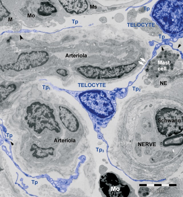
Fig 10
Telocytes form an interstitial network in human dermis. Digitally enhanced (blue colour) TEM image shows telocytes (blue) with telopodes (Tp). Telocytes are connected by junctions (black arrows; see d...
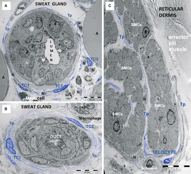
Fig 11
Telocytes form a sheath around human sweat gland. TEM micrographs show telocytes (TC1u2013TC3) with their telopodes (Tp) (A) enfolding the sweat gland, (B) its excretory duct and (C) arrector pili mus...
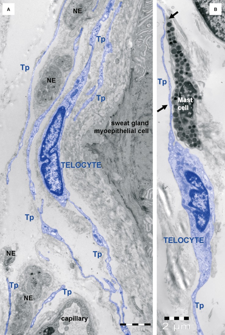
Fig 12
Telocyte layers in human skin appendages. (A) Telocytes (blue) form layers around an eccrine sweat gland mixed with nerve endings (NE). (B) Telocytes are typically associated with mast cells. Tp u2013...
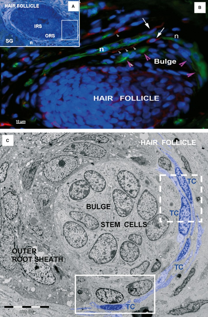
Fig 13
Telocytes around human hair follicle and adjacent sebaceous gland. (A) Light microscopy image of toluidine-blue coloured, thin section shows a hair follicle and an adjacent sebaceous gland (SG). IRS: ...
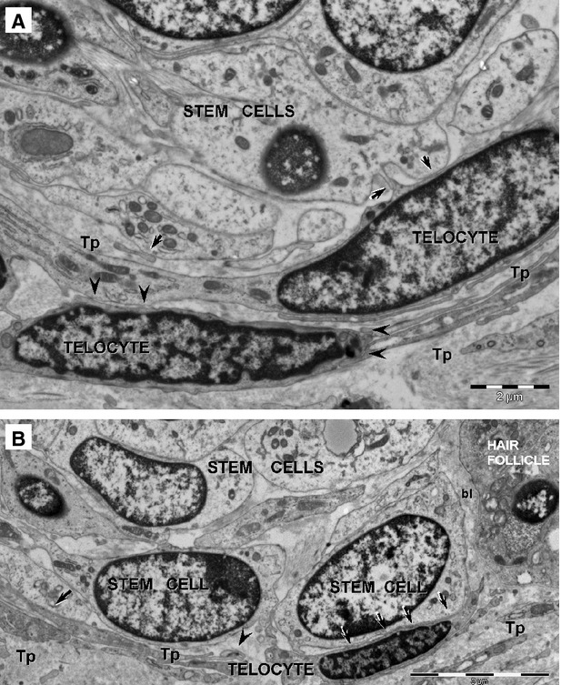
Fig 14
Telocytes bordering stem cells. High magnification TEM images of the boxed areas in Fig. 13 C. The dashed-line box is magnified in (A); the continuous-line box is magnified in (B). Two layers of inter...
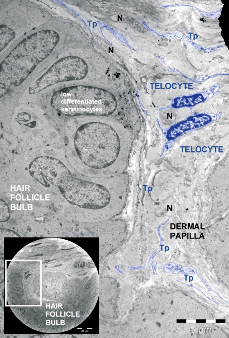
Fig 15
Telocytes associated with nerve endings. TEM image shows telocytes and numerous telopodes (Tp) next to nerve endings (N) that are present in the dermal papilla of a hair follicle. The inset image show...
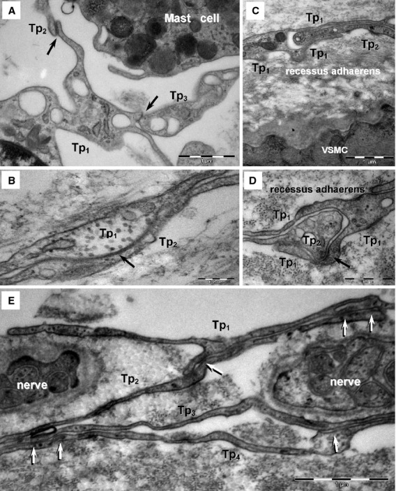
Fig 16
Homocellular junctions between telocytes in human skin (TEM). (A) Three telopodes (Tp 1 u2013Tp 3 ) are connected by small adhaerens junctions (arrows). (B) A gap junction is visible between two telop...
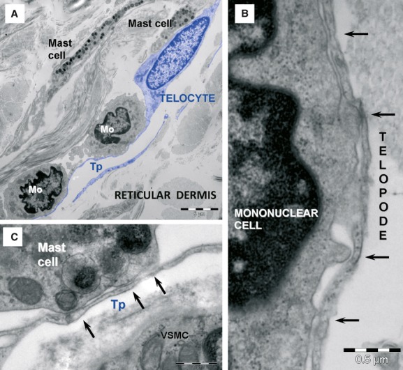
Fig 17
Heterocellular contacts between telocytes and other interstitial cells. (A) TEM image shows telocytes (coloured blue), mononuclear cells (Mo) and mast cells. (B) High magnification of the contact area...
Figure images are served from the NIH/NLM PubMed Central Open Access Subset or Europe PMC; copyright remains with the publishers and authors.
💬 Discussion
0 commentsNo comments yet. Be the first to start a discussion!
Leave a Comment