Abstract
With faithful sample preservation and direct imaging of fully hydrated biological material, cryo-electron tomography provides an accurate representation of molecular architecture of cells. However, detection and precise localization of macromolecular complexes within cellular environments is aggravated by the presence of many molecular species and molecular crowding. We developed a template-free image processing procedure for accurate tracing of complex networks of densities in cryo-electron tomograms, a comprehensive and automated detection of heterogeneous membrane-bound complexes and an unsupervised classification (PySeg). Applications to intact cells and isolated endoplasmic reticulum (ER) allowed us to detect and classify small protein complexes. This classification provided sufficiently homogeneous particle sets and initial references to allow subsequent de novo subtomogram averaging. Spatial distribution analysis showed that ER complexes have different localization patterns forming nanodomains. Therefore, this procedure allows a comprehensive detection and structural analysis of complexes in situ.
🔬 Techniques
🧬 Organisms
🧪 Sample Preparation
🔬 Cell Lines
🏭 Microscope Brands
📷 Detectors
💾 Data Repositories
🏛️ Research Organizations (ROR)
Affiliated research institutions:
📊 Figures
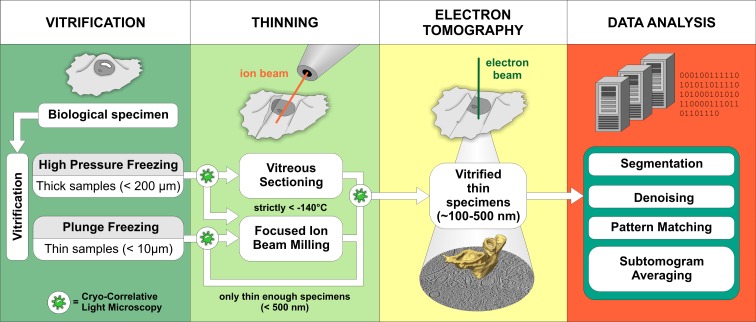
Figure 1.
Schematic representation of the cryo-ET workflow. Vitrification. The sample type usually dictates the vitrification method. Samples that are thin enough to be directly investigated by EM are commonly ...
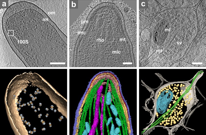
Figure 2.
Cryo-ET of intact cells. (a) Intact E. coli. Top: tomographic slice. Bottom: isosurface representation with positions of template-matched ribosome pairs: om, outer membrane; im, inner membrane; inset ...
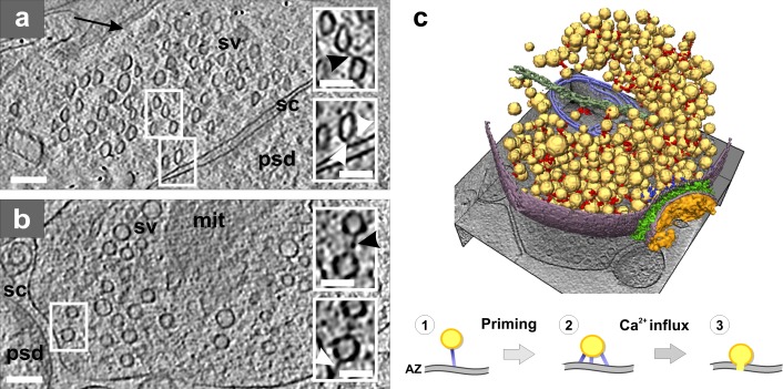
Figure 3.
Presynaptic cytomatrix. (a) Tomographic slice of a synapse from a high-pressure frozen organotypic hippocampal slice, thinned by vitreous sectioning. Synaptic vesicles are compressed along the cutting...
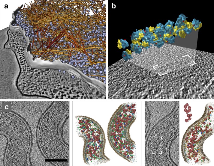
Figure 4.
Image processing: segmentation and template matching. (a) Automated segmentation of actin filaments in Dictyostelium discoideum : actin filaments (yellow to red); ribosomes (blue). Reproduced with per...
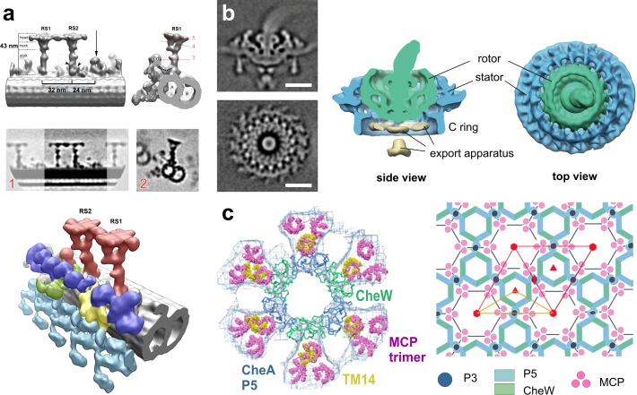
Figure 5.
Image processing: subtomogram averaging. (a) Isolated C. reinhardtii axoneme. Top row: surface renderings of the density map obtained by averaging radial spokes (RS). Middle row: tomographic slices of...
Figure images are served from the NIH/NLM PubMed Central Open Access Subset or Europe PMC; copyright remains with the publishers and authors.
💬 Discussion
0 commentsNo comments yet. Be the first to start a discussion!
Leave a Comment