Abstract
Antimicrobial peptides play an important role in host defense against microbial pathogens. Their high cationic charge and strong amphipathic structure allow them to bind to the anionic microbial cell membrane and disrupt the membrane bilayer by forming pores or channels. In contrast to the classical pore-forming peptides, studies on histatin-5 (Hst-5) have suggested that the peptide is transported into the cytoplasm of Candida albicans in a non-lytic manner, and cytoplasmic Hst-5 exerts its candicidal activities on various intracellular targets, consistent with its weak amphipathic structure. To understand how Hst-5 is internalized, we investigated the localization of FITC-conjugated Hst-5. We find that Hst-5 is internalized into the vacuole through receptor-mediated endocytosis at low extracellular Hst-5 concentrations, whereas under higher physiological concentrations, Hst-5 is translocated into the cytoplasm through a mechanism that requires a high cationic charge on Hst-5. At intermediate concentrations, two cell populations with distinct Hst-5 localizations were observed. By cell sorting, we show that cells with vacuolar localization of Hst-5 survived, while none of the cells with cytoplasmic Hst-5 formed colonies. Surprisingly, extracellular Hst-5, upon cell surface binding, induces a perturbation on the cell surface, as visualized by an immediate and rapid internalization of Hst-5 and propidium iodide or rhodamine B into the cytoplasm from the site using time-lapse microscopy, and a concurrent rapid expansion of the vacuole. Thus, the formation of a spatially restricted site in the plasma membrane causes the initial injury to C. albicans and offers a mechanism for its internalization into the cytoplasm. Our study suggests that, unlike classical channel-forming antimicrobial peptides, action of Hst-5 requires an energized membrane and causes localized disruptions on the plasma membrane of the yeast. This mechanism of cell membrane disruption may provide species-specific killing with minimal damage to microflora and the host and may be used by many other antimicrobial peptides.
🔬 Techniques
🔭 Microscopes
🧬 Organisms
💻 Software
✨ Fluorophores
🧪 Sample Preparation
🏭 Microscope Brands
🧪 Reagent Suppliers
🔎 Objectives
💻 Software Details
🏛️ Research Organizations (ROR)
Affiliated research institutions:
📋 Methods
Yeast strains and growth conditions
The following fungal strains were used in this study: C. albicans SC5314 (wild-type clinical isolate); C. albicans vps36 (BWP17 Cavps36 Δ:: UAU1 / Cavps36 Δ:: URA3 ) and C. albicans snf7 (BWP17 Casnf7 Δ:: UAU1 / Casnf7 Δ:: URA3 ) (gifts from A. Mitchell, [67] ); C. albicans COU46 (CAI4 Camyo5 :: hisG / Camyo5 :: hisG ) (gift from M. Whiteway, [68] ); C. albicans SPA2-GFP (BWP17 SPA2/SPA2-GFP-URA3 ); C. glabrata BG2 (gift from B. Cormack, wild-type clinical isolate, [69] ); and S. cerevisiae BY4741 ( MATa , leu2 Δ 0 , met15 Δ 0 , and ura3 Δ 0 ). All of the yeast strains were maintained on YPD plates [1% (w/v) yeast extract, 2% (w/v) peptone, and 2% (w/v) glucose]. Prior to Hst-5 localization assays, the cells were grown overnight at 30°C in 5 ml YPD broth. A 1/50 dilution of the overnight culture was suspended in fresh 5 ml YPD and grown for an additional 4 hours at 30°C to obtain a mid-log phase culture at which time the optical density was determined (OD 595 of 1.0 = 3×10 7 cells/ml) using a Beckman Coulter DU 800 spectrophotometer to obtain a cell population of 10 6 cells/ml. Peptides Unconjugated Hst-5 and FITC- and biotin-labeled Hst-5 ( DSHAKRHHGYKRKFHEKHHSHRGY ) and FITC-labeled Hst-5 m68 ( DSHAKRHHGYKR E FHEKHHSH G GY ) were synthesized and purified by Genemed Synthesis, Inc. (San Francisco, CA). The identity and purity of the peptides were confirmed by mass spectrometry. Both FITC-Hst-5 and biotin-Hst-5 have been shown to have similar levels of candicidal activity when compared against unlabeled Hst-5 [10] , [13] , [49] . Hst-5 localization studies The intracellular localization of FITC-Hst-5 (5, 10, 20, and 50 µM) and FITC-m68 (5, 10, 20, 50, and 200 µM) was investigated either alone or in a double-labeling experiment using FM4-64 (Molecular Probes, Inc. Eugene, OR). Yeast cells in 50 µl (∼10 6 cells/ml) were incubated for 30 minutes at 30°C with 10 µM FITC-Hst-5 and 10 µM FM4-64; the cells were then washed twice with 10 mM NaN 3 and 10 mM NaF in 20 mM PBS buffer, and analyzed immediately by wide-field fluorescence microscopy. To depolarize F-actin, cells were treated with either 5 µM cytochalasin A or 50 µM latrunculin A (Sigma, St. Louis, MO) for 1 hour at room temperature. The control cells (wild-type and myo5 ) were treated with the equivalent volume of the DMSO solvent (0.5%). The cells were then exposed to 5 µM of FITC-Hst-5 for 30 minutes at 30°C. The cells were then washed twice with buffer containing NaN 3 and NaF and analyzed by wide-field fluorescence microscopy and flow cytometry. For live cell imaging, 300 µl of a 100 µg/ml solution of concanavalin A (MP Biomedicals, LLC. Solon, Ohio) was coated onto a sterile 0.17 mm glass bottom dish (WillCo Wells BV, Amsterdam, Denmark). The wells were incubated for 1 hour at room temperature and then washed three times with water. A 300 µl buffer suspension of ∼2×10 6 C. albicans cells were aliquoted onto the well and incubated at room temperature. After settling and binding for 15 minutes unbound cells were washed away [70] . Buffer containing 50 µM of either unconjugated Hst-5 or FITC-Hst-5 and 5 µg/ml PI or RB was added to the cells and uptake of fluorescence was followed by time-lapse confocal microscopy. To determine the position of site specific breach by Hst-5 in relation to known cellular markers, cells were either observed using 2 µg/ml calcofluor white (Sigma, St. Louis, MO) and 5 µg/ml PI in PBS buffer or the C. albicans Spa2-GFP strain was grown in the presence of PI and 2% fetal calf serum in water. Buffer containing 50 µM biotin-Hst-5 was added to the cells and uptake of PI was followed by time-lapse wide-field fluorescence microscopy at room temperature and 37°C, respectively.
Show full methods section
Yeast strains and growth conditions
The following fungal strains were used in this study: C. albicans SC5314 (wild-type clinical isolate); C. albicans vps36 (BWP17 Cavps36 Δ:: UAU1 / Cavps36 Δ:: URA3 ) and C. albicans snf7 (BWP17 Casnf7 Δ:: UAU1 / Casnf7 Δ:: URA3 ) (gifts from A. Mitchell, [67] ); C. albicans COU46 (CAI4 Camyo5 :: hisG / Camyo5 :: hisG ) (gift from M. Whiteway, [68] ); C. albicans SPA2-GFP (BWP17 SPA2/SPA2-GFP-URA3 ); C. glabrata BG2 (gift from B. Cormack, wild-type clinical isolate, [69] ); and S. cerevisiae BY4741 ( MATa , leu2 Δ 0 , met15 Δ 0 , and ura3 Δ 0 ). All of the yeast strains were maintained on YPD plates [1% (w/v) yeast extract, 2% (w/v) peptone, and 2% (w/v) glucose]. Prior to Hst-5 localization assays, the cells were grown overnight at 30°C in 5 ml YPD broth. A 1/50 dilution of the overnight culture was suspended in fresh 5 ml YPD and grown for an additional 4 hours at 30°C to obtain a mid-log phase culture at which time the optical density was determined (OD 595 of 1.0 = 3×10 7 cells/ml) using a Beckman Coulter DU 800 spectrophotometer to obtain a cell population of 10 6 cells/ml. Peptides Unconjugated Hst-5 and FITC- and biotin-labeled Hst-5 ( DSHAKRHHGYKRKFHEKHHSHRGY ) and FITC-labeled Hst-5 m68 ( DSHAKRHHGYKR E FHEKHHSH G GY ) were synthesized and purified by Genemed Synthesis, Inc. (San Francisco, CA). The identity and purity of the peptides were confirmed by mass spectrometry. Both FITC-Hst-5 and biotin-Hst-5 have been shown to have similar levels of candicidal activity when compared against unlabeled Hst-5 [10] , [13] , [49] . Hst-5 localization studies The intracellular localization of FITC-Hst-5 (5, 10, 20, and 50 µM) and FITC-m68 (5, 10, 20, 50, and 200 µM) was investigated either alone or in a double-labeling experiment using FM4-64 (Molecular Probes, Inc. Eugene, OR). Yeast cells in 50 µl (∼10 6 cells/ml) were incubated for 30 minutes at 30°C with 10 µM FITC-Hst-5 and 10 µM FM4-64; the cells were then washed twice with 10 mM NaN 3 and 10 mM NaF in 20 mM PBS buffer, and analyzed immediately by wide-field fluorescence microscopy. To depolarize F-actin, cells were treated with either 5 µM cytochalasin A or 50 µM latrunculin A (Sigma, St. Louis, MO) for 1 hour at room temperature. The control cells (wild-type and myo5 ) were treated with the equivalent volume of the DMSO solvent (0.5%). The cells were then exposed to 5 µM of FITC-Hst-5 for 30 minutes at 30°C. The cells were then washed twice with buffer containing NaN 3 and NaF and analyzed by wide-field fluorescence microscopy and flow cytometry. For live cell imaging, 300 µl of a 100 µg/ml solution of concanavalin A (MP Biomedicals, LLC. Solon, Ohio) was coated onto a sterile 0.17 mm glass bottom dish (WillCo Wells BV, Amsterdam, Denmark). The wells were incubated for 1 hour at room temperature and then washed three times with water. A 300 µl buffer suspension of ∼2×10 6 C. albicans cells were aliquoted onto the well and incubated at room temperature. After settling and binding for 15 minutes unbound cells were washed away [70] . Buffer containing 50 µM of either unconjugated Hst-5 or FITC-Hst-5 and 5 µg/ml PI or RB was added to the cells and uptake of fluorescence was followed by time-lapse confocal microscopy. To determine the position of site specific breach by Hst-5 in relation to known cellular markers, cells were either observed using 2 µg/ml calcofluor white (Sigma, St. Louis, MO) and 5 µg/ml PI in PBS buffer or the C. albicans Spa2-GFP strain was grown in the presence of PI and 2% fetal calf serum in water. Buffer containing 50 µM biotin-Hst-5 was added to the cells and uptake of PI was followed by time-lapse wide-field fluorescence microscopy at room temperature and 37°C, respectively.
Fluorescence microscopy
Wide-field fluorescence images were obtained on either Zeiss Axioplan 2 or the inverted Zeiss Axio Observer.Z1 Microscope (Carl Zeiss MicroImaging, Inc. Thornwood, NY) fluorescent system, equipped with the AttoArc HBO 100 and the X-Cite series 120 mercury lamps, respectively. Images were taken using a 100× NA 1.4 objective. Both fluorescence microscopes were equipped with GFP, RFP, and DAPI filter sets. Data sets were obtained as 10–20 optical sections per wavelength spaced 0.2 µm apart along the Z-axis. Out of focus information was removed using a constrained iterative deconvolution algorithm. During the experiment cells were kept at either a constant 30° or 37°C using the TempModule S system on the microscope. Processing was done on a PC using the software packages Axiovision 3.1 and 4.6.3, as well as Photoshop (Adobe Systems Inc., Mountain View, CA). Confocal laser scanning microscopy was performed on an inverted LSM510 laser scanning microscope (Carl Zeiss, Göttingen, Germany) using a Plan-Apo 100×/1.4 NA lens. For the simultaneous detection of fluorescein-labeled peptides and the fluorochromes PI or RB, the 488-nm line of the argon ion laser and the light of a 543-nm helium neon laser were directed over an HFT UV/488/543/633 beam splitter, and the fluorescence was detected using an NFT 545 beam splitter in combination with a BP 500–550 band pass filter for fluorescein detection and an BP 565–615 band pass filter for PI and RB detection.
FACS analysis
The distribution of FITC-labeled Hst-5 over the cell population was investigated by using a dual laser fluorescence-activated cell sorter (BD FACSCalibur System, Becton Dickinson, San Jose, CA). The results were analyzed with the software package CellQuest Pro (version 5.1.1) provided by Beckton Dickinson. Flow cytometric cell sorting and candidacidal activity of histatin-5 C. albicans cells were incubated for 30 minutes with 10 µM of FITC-Hst-5 at 30°C. The cells were then washed twice with 20 mM PBS buffer and under went flow cytometric cell sorting using the DAKO Cytomation MoFlo Flow Cytometer (DAKO, Glostrup, Denmark). The results were analyzed on a PC using the software package Summit (version 4.0) provided by DAKO. The cells were sorted by gating the two peaks of the histogram representing vacuolar and cytoplasmic localization of Hst-5. Cellular localization of Hst-5 was confirmed with fluorescence microscopy. The sorted cells were then plated onto YPD plates and incubated overnight at 30°C (data as a mean±1SD of triplicate cultures).
Determination of charge for Hst-5 and m68
Determination of charge for Hst-5 and m68 were done using PROTEIN CALCULATOR v3.3 ( www.scripps.edu/∼cdputnam/protcalc2.html ). The pKa values for the individual amino acids are from Stryer Biochemistry, 3 rd edition. The software was designed by Chris Putnam at the Scripps Research Institute cdputnam@scripps.edu .
Supporting Information Figure S1 Unlabeled Hst-5 induced internalization of the fluorchrome propidium iodide from spatially restricted sites on the cell surface. 50 µM Hst-5 (unconjugated) was added to the buffer containing PI, and uptake of fluorescence was followed by time-lapse confocal microscopy at room temperature, with frames recorded every 9 seconds for 7 minutes and 30 seconds. Six frames recorded at 9, 27, 45, 90, and 297 seconds are shown. (2.00 MB TIF) Click here for additional data file. Video S1 Cytoplasmic translocation of FITC-Hst-5 into the yeast C. albicans . The uptake of fluorescence after the addition of 50 µM FITC-Hst-5 to C. albicans cells. The images were recorded by time-lapse confocal microscopy at room temperature with frames recorded every 9 seconds for a total of 7 minutes and 30 seconds. (6.42 MB MOV) Click here for additional data file. Video S2 Rapid vacuole expansion and deformation of C. albicans after the addition of Hst-5. The morphological change to the yeast C. albicans after the addition of 50 µM Hst-5. The images were recorded by time-lapse microscopy at room temperature with frames recorded every 15 seconds for a total of 15 minutes. (2.20 MB MOV) Click here for additional data file. Video S3 Cytoplasmic translocation of propidium iodide into the yeast C. albicans . The uptake of PI after the addition of 50 µM FITC-Hst-5 to C. albicans cells. The images were recorded by time-lapse confocal microscopy at room temperature with frames recorded every 9 seconds for a total of 7 minutes and 30 seconds. (6.42 MB MOV) Click here for additional data file. Video S4 Localization of FITC-Hst-5 and propidium iodide using three-dimensional imaging. Buffer containing 50 µM FITC-Hst-5 and 5 µg/ml PI was added to the cells, and uptake of fluorescence was followed by wide-field time-lapse microscopy. The three-dimensional image was acquired from deconvolution using fifteen 0.2 micron optical sections. (2.10 MB MOV) Click here for additional data file.
📊 Figures
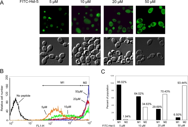
Figure 1
Concentration-dependent localizations of FITC-Hst-5.
C. albicans cells were incubated for 30 minutes with 5, 10, 20, and 50 u00b5M FITC-Hst-5. (A) Fluorescent and DIC images of cells with FITC-Hst-5 at the indicated concentrations and (B) flow cytometry...
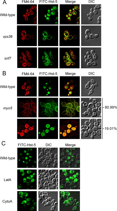
Figure 2
Vacuolar localization of FITC-Hst-5 is dependent on receptor-mediated endocytic pathway.
(A) C. albicans wild-type, vps36 , and snf7 cells were incubated for 30 minutes with 10 u00b5M FITC-Hst-5 and 10 u00b5M FM4-64 at 30u00b0C. (B) C. albicans wild-type and myo5 cells were incubated for ...
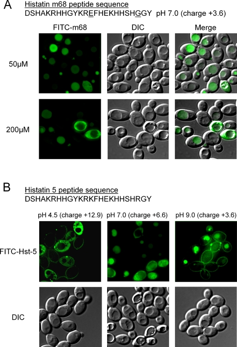
Figure 3
The cationic charge of Hst-5 is important for its uptake into the cytoplasm.
(A) C. albicans cells were incubated with 50 u00b5M and 200 u00b5M FITC-Hst-5 m68 for 30 minutes at 30u00b0C. (B) C. albicans yeast cells were treated with 20 u00b5M FITC-Hst-5 for 30 minutes at 30u00...
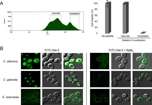
Figure 4
Cytoplasmic localization of Hst-5 is linked to its killing activity.
(A) C. albicans cells were incubated for 30 minutes with 10 u00b5M FITC-Hst-5 at 30u00b0C. The cells were sorted by gating the two peaks of the histogram representing vacuolar and cytoplasmic localiza...
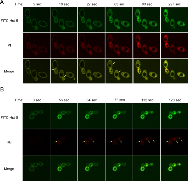
Figure 5
Hst-5 causes a single perturbation on the cell surface of C. albicans .
(A) 50 u00b5M FITC-Hst-5 was added to the buffer containing PI, and uptake of fluorescence was followed by time-lapse confocal microscopy at room temperature with frames recorded every 9 seconds for 7...
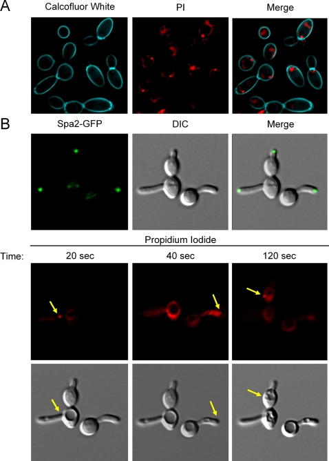
Figure 6
The Hst-5 induced perturbation of the membrane does not co-localize with a specific extracellular region.
(A) 50 u00b5M biotin-Hst-5 was added to C. albicans in buffer containing PI and calcofluor white. Uptake of PI was followed by time-lapse fluorescence microscopy at room temperature with frames record...
Figure images are served from the NIH/NLM PubMed Central Open Access Subset or Europe PMC; copyright remains with the publishers and authors.
💬 Discussion
0 commentsNo comments yet. Be the first to start a discussion!
Leave a Comment