Abstract
Single molecule localization microscopy (SMLM) techniques allow for sub-diffraction imaging with spatial resolutions better than 10 nm reported. Much has been discussed relating to different variations of SMLM and all-inclusive microscopes can now be purchased, removing the need for in-house software or hardware development. However, little discussion has occurred examining the reliability and quality of the images being produced, as well as the potential for overlooked preparative artifacts. As a result of the up to an order-of-magnitude improvement in spatial resolution, substantially more detail is observed, including changes in distribution and ultrastructure caused by the many steps required to fix, permeabilize, and stain a sample. Here we systematically investigate many of these steps including different fixatives, fixative concentration, permeabilization concentration and timing, antibody concentration, and buffering. We present three well-optimized fixation protocols for staining microtubules, mitochondria and actin in a mammalian cell line and then discuss various artifacts in relation to images obtained from samples prepared using the protocols. The potential for such errors to go undetected in SMLM images and the complications in defining a 'good' image using previous parameters applied to confocal microscopy are also discussed.
🔬 Techniques
💻 Software
✨ Fluorophores
🧪 Sample Preparation
🔬 Cell Lines
🏭 Microscope Brands
🧪 Reagent Suppliers
📷 Detectors
💻 Software Details
🏛️ Research Organizations (ROR)
Affiliated research institutions:
📋 Methods
Optimized Protocols for Paraformaldehyde, Glutaraldehyde and Methanol Fixation The main outcome of the research presented here is the optimization of several protocols using common fixatives for SMLM. Moreover, the crucial steps in the fixation process are pinpointed and discussed in relation to corresponding SMLM images containing artifacts. For all of these fixation protocols to progress well the chambered coverglass must be removed from the incubator and placed immediately on a pre-warmed 37°C surface with the fixation steps proceeding without delay.
Development of well optimized protocols yield exemplary SMLM images of microtubules, mitochondria and actin In the development of the optimized protocols we focussed primarily on microtubules (MTs) and mitochondria (MC) because they have very well defined structures. MTs are polymeric and expected to measure approximately 60–65 nm in width when immunostained and imaged with a localization precision of
📊 Figures
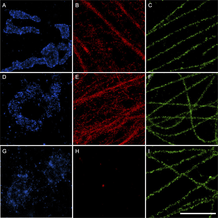
Figure 1
Optimized protocols using paraformaldehyde, glutaraldehyde and methanol preserve ultrastructure for SMLM imaging of mitochondria, actin and microtubules.
(Au2013C) COS-7 cells fixed using the optimized 3.7% paraformaldehyde protocol and stained using Alexa Fluor 647 in conjunction with (A) anti-Tom20, a protein component of the import receptor on the o...
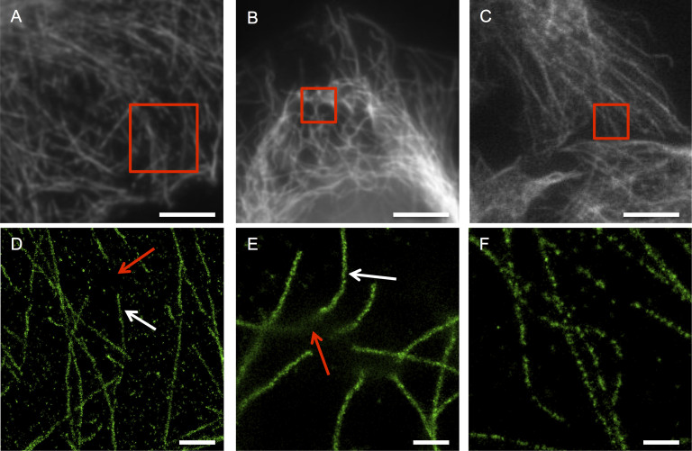
Figure 2
SMLM microtubule images of sub-optimally fixed cells reveal sub-diffraction artifacts not observable in epifluorescence images.
(Au2013C) Epifluorescence images of COS-7 cells stained for tubulin using Alexa Fluor 647 after fixation with PFA (A) for a shorter period than optimal, (B) at a lower concentration than optimal, and ...
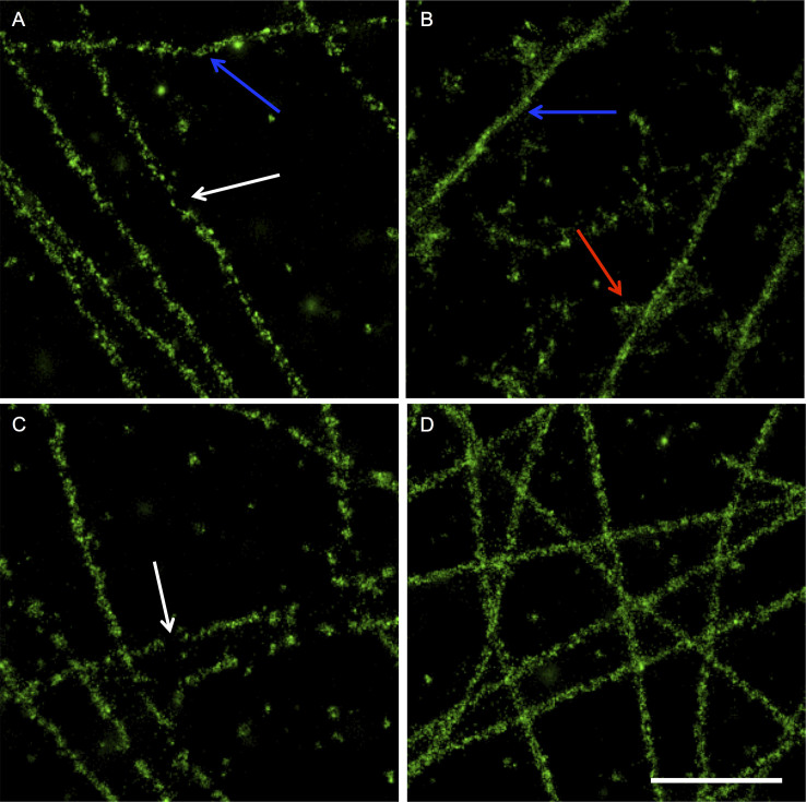
Figure 3
SMLM images of microtubules prepared using standard protocols but with careful initial preparation and application of the fixative solutions show some sub-diffraction artifacts but preserve much of the filamentous architecture.
(Au2013D) COS-7 cells stained for tubulin using Alexa Fluor 647 after fixation with (A) -20u00b0C methanol following a PBS wash, (B) u221220u00b0C methanol allowed to equilibrate to room temperature d...
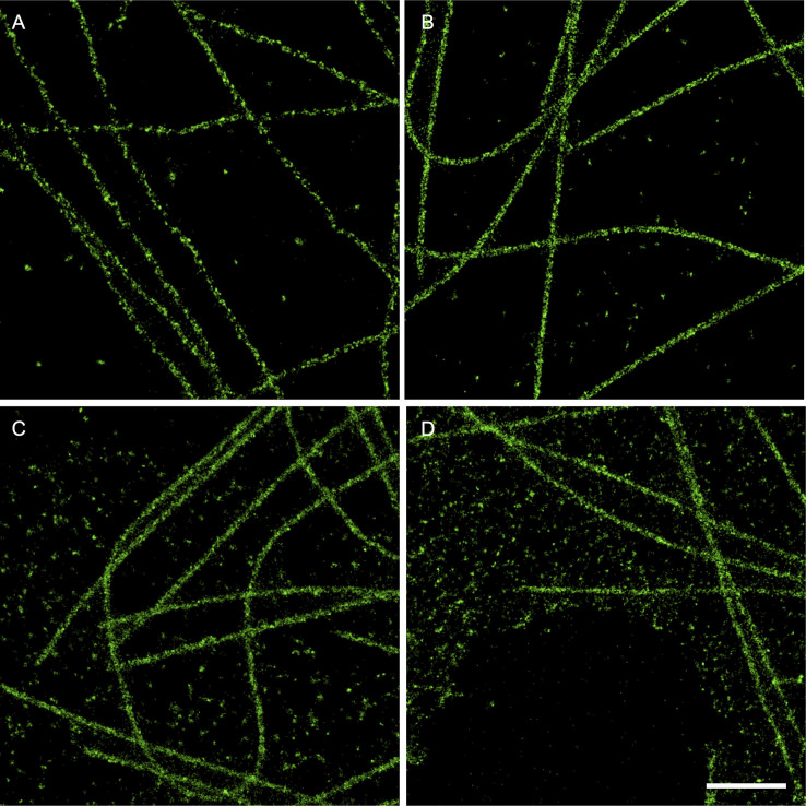
Figure 4
The timing of permeabilization of cells significantly affects the distribution of fluorophores within the cell, resulting in differences in the u2018cleanlinessu2019 of the image as well as the degree to which the image is representative of the biologically native structure and distribution.
(Au2013D) COS-7 cells stained for tubulin using Alexa Fluor 647 after (A) fixation with u221220u00b0C methanol, (B) pre-extraction with 0.3% Triton X-100 in HEPES buffer before 3% glutaraldehyde fixat...
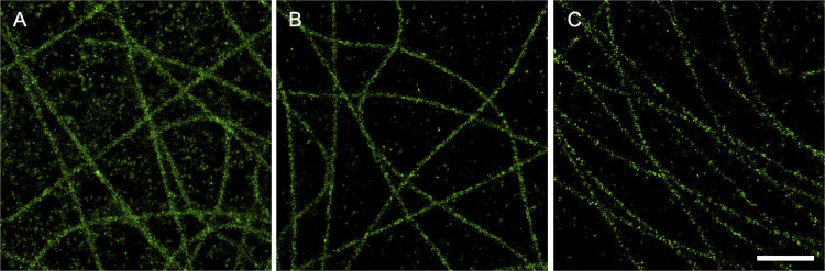
Figure 5
Varying antibody concentration affects apparent microtubule width, non-filamentous stain, and filament continuityu2014all of which can affect spatial resolution.
(Au2013C) COS-7 cells fixed using the optimized glutaraldehyde protocol and then stained for tubulin using mouse anti-u03b2-tubulin and Alexa Fluor 647 conjugated rabbit-anti-mouse. (A) Both primary a...
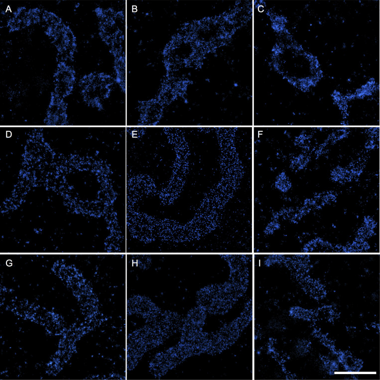
Figure 6
Varying fixative and antibody concentration affects the apparent clustering distribution of import receptors on the mitochondrial membrane.
(Au2013I) COS-7 cells stained using Alexa Fluor 647 primary/secondary antibodies against the Tom20 protein subunit of the import receptor on the outer mitochondria membrane. Cells were stained with a ...
Figure images are served from the NIH/NLM PubMed Central Open Access Subset or Europe PMC; copyright remains with the publishers and authors.
💬 Discussion
0 commentsNo comments yet. Be the first to start a discussion!
Leave a Comment