Abstract
Intravascular neutrophils and platelets collaborate in maintaining host integrity, but their interaction can also trigger thrombotic complications. We report here that cooperation between neutrophil and platelet lineages extends to the earliest stages of platelet formation by megakaryocytes in the bone marrow. Using intravital microscopy, we show that neutrophils "plucked" intravascular megakaryocyte extensions, termed proplatelets, to control platelet production. Following CXCR4-CXCL12-dependent migration towards perisinusoidal megakaryocytes, plucking neutrophils actively pulled on proplatelets and triggered myosin light chain and extracellular-signal-regulated kinase activation through reactive oxygen species. By these mechanisms, neutrophils accelerate proplatelet growth and facilitate continuous release of platelets in steady state. Following myocardial infarction, plucking neutrophils drove excessive release of young, reticulated platelets and boosted the risk of recurrent ischemia. Ablation of neutrophil plucking normalized thrombopoiesis and reduced recurrent thrombosis after myocardial infarction and thrombus burden in venous thrombosis. We establish neutrophil plucking as a target to reduce thromboischemic events.
🔬 Techniques
🔭 Microscopes
✨ Fluorophores
🧪 Sample Preparation
🔬 Cell Lines
🏭 Microscope Brands
🧪 Reagent Suppliers
📷 Detectors
🔎 Objectives
💻 Software Details
🏷️ Research Resource Identifiers (RRIDs)
Verified research resources used in this paper:
🏛️ Research Organizations (ROR)
Affiliated research institutions:
📋 Methods
Key resources table REAGENT or RESOURCE SOURCE IDENTIFIER Antibodies Ultra-LEAF
Purified anti-mouse Ly-6G/Ly-6C (clone RB6-8C5), neutrophil depletion Biolegend Cat#108453; RRID: AB_2616681 Ultra-LEAF™ Purified Rat IgG2b, κ Isotype Ctrl (clone RTK4530) Biolegend Cat# 400671; RRID: AB_11147763 Pacific Blue™ anti-mouse Ly-6G/Ly-6C (clone RB6-8C5) Biolegend Cat#108430; RRID: AB_893556 PE anti-mouse CD115 (clone AFS98) Biolegend Cat#135506; RRID: AB_1937253 PE anti-mouse Ly-6G antibody (clone 1A8) Biolegend Cat#127608; RRID: AB_1186099 APC anti-mouse/rat CD42d (clone 1C2) Biolegend Cat#148506; RRID: AB_2564602 Pacific Blue™ anti-mouse Ly-6G Antibody Biolegend Cat#127612; RRID: AB_2251161 PerCP/Cyanine5.5 anti-mouse CD154 Antibody Biolegend Cat#106514; RRID: AB_2563498 APC/Cyanine7 anti-mouse CD63 Antibody Biolegend Cat#143908; RRID: AB_2565498 Pacific Blue™ anti-mouse/rat CD29 Antibody Biolegend Cat#102224; RRID: AB_2128079 FITC anti-mouse CD3 complex (clone 17A2) BD Bioscience Cat#555274; RRID: AB_395698 FITC anti-mouse CD61 (clone 2C9.G2) BD Bioscience Cat#553346; RRID: AB_10895806 BV711 Rat Anti-Mouse CD62P BD Bioscience Cat#740693; RRID: AB_2740377 PE-Cyanine5 anti Human/Mouse CD45R (clone RA3-6B2) ebioscience Cat#15–045282; RRID: AB 468755 FITC anti-mouse CD41a (clone MWReg30) ebioscience Cat#11-0411-85; RRID: AB_763483 Biotin anti-mouse CD41a (clone MWReg30) ebioscience Cat#13-0411-82; RRID: AB_763484 PE anti-mouse CXCR4 (clone 2B11) ebioscience Cat#12-9991-82; RRID: AB_891391 Streptavidin eFluor 450 ebioscience Cat#48-4317-82,RRID: AB_10359737 Streptavidin PE ebioscience Cat#12-4317-87 Biotin anti-mouse VE-cadherin (clone eBioBV13) ebioscience Cat#13-1441-82; RRID: AB_1234997 GFP recombinant rabbit monoclonal antibody Invitrogen Cat# G10362 ; RRID: AB_2536526 Goat anti-Rat IgG (H + L) Cross-Adsorbed Secondary Antibody, Alexa Fluor 647 Invitrogen Cat#A-21247; RRID: AB_141778 F(ab')2-Goat anti-Rabbit IgG (H + L) Cross-Adsorbed Secondary Antibody, Alexa Fluor 488 Invitrogen Cat#A-11070; RRID: AB_2534114 DyLight 649 anti-mouse CD42b emfret Cat# M040- Antibodies for In Vivo Mouse Platelet Labeling emfret Cat#X488; RRID: AB_2890921 Antibodies for In Vivo Mouse Platelet Labeling emfret Cat#X649; RRID: AB_2861336 Integrin alphaIIb beta3 (GPIIb/IIIa, CD41/CD61) emfret Cat#M023-2; RRID: AB_2833084 Rabbit Anti-Myeloperoxidase Polyclonal Antibody Abcam Cat#ab9535; RRID: AB_307322 Alexa 488 anti-mouse GPIX p0p6-derivative Stegner lab N/A Mouse GPVI Alexa Fluor® 647-conjugated Antibody R&D systems Cat# FAB6758R Human/Mouse CXCL12/SDF-1 Antibody R&D systems Cat#MAB350; RRID: AB_2088149 Phospho-p44/42 MAPK (Erk1/2) (Thr202/Tyr204) antibody Cell Signaling Cat#4370,RRID: AB_2315112 p44/42 MAPK (Erk1/2) (137F5) Rabbit mAb antibody Cell Signaling Cat#4695; RRID: AB_390779 Phospho-Myosin Light Chain 2 (Ser19) Antibody Cell Signaling Cat#3671; RRID: AB_330248 Myosin Light Chain 2 Antibody Cell Signaling Cat#3672; RRID: 10692513 Goat anti-Rabbit IgG (H + L) Secondary Antibody, HRP Thermo scientific Cat#31460; RRID: AB_228341 MC-21(anti-CCR2) antibody Courtesy Matthias Mack N/A Experimental models: Organisms/strains C57BL/6J Jackson Laboratory N/A Mrp8-cre Passegué et al., 2004 B6.Cg-Tg( S100A8-cre ,-EGFP)1Ilw Rosa26-Confetti Livet et al., 2007 Gt(ROSA)26Sor tm1(CAG−Brainbow2.1)Cle/J Rosa26-iDTR Buch et al., 2005 C57BL/6-Gt(ROSA)26Sor tm1(HBEGF)Awai/J Lyz2 -eGFP Faust et al., 2000 B6.129P- Lyz2 tm1(EGFP)1.1Graf Cxcl 12 fl/fl Greenbaum et al., 2013 B6(FVB)- Cxcl 12 tm1.1Link /J Pf4-cre Tiedt et al., 2007 C57BL/6-Tg( Pf4 -icre)Q3Rsko/J Cxcr4 fl/fl Nie et al., 2004 B6.129P2- Cxcr4 tm2Yzo /J Selplg −/− Yang et al., 1999 B6.Cg- Selplg tm1Fur /J Catchup Hasenberg et al., 2015 B6.Ly6g tm1.1(cre)Gunz Cyba mt/mt Nakano et al., 2008 A.B6 Tyr + - Cyba nmf333 /J Cd19-cre Demircik et al., 2013 B6.129P2(C)-Cd19 tm1(cre)Cgn /J Cd3ε −/− Sommers et al., 2000 B6; 129-Cd3e tm1Lov /J Rag1 −/− Mombaerts et al., 1992 B6.129S7- Rag1 tm1Mom /J Chemicals and recombinant proteins Thiazole Orange Sigma-Aldrich Cat# 390062 2′,7′-Dichlorodihydrofluorescein diacetate Sigma-Aldrich Cat# D6883 EZ-Link™ Sulfo-NHS-Biotin Thermo Fisher Cat# 27217 Phorbol 12-myristate 13-acetate Sigma-Aldrich Cat# P8139 Mouse recombinant thrombopoietin Immunotools Cat# 12343615 Mouse recombinant SDF-1 alpha Immunotools Cat# 12343365 Apocynin Sigma-Aldrich Cat# 10809 Qtracker705 Vascular labels Thermo Fisher Cat# Q21061MP AMD3100 Tocris Cat# 3299 U46619 Cayman Cat#16450 Thrombin Chrono-Log Cat# P/N386 Proteome Profiler Mouse XL Cytokine Array R&D systems Cat# ARY028 ML-7 hydrochloride MedChemExpress Cat#HY-15417 FR180204 MedChemExpress Cat# HY-12275 Critical commercial assays Neutrophil Isolation Kit, mouse Miltenyi Biotec Cat# 130-097-658 μ-Slide 4 Well Ibidi Cat# 80426 Pan T Cell Isolation Kit II, mouse Miltenyi Biotec Cat# 130-095-130 Monocyte Isolation Kit (BM), mouse Miltenyi Biotec Cat# 130-100-629 Pan B Cell Isolation Kit II, mouse Miltenyi Biotec Cat# 130-095-813 Software and algorithms Imaris Bitplane RRID: SCR_007370 Flowjo vX Treestar RRID: SCR_008520 Prism Graphpad RRID: SCR_002798 ImageJ NIH RRID: SCR_003070 Resource availability All data are available in the main text or the supplementary materials. Lead contact Further information and requests for resources and reagents should be directed to and will be fulfilled by the lead contact, Tobias Petzold ( Tobias.Petzold@med.uni-muenchen.de ).
Show full methods section
Key resources table REAGENT or RESOURCE SOURCE IDENTIFIER Antibodies Ultra-LEAF
Purified anti-mouse Ly-6G/Ly-6C (clone RB6-8C5), neutrophil depletion Biolegend Cat#108453; RRID: AB_2616681 Ultra-LEAF™ Purified Rat IgG2b, κ Isotype Ctrl (clone RTK4530) Biolegend Cat# 400671; RRID: AB_11147763 Pacific Blue™ anti-mouse Ly-6G/Ly-6C (clone RB6-8C5) Biolegend Cat#108430; RRID: AB_893556 PE anti-mouse CD115 (clone AFS98) Biolegend Cat#135506; RRID: AB_1937253 PE anti-mouse Ly-6G antibody (clone 1A8) Biolegend Cat#127608; RRID: AB_1186099 APC anti-mouse/rat CD42d (clone 1C2) Biolegend Cat#148506; RRID: AB_2564602 Pacific Blue™ anti-mouse Ly-6G Antibody Biolegend Cat#127612; RRID: AB_2251161 PerCP/Cyanine5.5 anti-mouse CD154 Antibody Biolegend Cat#106514; RRID: AB_2563498 APC/Cyanine7 anti-mouse CD63 Antibody Biolegend Cat#143908; RRID: AB_2565498 Pacific Blue™ anti-mouse/rat CD29 Antibody Biolegend Cat#102224; RRID: AB_2128079 FITC anti-mouse CD3 complex (clone 17A2) BD Bioscience Cat#555274; RRID: AB_395698 FITC anti-mouse CD61 (clone 2C9.G2) BD Bioscience Cat#553346; RRID: AB_10895806 BV711 Rat Anti-Mouse CD62P BD Bioscience Cat#740693; RRID: AB_2740377 PE-Cyanine5 anti Human/Mouse CD45R (clone RA3-6B2) ebioscience Cat#15–045282; RRID: AB 468755 FITC anti-mouse CD41a (clone MWReg30) ebioscience Cat#11-0411-85; RRID: AB_763483 Biotin anti-mouse CD41a (clone MWReg30) ebioscience Cat#13-0411-82; RRID: AB_763484 PE anti-mouse CXCR4 (clone 2B11) ebioscience Cat#12-9991-82; RRID: AB_891391 Streptavidin eFluor 450 ebioscience Cat#48-4317-82,RRID: AB_10359737 Streptavidin PE ebioscience Cat#12-4317-87 Biotin anti-mouse VE-cadherin (clone eBioBV13) ebioscience Cat#13-1441-82; RRID: AB_1234997 GFP recombinant rabbit monoclonal antibody Invitrogen Cat# G10362 ; RRID: AB_2536526 Goat anti-Rat IgG (H + L) Cross-Adsorbed Secondary Antibody, Alexa Fluor 647 Invitrogen Cat#A-21247; RRID: AB_141778 F(ab')2-Goat anti-Rabbit IgG (H + L) Cross-Adsorbed Secondary Antibody, Alexa Fluor 488 Invitrogen Cat#A-11070; RRID: AB_2534114 DyLight 649 anti-mouse CD42b emfret Cat# M040- Antibodies for In Vivo Mouse Platelet Labeling emfret Cat#X488; RRID: AB_2890921 Antibodies for In Vivo Mouse Platelet Labeling emfret Cat#X649; RRID: AB_2861336 Integrin alphaIIb beta3 (GPIIb/IIIa, CD41/CD61) emfret Cat#M023-2; RRID: AB_2833084 Rabbit Anti-Myeloperoxidase Polyclonal Antibody Abcam Cat#ab9535; RRID: AB_307322 Alexa 488 anti-mouse GPIX p0p6-derivative Stegner lab N/A Mouse GPVI Alexa Fluor® 647-conjugated Antibody R&D systems Cat# FAB6758R Human/Mouse CXCL12/SDF-1 Antibody R&D systems Cat#MAB350; RRID: AB_2088149 Phospho-p44/42 MAPK (Erk1/2) (Thr202/Tyr204) antibody Cell Signaling Cat#4370,RRID: AB_2315112 p44/42 MAPK (Erk1/2) (137F5) Rabbit mAb antibody Cell Signaling Cat#4695; RRID: AB_390779 Phospho-Myosin Light Chain 2 (Ser19) Antibody Cell Signaling Cat#3671; RRID: AB_330248 Myosin Light Chain 2 Antibody Cell Signaling Cat#3672; RRID: 10692513 Goat anti-Rabbit IgG (H + L) Secondary Antibody, HRP Thermo scientific Cat#31460; RRID: AB_228341 MC-21(anti-CCR2) antibody Courtesy Matthias Mack N/A Experimental models: Organisms/strains C57BL/6J Jackson Laboratory N/A Mrp8-cre Passegué et al., 2004 B6.Cg-Tg( S100A8-cre ,-EGFP)1Ilw Rosa26-Confetti Livet et al., 2007 Gt(ROSA)26Sor tm1(CAG−Brainbow2.1)Cle/J Rosa26-iDTR Buch et al., 2005 C57BL/6-Gt(ROSA)26Sor tm1(HBEGF)Awai/J Lyz2 -eGFP Faust et al., 2000 B6.129P- Lyz2 tm1(EGFP)1.1Graf Cxcl 12 fl/fl Greenbaum et al., 2013 B6(FVB)- Cxcl 12 tm1.1Link /J Pf4-cre Tiedt et al., 2007 C57BL/6-Tg( Pf4 -icre)Q3Rsko/J Cxcr4 fl/fl Nie et al., 2004 B6.129P2- Cxcr4 tm2Yzo /J Selplg −/− Yang et al., 1999 B6.Cg- Selplg tm1Fur /J Catchup Hasenberg et al., 2015 B6.Ly6g tm1.1(cre)Gunz Cyba mt/mt Nakano et al., 2008 A.B6 Tyr + - Cyba nmf333 /J Cd19-cre Demircik et al., 2013 B6.129P2(C)-Cd19 tm1(cre)Cgn /J Cd3ε −/− Sommers et al., 2000 B6; 129-Cd3e tm1Lov /J Rag1 −/− Mombaerts et al., 1992 B6.129S7- Rag1 tm1Mom /J Chemicals and recombinant proteins Thiazole Orange Sigma-Aldrich Cat# 390062 2′,7′-Dichlorodihydrofluorescein diacetate Sigma-Aldrich Cat# D6883 EZ-Link™ Sulfo-NHS-Biotin Thermo Fisher Cat# 27217 Phorbol 12-myristate 13-acetate Sigma-Aldrich Cat# P8139 Mouse recombinant thrombopoietin Immunotools Cat# 12343615 Mouse recombinant SDF-1 alpha Immunotools Cat# 12343365 Apocynin Sigma-Aldrich Cat# 10809 Qtracker705 Vascular labels Thermo Fisher Cat# Q21061MP AMD3100 Tocris Cat# 3299 U46619 Cayman Cat#16450 Thrombin Chrono-Log Cat# P/N386 Proteome Profiler Mouse XL Cytokine Array R&D systems Cat# ARY028 ML-7 hydrochloride MedChemExpress Cat#HY-15417 FR180204 MedChemExpress Cat# HY-12275 Critical commercial assays Neutrophil Isolation Kit, mouse Miltenyi Biotec Cat# 130-097-658 μ-Slide 4 Well Ibidi Cat# 80426 Pan T Cell Isolation Kit II, mouse Miltenyi Biotec Cat# 130-095-130 Monocyte Isolation Kit (BM), mouse Miltenyi Biotec Cat# 130-100-629 Pan B Cell Isolation Kit II, mouse Miltenyi Biotec Cat# 130-095-813 Software and algorithms Imaris Bitplane RRID: SCR_007370 Flowjo vX Treestar RRID: SCR_008520 Prism Graphpad RRID: SCR_002798 ImageJ NIH RRID: SCR_003070 Resource availability All data are available in the main text or the supplementary materials. Lead contact Further information and requests for resources and reagents should be directed to and will be fulfilled by the lead contact, Tobias Petzold ( Tobias.Petzold@med.uni-muenchen.de ).
Materials availability
This study did not generate new unique reagents. All reagents generated or used in this study are available on request from the lead contact with a completed Materials Transfer Agreement. Information on reagents used in this study is available in the key resources table.
Experimental model and subject details
Mouse strains C57BL/6J, Pf4-cre (C57BL/6-Tg( Pf4 -cre)Q3Rsko/J), Rosa26-Confetti (Gt(ROSA)26Sortm1(CAG-Brainbow2.1)Cle/J), Lyz2 -eGFP (B6.129P-Lyz2tm1(EGFP)1.1Graf), Cxcl12 fl/fl (B6(FVB)- Cxcl12 tm1.1Link/J), Cybb (B6.129S-Cybbtm1Din/J) mice were purchased from Jackson Laboratory and were kept under pathogen free conditions. Rag-1 deficient mice, Mrp8-cre mice (B6.Cg-Tg(S100A8-cre,-EGFP)1Ilw), Cxcr4 fl/fl (B6.129P2-Cxcr4tm2Yzo/J) mice or Rosa26-iDTR (C57BL/6-Gt(ROSA26)Sortm1(HBEGF)Awai/J) mice were kindly provided by Prof. Andres Hidalgo and crossed to generate Mrp8-cre /cxcr4 fl/fl and Mrp8-cre /Rosa26iDTR mice. Catchup mice ( Hasenberg et al., 2015 ) were a gift from Prof. Matthias Gunzer. Cyba mt/mt (A.B6 Tyr + -Cybanmf333/J) mice were provided by Prof. Agnes Görlach. Selplg −/− deficient mice were kindly provided Prof. Vestweber ( Xia et al., 2002 ). CD3e deficient mice ( Sommers et al., 2000 ) were kindly supported by Prof. Marc Schmidt-Supprian. Mx1-Cre integrin Itgb1 fl/fl and Kindlin-3 −/− mice were kindly provided by Dr. Markus Moser. CD19-Cre/Rosa26iDTR mice were kindly provided by Dr. Wenyan He. To generate Pf4-cre (+)/ Confetti −/− / Lyz2 -eGFP mice, Rosa26-Confetti mice were crossed with Pf4-cre (and subsequently with Lyz2 -eGFP mice. All mouse strains were backcrossed and maintained on a C57BL/6 background. If not otherwise indicated, mouse of same sex and age were paired and assigned into experimental groups. All experiments performed on mice were approved by Bavarian local legislation on animal protection.
Methods Primary-fetal-liver-cell-derived megakaryocyte culture
Murine fetal liver cells were harvested on embryonic day 13.5–14.5 as described before ( Zhang et al., 2012 ). Suspended cells were cultured in DMEM medium (10% fetal bovine serum+1% penicillin/streptomycin) containing 70 ng/ml thrombopoietin (TPO, ImmunoTools) for 5 days at 37°C and 5%CO 2 . On day five, megakaryocytes were enriched using a bovine serum albumin (BSA) gradient method.
BM-derived MK isolation
Femur and humerus bones were isolated and BM was flushed with 2% FCS/PBS. Cells were resuspended as single cell population to pass a 70 μm cell strainer. Cells were centrifuged, resuspended in DMEM medium and supplemented with 10% FBS and 1% penicillin/streptomycin. Cells were cultured in 6-well plates in DMEM medium with 10% FBS and 1% penicillin/streptomycin, 70 ng TPO and 15 U/ml heparin for 5 days at 37°C (5% CO 2 ). MKs were enriched using a BSA gradient (3%–1.5%–0% BSA in PBS) over 30 min twice.
Leukocyte isolation
Murine neutrophils were isolated from large bones by flushing the BM with PBS supplemented with 2% FCS. BM cells were resuspended with a 20-gauge syringe needle and filtered by 40 μm strainer, followed by centrifugation at 300 x g for 5 min at room temperature (RT). Erythrocyte lysis was performed by addition of lysis buffer (155 mM NH4Cl2, 10 mM KHCO3, 0.1 mM EDTA, pH 7.3). Lysis reaction was stopped after 5 min by addition of PBS containing 2 mM EDTA. Neutrophils were enriched using a murine neutrophil enrichment kit (Miltenyi Biotec) according to the manufacturer’s instruction. B-cells, T-cells and monocytes were harvested from spleen (B-cells and T-cells) or BM. Murine spleen was cut and grinded into small pieces by scalpel and syringe. Then cells were resuspended into single cells suspension by 20-gauge syringe needle with 2% FCS/PBS. Afterward, B-cells and T-cells were isolated and enriched by isolation kit (Miltenyi Biotec) following the manufacturer’s instructions. Monocytes were isolated in analogy to neutrophils. For monocyte isolation from the BM an murine monocyte isolation kit (Miltenyi Biotec) was used. Co-culture For co-culture assays 2.5 x 10 4 BM-derived megakaryocytes or fetal liver cells (FLC) (for stainings) were seeded in 6-wells dishes coated with 100 μg/ml fibrinogen and cultured in medium [40% (DMEM+10% fetal bovine serum+1% Penicillin/Streptomycin) with 60% Leibovitz L15 medium] for 6 hours at 37°C and 5%CO 2 . After 30 min 2,5 x 10 5 neutrophils were added to the MK culture. MK-leukocyte ratios were adapted according to their neutrophil count ratio (100% neutrophils, 75% B-cells, 24% monocytes, 12% T-cells). If indicated, blocking antibody treatment was started immediately before co-culturing. If indicated, neutrophils were pre-incubated with cytochalasin D (100 nM) or blebbistatin (50 nM) for 30 min at 37°C before being added into the co-culture with MKs. After 6 h supernatants were collected and fixed with 1% paraformaldehyde (PFA) for 10 min at RT. Released platelet particles (PP) were centrifuged and stained with directly fluorescently labelled antibodies for CD42d-APC, Ly6G-PE and CD61-FITC before being analyzed by flow cytometry. Co-culture (immunoblot) Co-culture experiments were performed as described above. MKs were incubated with 2.5 x 10 6 neutrophils in the presence of 30 nM apocynin or 30 nM H 2 O 2 for 6 hours. To isolate a clean MK population, neutrophils were flushed away by repetitive washing steps with 1x cold Tris-buffered saline. Successful removal was confirmed by microscopy. Next, adherent megakaryocytes were scraped off before homogenized in cold RIPA buffer containing 50 nM protease and phosphatase inhibitors (Thermo Fischer). Samples (10 μg) were prepared and subjected to immunoblotting. Following the blot step, the PVDF membrane was incubated with the following antibodies: p-ERK, t-ERK, p-MLC and t-MLC. Thereafter, membranes were incubated with HRP coupled secondary antibody (1:5000 or 1:30000 respectively). Chemiluminescence signals were detected on X-ray film (Fuji) after addition of chemiluminescent detection reagent to the membrane (ECL, Merck). Relative signal strengths were determined by densitometry using ImageJ software.
Blood count analysis
Murine blood was drawn by intracardial puncture from anesthetized [isoflurane (DeltaSelect), fentanyl i.p. (0.05 mg/kg body weight; CuraMed Pharma)] mice and collected in a tube containing acid citrate dextrose (39 mM citric acid, 75 mM sodium citrate, 135 mM dextrose; ACD) buffer. Blood counts were analyzed on a hematology system blood counter (IDEXX ProCyte DX) or Gallios flow cytometer (Beckman) / BD FACSCantoII flow cytometer (BD) after addition of specific labelling antibodies (i.e. CD41) and fluorescent bead calibration. Therefore, 2 μL murine blood was stained with the respective antibody (dilution 1:100) for 20 min at RT in the dark before 4 μL multifluorescent counting beads were added and flow cytometry analysis was performed.
Flow cytometry
Erythrocyte-lysed whole blood or bone marrow cells were surface-stained in PBS for 30 min at 4°C. Multiparametric flow cytometric analyses were performed on Gallios flow cytometer (Beckman) or BD FACSCantoII flow cytometer (BD). Dead cells were excluded by FSC, SSC and 49,6-diamino-2-phenylin-dole (DAPI) stain. Neutrophils were gated as Gr-1 + (RB6-8C5)/ CD115 - (AFS98) events; monocytes, B-cells or T-cells were gated as Gr-1 + / CD115 + , CD45R + (RA3-6B2) or CD3 + (17A2) events respectively. Mature MKs were gated by as CD41 + /CD42d + double positive population. MK ploidy was quantified following propidium iodide staining in MKs.
Quantification of reticulated platelets
Reticulated platelet counts were determined by analyzing fixed (1% PFA for 10 min) citrate blood. Human reticulated platelets were stained with CD41-PE (1:100) and thiazole orange (250 ng/ml) for 20 min, before being analyzed by flow cytometry. Murine reticulated platelets were detected following a double staining with CD42d-PE antibody and thiazole orange (1 μg/ml). Neutrophil depletion For neutrophil depletion 50 μg Gr-1 antibody (RB6-8C5 clone) was injected by tail vein or 3 times every second day. For diphtheria toxin (DT)-induced neutrophil depletion, Mrp8-cre /Rosa26-iDTR mice received 10 ng/g body weight DT over 5 consecutive days by i.p. injection. Monocyte and B cell depletion To deplete monocytes in vivo , 10–12 weeks old, C57BL/6 male mice received (i.p.) 5 consecutive days anti-CCR2 (MC-21) or isotype control antibody, 20 μg/per mouse daily. For B-lymphocyte depletion, Cd19- cre /iDTR mice received (i.p.) 3 consecutive days diphtheria toxin, 10 ng/g body weight per day. N-acetylcysteine (NAC) treatment in vivo 10–12 weeks old mice were assigned to receive 200 mg/kg body weight N-acetylcysteine or 0.9%NaCl (vehicle) i.p. over 5 days. At treatment day five mice were euthanized and blood and bones were harvested for further analysis.
Platelet lifespan measurement
To quantify platelet lifespan a biotin pulse labelling assay was performed. Therefore 30 mg/kg body weight Sulfo-NHS-biotin was injected i.v. At the indicated time points 20 μL blood was collected in ACD buffer after tail vein puncture. Whole blood was stained with 1:100 streptavidin-PE (SA-PE) to stain biotin labelled cells in addition to a CD42d-APC labelling to identify the total platelet fraction.
Cell labeling for in vivo visualization
To visualize neutrophils and MKs simultaneously in vivo , 5 μg Ly6G-PE antibody (Biolegend) or 15 μg anti-GPIX-Alexa488 derivative antibody was administered i.v.
Intravital multiphoton imaging
Preparation of the calvarium was performed as described before ( 13 ) ( Zhang et al., 2012 ). Briefly 10–18 weeks old mice received anesthesia with 5.0 Vol. % isoflurane and 2% oxygen, followed by an i.p. injection of MMF solution (90 μL Midazolam (0.5 mg/kg), 15 μL Meditomidin (0.05 mg/kg) and 90 μL Fentanyl (5 mg/kg)) with repetitive injections every 45 min. A PE-10 polyethylene catheter was placed into the murine tail vein for fluids and reagents administration. Mice were immobilized using custom made stage on a heating pad to maintain body temperature. A LaVison Biotech intravital multiphoton microscope system, based on Ti:sa laser and OPO laser, equipped with a 16X water immersion objective (NA 0.95; Carl Zeiss, Germany) was used to visualize the mouse calvarium and to acquire images. For visualizing of Pf4-Cre (+) /Confetti/ Lyz2 -eGFP mice following laser settings were used: Ti:sa laser (wavelength 800 nm) and an OPO system (wavelength 1050 nm). For clarity MKs are shown single colored in red representing the brightest color in this imaging setup. For the dual antibody labeling setup, a Ti:sa laser (wavelength 840 nm) was used. The vasculature was visualized after i.v. injection of 15 μL Qtracker705 following excitation by Ti:sa laser (wavelength 800 nm). For 4-dimensional image acquisition, image stacks were acquired by capturing a region of interest of 405 × 405μm 2 with 2 μm thick imaging layers over a total depth of 40–50 μm every 60 seconds over 1 h. Original image data was processed by IMARIS software (Bitplane).
Time lapse imaging of MK-neutrophil interactions in vitro
Neutrophil plucking in co-culture was visualized over a period of at least 3 hours by a Brightfield and Phase Contrast Microscopy (Zeiss, Axiovert 200) with time-lapse images acquisition every 60 seconds. ImageJ software was used for movie generation. Adoptive neutrophil transfer Neutrophils were isolated from the BM as described before. Next, 2.5 x 10 6 neutrophils were injected i.v. into the recipient mice. After one hour, recipient mice were anaesthetized, sacrificed and perfused with PBS and 4% PFA. Bones were embedded in tissue-tek before stored at −20°C. Adoptive neutrophil transfer in Mrp8-cre (+)/iDTR mice Neutropenia was induced in Mrp8-cre (+) iDTR mice as described before. Neutrophil plucking was visualized by multiphoton intravital microscopy over one hour before and after transfer of 2.5 x 10 6 neutrophils labeled with Ly6G-PE (1: 50, clone 1A8).
Bone whole-mount staining
Frozen, tissue-tec embedded bones (i.e. tibia, humeri) were cut horizontally using a CryoStar NX70 cryotome (Lecia). Bones were immersed in a small tube with blocking buffer (10% goat serum, 3%BSA, 0.5% Triton-X100) at 4°C overnight. Bones were then incubated in the staining solution containing an anti-CD144-biotin and an anti-GFP antibody for 6–8 hours at 4°C. For the secondary antibody staining, bones were washed and incubated in a secondary antibody solution (i.e. streptavidin eFluor450, goat anti-rabbit Alexa488 and CD41-PE) at 4°C overnight. For confocal 3D imaging, bones were immobilized in a plastic sample holder. 3D images were acquired using an inverted Zeiss LSM 880 Airyscan confocal microscope with a plan-Apochromat 100x/1.46 oil-immersion objective and auto-imaging system (Zeiss). Picture analysis was done by using IMARIS software (Bitplane). Immunofluorescent staining For MK staining, fetal liver-derived MKs were seeded on coverslips coated with 100 ng/mL fibrinogen for 6 hours in DMEM medium (10% FCS and 1% penicillin/streptomycin) at 37°C and 5% CO 2 . Cells then were fixed with 4% PFA for 10 min. After a three-times PBS washing step, MKs were blocked and permeabilized with a blocking solution containing 10% goat serum and 0.5% Triton-X100 for 1 hour at RT. SDF-1 was stained with anti-mouse SDF-1 alpha purified antibody (R&D systems). MKs and nuclei were labeled by an anti-mouse CD41-FITC antibody and Hoechst 33342, respectively.
ROS quantification
ROS amounts were determined in neutrophils isolated from murine BM. Neutrophils were stimulated with 50 nM phorbol 12-myristate 13-acetate (PMA) or 200 ng/mL SDF-1 + 50 nM PMA for 1 h at RT. Cells were subsequently stained with 10 nM 2′,7′-Dichlorodihydrofluorescein diacetate (DCFDA) for 30 min before flow cytometric analysis. For negative control, equal number of neutrophils received same DCFDA staining procedure without prior stimulation. ROS visualization in vitro Co-cultured cells were stained with 10 nM ′,7′-Dichlorodihydrofluorescein diacetate (DCFDA) for 30 min before being fixed (4% PFA), three times washed with PBST(0.5% tween 20), and stained with Ly6G-PE (1A8) for 30 min. Nuclei were counterstained with DAPI for 5 min at RT. ROS fluorescence was imaged using a Zeiss Axio Observer fluorescent microscopy equipped with 20X objective. Images were analyzed by IMARIS software.
Murine BM cytokine profile analysis
After induction of neutropenia by Gr-1 or control antibody treatment, two tibia and femur bones were isolated. Bones were centrifuged and flushed with 400 μL PBS as described before to wash out BM interstitial fluid. Cytokine amounts were analyzed using a murine proteome cytokine array kit (R&D system, cat#ARY028) according to the manufacture’s instruction. Briefly, 200 μL BM samples were mixed with 15 μL detection cocktail antibody and incubated on a membrane for 1 hour at RT. Next, membranes were incubated with streptavidin-HRP for 30 min at RT. For signal detection, ECL chemiluminescence substrate was added and specific chemiluminescence signals on the membrane were visualized by X-ray film exposure. Relative signal strengths (mean gray intensity compared to reference signal) of each dot were determined by densitometry using ImageJ software. Acute myocardial infarction mouse model Male 12- to 20-week old mice were subjected to 1 hour occlusive ligation of left anterior descending (LAD) coronary artery or a sham operation without ligation. Briefly, anesthetized mice were placed on the heat pad to maintain body temperature. Afterward trachea was exposed, a tube connected with ventilator was placed into the trachea to keep mouse respiration rate (RR) at 150/min. Chest cavity was opened at the site between 3 rd and 4 th ribs and the LAD artery was exhibited by carefully removing pericardium. Thereafter an 8-0 silk suture was slowly passed under the LAD 2–3 mm below left atria avoiding to tear heart tissue. The ligature was closed for one hour. For sham-operated mice, the suture was slowly pulled under the LAD without ligation. After one hour, the ligature was carefully released from the LAD. The chest cavity, muscle and skin layer were closed with a 5-0 suture afterward. All operated mice were kept in a clean cage with heat pad for 48 hours and thereafter used for further investigations. Ferric(III) chloride-induced thrombus formation in the murine carotid artery model In anesthetized mice the right common carotid artery was surgically prepared and vessel injury was induced by application of FeCl 3 (1 μL of 10% FeCl 3 soaked in 1 mm 2 Whatman paper) for 2 min. For intravital microscopy, platelets were labelled by injection of a fluorescently labelled non-blocking platelet antibody (anti-mouse-GPIb-X488, Emfret, 0.5 μg/g body weight) before application of FeCl 3 . Vessel injury-induced thrombus formation was monitored and imaged for 60 min by in vivo video fluorescence microscopy (Axio Zoom.V16 Zeiss microscope equipped with a Zeiss AxioCam). Vessel occlusion was defined as complete cessation of blood flow for at least 10 seconds. Thrombus size was calculated from single images using ImageJ software. Platelet recruitment into Fe(III) chloride-induced mesenteric artery thrombosis Single cell imaging of platelet recruitment into thrombi in vivo was investigated in murine mesenteric arterioles after vessel injury using FeCl 3 . In anesthetized mice the mesentery was surgically prepared and mounted on a custom-made stage for intravital microscopy. After the surgical preparation of the mouse mesentery, 3–5 arterioles were chosen and 1% FeCl 3 was applied for 10 seconds. To selectively identify freshly released platelets a fluorescently labelled non-blocking platelet antibody (anti-mouse-GPIb-X649, Emfret, 0.5ul/g body weight) was applied 12 hours before the experiment and the same antibody but with different fluorochrome (anti-mouse-GPIb-X488, Emfret, 0.5 μg/g body weight) was applied shortly before the experiment. Accordingly, newly produced platelets within the last 12 hours were single positive while all other platelets were positively stained for both fluorochromes. Intravital fluorescence microscopy was performed using a confocal microscope Zeiss LSM 880 with airyscan microscope. Images were recorded digitally with a Zeiss AxioCam) and analyzed with ImageJ software. Venous thrombosis model by flow restriction in the vena cava inferior After a median laparotomy the vena cava inferior (IVC) was exposed and a space holder was positioned followed by a narrowing ligature. Subsequently, the wire was removed to avoid complete vessel occlusion. Side branches were not ligated or manipulated. All groups were age, sex, and weight matched. Mice with bleedings or any injury of the IVC during surgery were excluded from further analysis. For thrombus weight measurement after 48 hours, the IVC was excised just below the renal veins and proximal to the confluence of the common iliac veins.
LPS model
Inflammation was induced by a single intraperitoneal injection of 0.1 mg/kg (bodyweight) LPS as done before ( Aslam et al., 2006 ).
Generation of bone marrow chimera
Bone marrow chimeras were generated by injecting 6x10 6 bone marrow cells isolated from Cybb −/− deficient mice ( Pollock et al., 1995 ) into lethally irradiated 8 weeks old CD45.1 recipient mice as done before ( Petzold et al., 2013 ).
Human samples Acute
ST-elevation myocardial infarction (symptom onset
📊 Figures
Figureu00a01
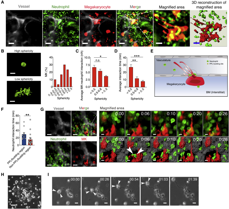
Neutrophil plucking on PPL-forming MKs (A) Two-photon imaging of thrombopoiesis inside BM in dual reporter mice ( Pf4-cre (+)/ Confetti u2212/u2212 / Lyz2 -eGFP). Representative images from 4 independ...
Figureu00a02
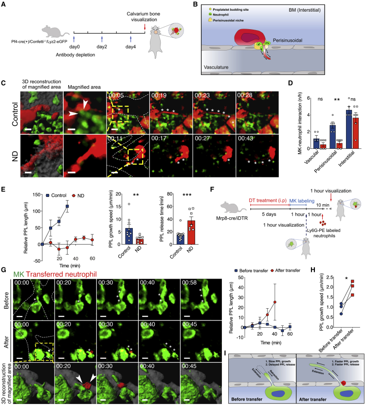
Neutrophil plucking accelerates PPL growth and release during thrombopoiesis (Au2013E) Analysis of MK-neutrophil interaction in Gr-1-treated neutropenic and control-antibody-treated dual reporter mice...
Figureu00a03
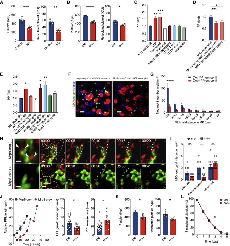
Neutrophil-expressed CXCR4 regulates neutrophil plucking and platelet production (A and B) Quantification of platelet and reticulated platelet counts in two murine models of neutropenia. (A) Platelet ...
Figureu00a04
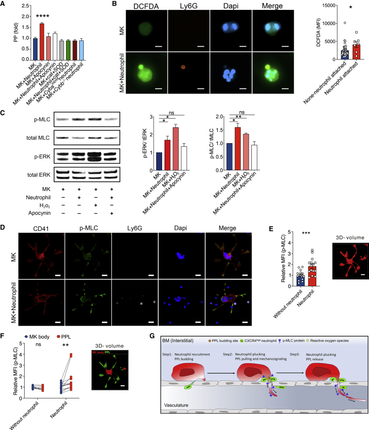
Neutrophil-derived reactive oxygen species drive thrombopoiesis (A) PP release from inu00a0vitro MK cultures following co-culture with neutrophils in the presence or absence of NADPH inhibitor apocyni...
Figureu00a05
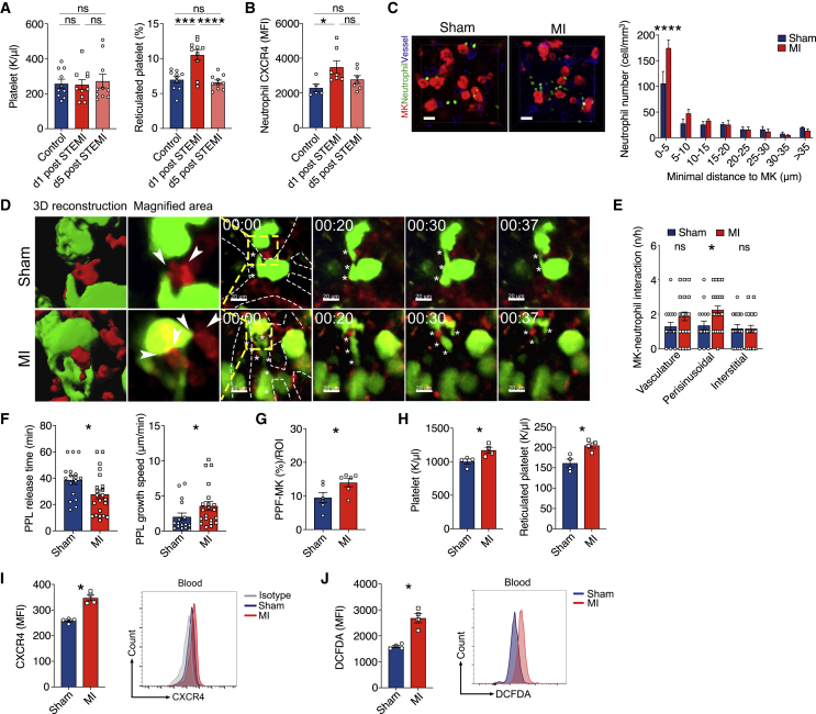
Myocardial infarction triggers thrombopoiesis by neutrophil plucking (A and B) Patients admitted because of acute ST-elevation myocardial infarction (STEMI) with symptom onset <12u00a0h undergoing ...
Figureu00a06
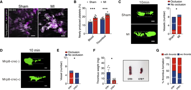
Genetic ablation of neutrophil CXCR4 prevents secondary ischemic events and reduces thrombus burden in cardiovascular diseases (A and B) Fe-(III) chloride, mesenteric artery thrombosis in I/R and sham...
Figure images are served from the NIH/NLM PubMed Central Open Access Subset or Europe PMC; copyright remains with the publishers and authors.
💬 Discussion
0 commentsNo comments yet. Be the first to start a discussion!
Leave a Comment