Abstract
The well-known saying of "Seeing is believing" became even more apt in biology when stimulated emission depletion (STED) nanoscopy was introduced in 1994 by the Nobel laureate S. Hell and coworkers. We presently stand at a juncture. Nanoscopy represented a revolution in fluorescence microscopy but now is a mature technique applied to many branches of biology, physics, chemistry, and materials science. We are currently looking ahead to the next generation of optical nanoscopes, to the new key player that will arise in the forthcoming years. This article gives an overview of the various cutting-edge implementations of STED nanoscopy and tries to shine a light into the future: imaging everything faster with unprecedented sensitivity and label-free.
🔬 Techniques
🔭 Microscopes
✨ Fluorophores
🧪 Sample Preparation
🔬 Cell Lines
🏭 Microscope Brands
🔎 Objectives
🏛️ Research Organizations (ROR)
Affiliated research institutions:
📊 Figures
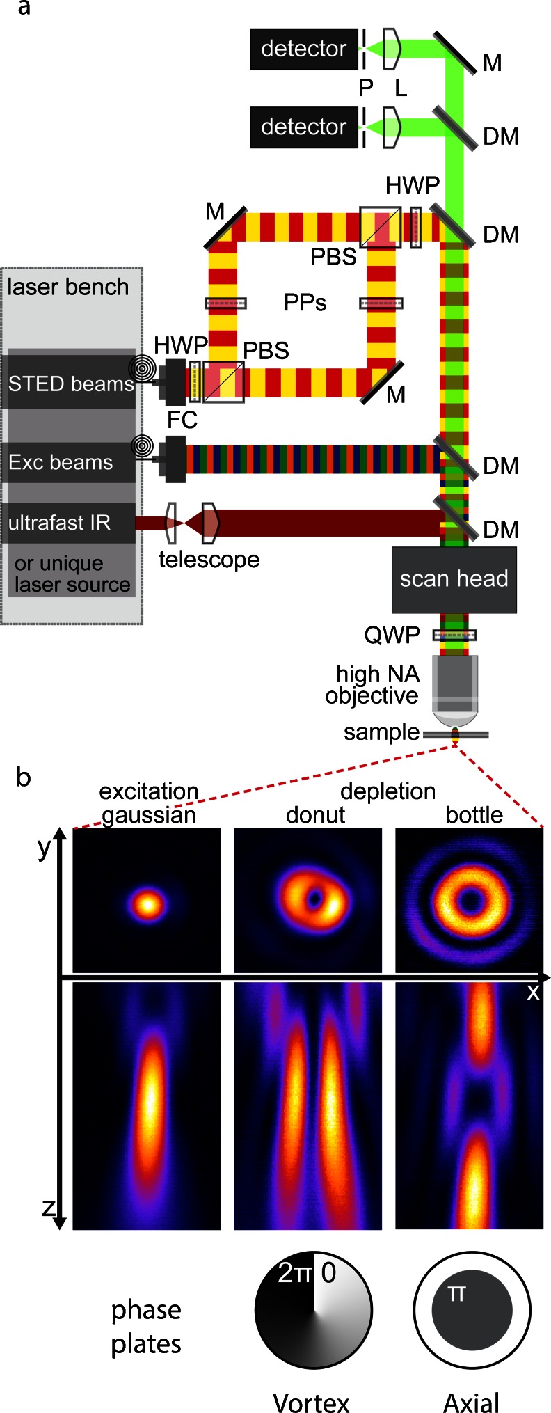
Fig. 1
a Generalized stimulated emission depletion ( STED ) set-up. We show several laser beams in order to take into account the multicolor STED set-ups and to emphasize the huge laser availability and the ...
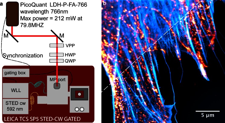
Fig. 2
Customized commercial STED set-up and example of the image that it can acquire. a Representation of a set-up that is essentially constituted by a commercial Leica TCS SP5 gated STED-CW whereby a power...
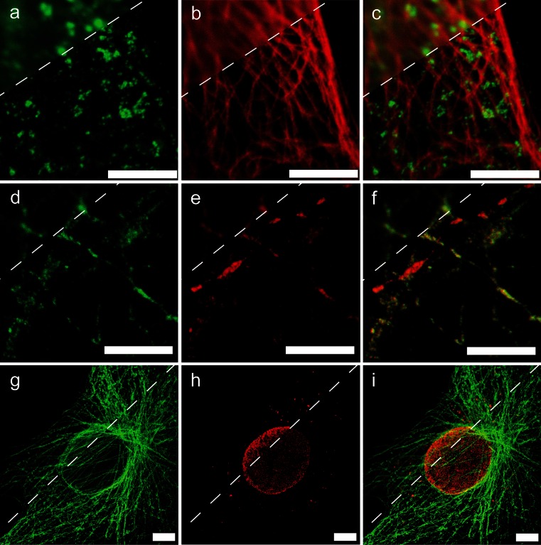
Fig. 3
Three examples of multicolor STED imaging. au2013f Images were acquired by a custom-made pulsed STED microscope based on a supercontinuum laser source (Galiani et al. 2012b ). gu2013i Images were acqu...
Figure images are served from the NIH/NLM PubMed Central Open Access Subset or Europe PMC; copyright remains with the publishers and authors.
💬 Discussion
0 commentsNo comments yet. Be the first to start a discussion!
Leave a Comment