Abstract
Cilia are organelles found on most eukaryotic cells, where they serve important functions in motility, sensory reception, and signaling. Recent advances in electron tomography have facilitated a number of ultrastructural studies of ciliary components that have significantly improved our knowledge of cilium architecture. These studies have produced nanometer-resolution structures of axonemal dynein complexes, microtubule doublets and triplets, basal bodies, radial spokes, and nexin complexes. In addition to these electron tomography studies, several recently published crystal structures provide insights into the architecture and mechanism of dynein as well as the centriolar protein SAS-6, important for establishing the 9-fold symmetry of centrioles. Ciliary assembly requires intraflagellar transport (IFT), a process that moves macromolecules between the tip of the cilium and the cell body. IFT relies on a large 20-subunit protein complex that is thought to mediate the contacts between ciliary motor and cargo proteins. Structural investigations of IFT complexes are starting to emerge, including the first three-dimensional models of IFT material in situ, revealing how IFT particles organize into larger train-like arrays, and the high-resolution structure of the IFT25/27 subcomplex. In this review, we cover recent advances in the structural and mechanistic understanding of ciliary components and IFT complexes.
🔬 Techniques
🧬 Organisms
✨ Fluorophores
🧪 Sample Preparation
📷 Detectors
💻 Software Details
🏛️ Research Organizations (ROR)
Affiliated research institutions:
📊 Figures
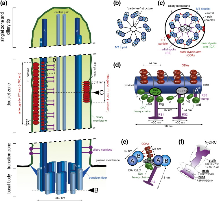
Fig.u00a01
Simplified representations of a cilium and various axonemal structures. (a) Longitudinal section through a motile cilium. The various regions of the cilium are indicated on the left (for detailed info...
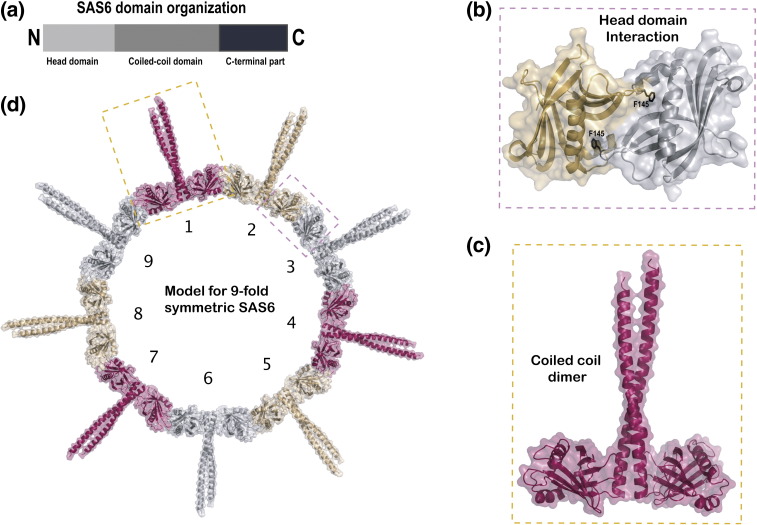
Fig.u00a02
SAS-6 crystal structures: molecular basis for the 9-fold symmetry of centrioles. (a) Schematic representation of the domain organization of SAS-6. The protein contains an N-terminal head domain, a cen...
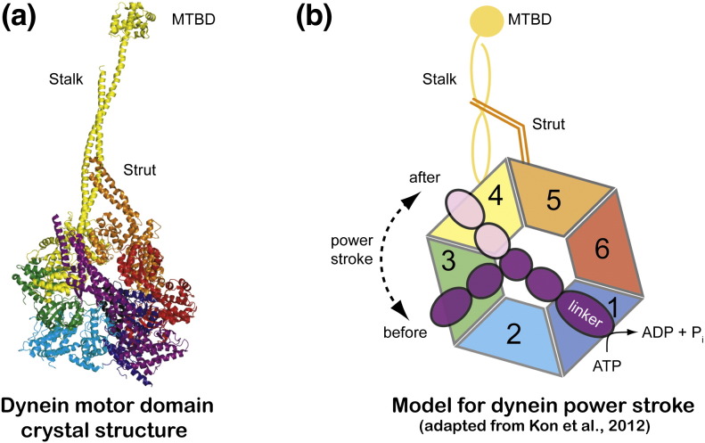
Fig.u00a03
Dynein motor domain crystal structure. (a) Crystal structure of the dynein motor domain from Dictyostelium discoideum shown in cartoon representation. The various structural elements such as stalk, st...
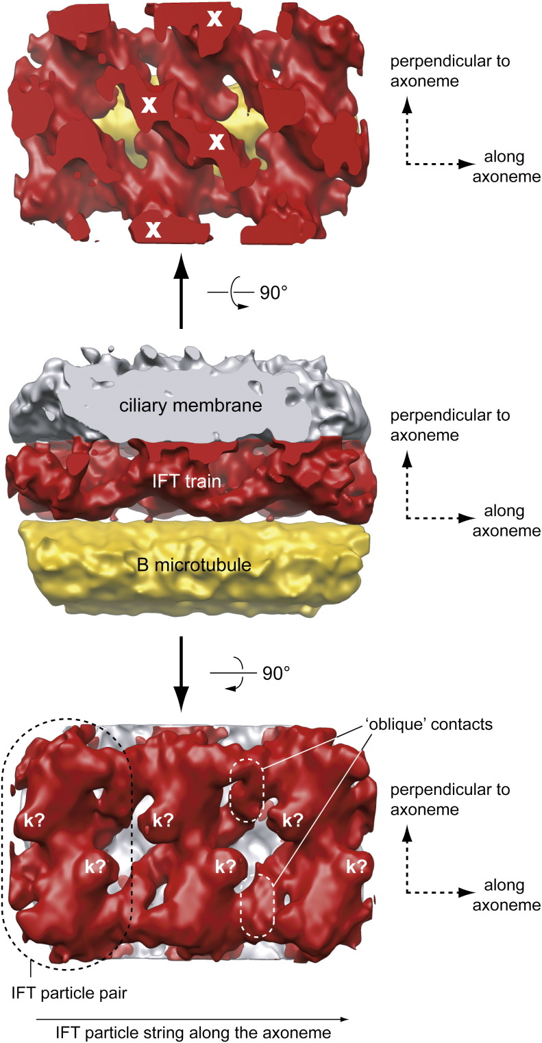
Fig.u00a04
EM structure of IFT trains in situ . Electron tomographic reconstruction of IFT trains using subtomographic averaging. The center image shows an IFT train (red) sandwiched between the flagellar membra...
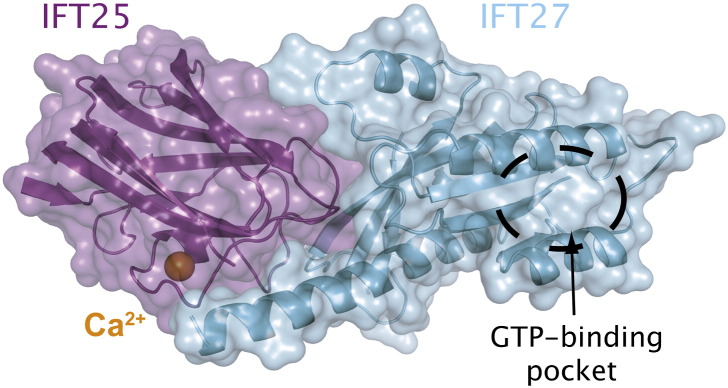
Fig.u00a05
Crystal structure of the IFT25/27 complex. Crystal structure at 2.6u00a0u00c5 resolution of the IFT25/27 complex from C. reinhardtii. IFT25 (purple) binds one divalent calcium ion (shown as an orange ...
Figure images are served from the NIH/NLM PubMed Central Open Access Subset or Europe PMC; copyright remains with the publishers and authors.
💬 Discussion
0 commentsNo comments yet. Be the first to start a discussion!
Leave a Comment