Abstract
Investigation of cell membrane structure and dynamics requires high spatial and temporal resolution. The spatial resolution of conventional light microscopy is limited due to the diffraction of light. However, recent developments in microscopy enabled us to access the nano-scale regime spatially, thus to elucidate the nanoscopic structures in the cellular membranes. In this review, we will explain the resolution limit, address the working principles of the most commonly used super-resolution microscopy techniques and summarise their recent applications in the biomembrane field.
🔬 Techniques
✨ Fluorophores
🧪 Sample Preparation
🔬 Cell Lines
🏭 Microscope Brands
🧪 Reagent Suppliers
🏛️ Research Organizations (ROR)
Affiliated research institutions:
📊 Figures
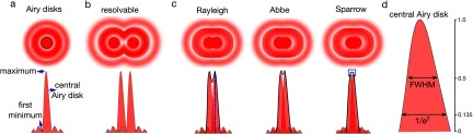
Figure 1.
Diffraction limit. (a) When light interacts with fringes of intricate objects (such as small structures in biological samples), it broadens to a diffraction pattern as it propagates (Airy disks). This...
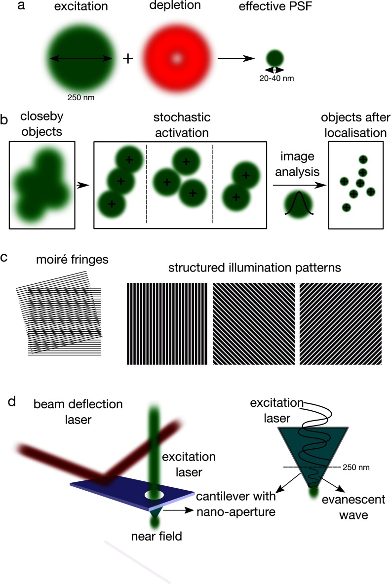
Figure 2.
Super-resolution techniques. (a) STED is based on the depletion of PSF peripheral signal by a donut-shaped depletion beam. (b) PALM/STORM uses the photo-controllable fluorophores to observe single flu...
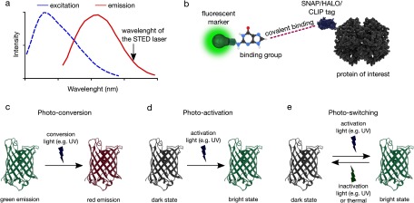
Figure 3.
Fluorescent labelling for super-resolution techniques. (a) Spectral requirement for STED-capable dyes (b) SNAP/HALO/CLIP tag labelling strategies where a fluorescent marker that is linked to a small b...
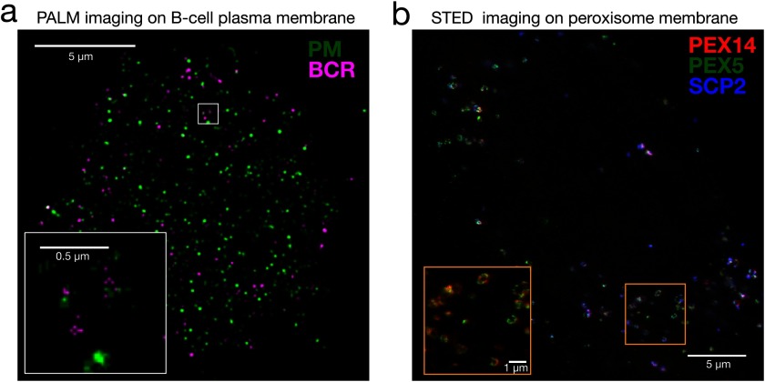
Figure 4.
Imaging membrane structures with super-resolution microscopy. (a) Dual-colour PALM image of a B cell chemically fixed 1u2009min after B cell receptor (BCR) clustering. Clustered BCR is shown in magent...
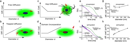
Figure 5.
Diffusion dynamics in the plasma membrane. (a) In free diffusion, the diffusion coefficient of the molecule stays constant with varying spot diameter, (b) in transient immobilisation, the diffusion co...
Figure images are served from the NIH/NLM PubMed Central Open Access Subset or Europe PMC; copyright remains with the publishers and authors.
💬 Discussion
0 commentsNo comments yet. Be the first to start a discussion!
Leave a Comment