Abstract
AbstractMultiple myeloma (MM) is incurable, so there is a significant unmet need for effective therapy for patients with relapsed or refractory disease. This situation has not changed despite the recent approval of the anti-CD38 antibody daratumumab, one of the most potent agents in MM treatment. The efficiency of daratumumab might be improved by combining it with synergistic anti-MM agents. We therefore investigated the potential of the histone deacetylase (HDAC) inhibitor ricolinostat to up-regulate CD38 on MM cells, thereby enhancing the performance of CD38-specific therapies. Using quantitative reverse transcription polymerase chain reaction and flow cytometry, we observed that ricolinostat significantly increases CD38 RNA levels and CD38 surface expression on MM cells. Super-resolution microscopy imaging of MM cells by direct stochastic optical reconstruction microscopy confirmed this rise with molecular resolution and revealed homogeneous distribution of CD38 molecules on the cell membrane. Particularly important is that combining ricolinostat with daratumumab induced enhanced lysis of MM cells. We also evaluated next-generation HDAC6 inhibitors (ACY-241, WT-161) and observed similar increase of CD38 levels suggesting that the upregulation of CD38 expression on MM cells by HDAC6 inhibitors is a class effect. This proof-of-concept illustrates the potential benefit of combining HDAC6 inhibitors and CD38-directed immunotherapy for MM treatment.
🔬 Techniques
🧪 Sample Preparation
🔬 Cell Lines
🏭 Microscope Brands
🧪 Reagent Suppliers
💾 Data Repositories
🏛️ Research Organizations (ROR)
Affiliated research institutions:
📋 Methods
Human subjects
Peripheral blood and bone marrow aspirates were obtained from healthy donors and MM patients after we obtained their written informed consent. Patient characteristics are depicted in Supplementary Table 1 . The research protocols were approved by the Institutional Review Boards of the University Hospital of Würzburg (UKW) and University Hospital Virgen del Rocío (HUVR). All procedures conformed to the Helsinki Declaration. Isolation of primary myeloma cells, regulatory T cells, and T cells We isolated peripheral blood mononuclear cells (PBMCs) and bone marrow mononuclear cells by density gradient centrifugation using Ficoll–Paque (Amersham Biosciences, Uppsala, Sweden). We isolated primary MM cells from bone marrow aspirates using CD138 immunomagnetic beads (Miltenyi Biotec, Bergisch-Gladbach, Germany). Regulatory T cells were isolated from PBMCs using a two-step immunomagnetic procedure (Miltenyi). T cells were also isolated from PBMCs using negative selection with immunomagnetic beads (Miltenyi). CD8+ and CD4+ T cells were activated by anti-CD3/CD28 bead stimulation (Thermo, Waltham, MA). Immunophenotyping The expression of CD38, BCMA, SLAMF7, CD55, and CD59 on MM cells was analyzed by flow cytometry using specifically conjugated mAbs and matched isotype controls. We stained with 7-aminoactinomycin D (7-AAD) to discriminate between living and dead cells. Flow cytometry analyses were performed on a FACSCanto II (Becton Dickinson, Heidelberg, Germany) and data was analyzed using FlowJo software (Treestar, OR, USA). Treatment with HDAC inhibitors and all-trans retinoic acid (ATRA) We cultured the MM cells in RPMI-1640 (Gibco, Darmstadt, Germany) supplemented with 10% fetal bovine serum at 1 × 10e5 cells/well in 96-well flat-bottom plates (Costar, Washington, DC). We reconstituted ricolinostat, ACY-241 and WT-161 in dimethylsulfoxide and added them to the medium at final concentrations of 1, 5, and 10 µM. We likewise reconstituted ATRA and Panobinostat in dimethylsulfoxide and added these compounds to the medium at final concentrations of 10 nM.
Show full methods section
Human subjects
Peripheral blood and bone marrow aspirates were obtained from healthy donors and MM patients after we obtained their written informed consent. Patient characteristics are depicted in Supplementary Table 1 . The research protocols were approved by the Institutional Review Boards of the University Hospital of Würzburg (UKW) and University Hospital Virgen del Rocío (HUVR). All procedures conformed to the Helsinki Declaration. Isolation of primary myeloma cells, regulatory T cells, and T cells We isolated peripheral blood mononuclear cells (PBMCs) and bone marrow mononuclear cells by density gradient centrifugation using Ficoll–Paque (Amersham Biosciences, Uppsala, Sweden). We isolated primary MM cells from bone marrow aspirates using CD138 immunomagnetic beads (Miltenyi Biotec, Bergisch-Gladbach, Germany). Regulatory T cells were isolated from PBMCs using a two-step immunomagnetic procedure (Miltenyi). T cells were also isolated from PBMCs using negative selection with immunomagnetic beads (Miltenyi). CD8+ and CD4+ T cells were activated by anti-CD3/CD28 bead stimulation (Thermo, Waltham, MA). Immunophenotyping The expression of CD38, BCMA, SLAMF7, CD55, and CD59 on MM cells was analyzed by flow cytometry using specifically conjugated mAbs and matched isotype controls. We stained with 7-aminoactinomycin D (7-AAD) to discriminate between living and dead cells. Flow cytometry analyses were performed on a FACSCanto II (Becton Dickinson, Heidelberg, Germany) and data was analyzed using FlowJo software (Treestar, OR, USA). Treatment with HDAC inhibitors and all-trans retinoic acid (ATRA) We cultured the MM cells in RPMI-1640 (Gibco, Darmstadt, Germany) supplemented with 10% fetal bovine serum at 1 × 10e5 cells/well in 96-well flat-bottom plates (Costar, Washington, DC). We reconstituted ricolinostat, ACY-241 and WT-161 in dimethylsulfoxide and added them to the medium at final concentrations of 1, 5, and 10 µM. We likewise reconstituted ATRA and Panobinostat in dimethylsulfoxide and added these compounds to the medium at final concentrations of 10 nM.
Antibody-dependent cellular cytotoxicity
(ADCC) assay with primary myeloma cells Ricolinostat-treated (5 µM, 48 h) primary MM cells were co-cultured with autologous PBMCs at an effector-to-target ratio of 3:1 in 96-well plates in the presence of solvent control, IgG1 isotype or daratumumab. After 24 h, the percentage of viable myeloma cells was determined by flow cytometry using 7-AAD to discriminate living from dead cells.
Statistical analysis
For statistical analyses, we used Prism Software (GraphPad, San Diego, CA). Shapiro–Wilk test was used to test for normality. Unpaired Student’s t -tests were used to analyze CD38 expression level. Two-way ANOVA testing was performed to analyze the functional data. Differences with a p -value < 0.05 were considered statistically significant.
Supplementary information Supplementary Information, clean version Supplemental Table 1 Supplemental Figure 1 Supplemental Figure 2 Supplemental Figure 3 Supplemental Figure 4 Supplemental Figure 5 Supplemental Figure 6 Supplemental Figure 7 Supplemental Figure 8 Supplemental Figure 9 Supplemental Figure 10 Supplemental Figure 11
📊 Figures
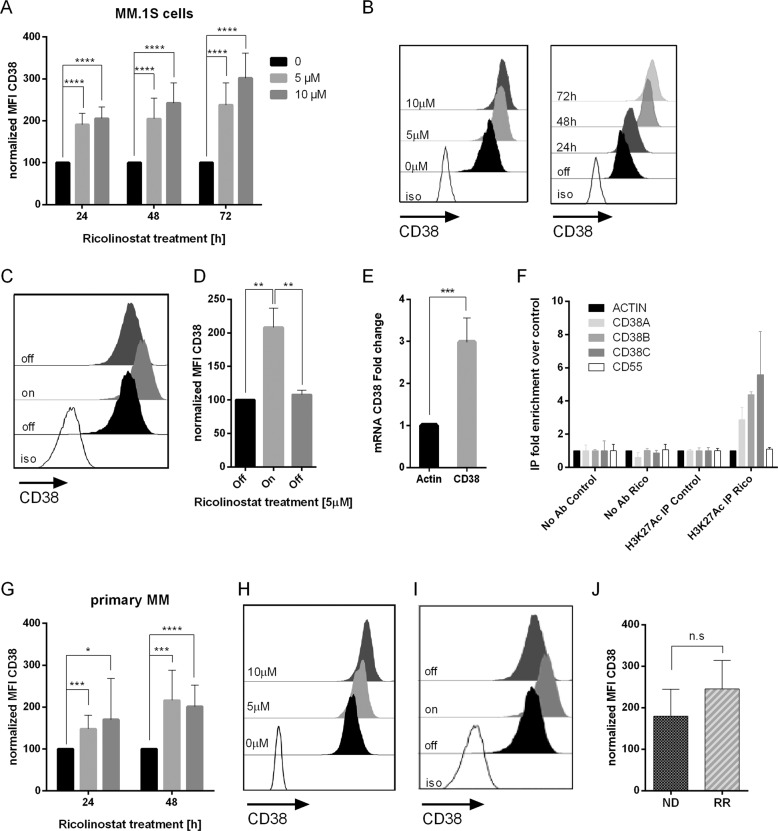
Fig. 1
Ricolinostat treatment leads to enhanced CD38 expression on myeloma cells.
a CD38 expression on MM.1S cells ( n =u200913 experiments) before and after treatment with ricolinostat at the given final concentrations (fc) for the given time intervals. Bar diagram shows CD38 expr...
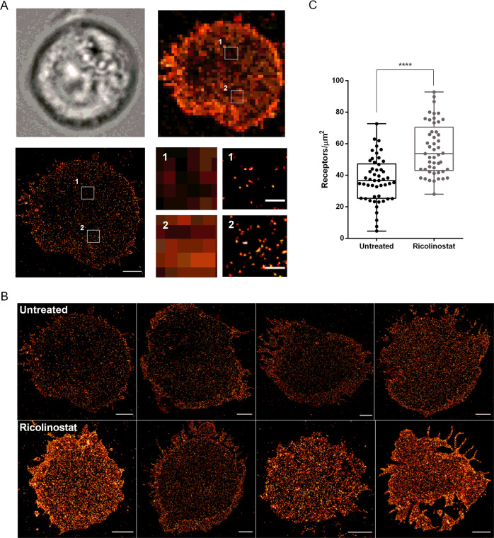
Fig. 2
Ricolinostat treatment leads to enhanced CD38 molecule density on MM.1S cells.
a MM.1S cells were visualized by transmitted light microscopy (upper left). Expression of CD38 was detected by conventional wide-field fluorescence microscopy (upper right) and d STORM (lower left). S...
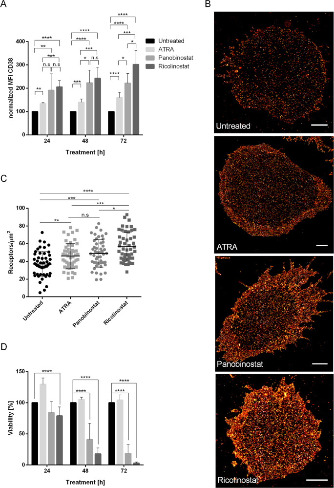
Fig. 3
Ricolinostat effect outperforms ATRA and panobinostat induction of CD38 elevation on myeloma cells.
MM.1S cells were treated with ATRA and panobinostat at fcs of 10u2009nM, ricolinostat at a fc of 5u2009u00b5M or left untreated. a Bar diagram shows CD38 expression on MM.1S cells after ATRA ( n =u200...
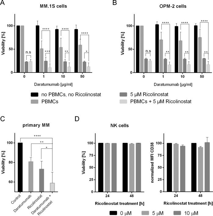
Fig. 4
Ricolinostat and daratumumab synergistically eliminate myeloma cells.
a , b ADCC against MM.1S ( a , n =u20098 experiments) and OPM-2 ( b , n =u20094 experiments) cells with and without ricolinostat treatment. Ricolinostat pre-treatment was performed for 48u2009h at 5u2...
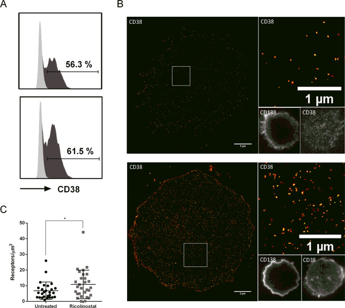
Fig. 5
Ricolinostat can induce CD38 expression on myeloma cells in daratumumab refractory patients.
a The histograms show flow cytometric analysis of CD38 expression on primary MM cells from a daratumumab refractory patient after overnight culture in the absence (upper graph) or presence (lower grap...
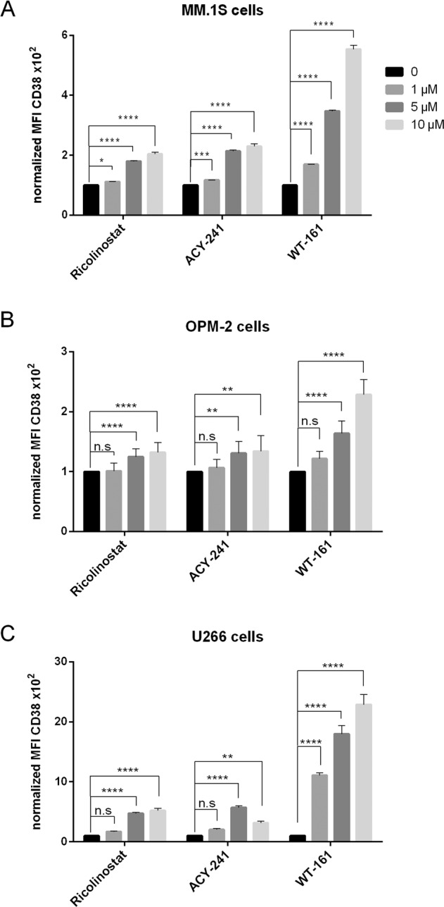
Fig. 6
Upregulation of CD38 expression on multiple myeloma cells by novel HDAC6 inhibitors is a class effect.
a u2013 c CD38 expression on MM.1S ( a , n =u20096 experiments), OPM2 ( b , n =u20096 experiments), and U266 ( c , n =u20093 experiments) cells before and after treatment with ricolinostat, ACY-241, a...
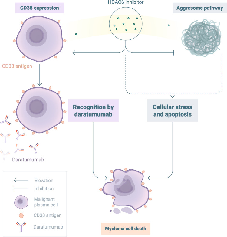
Fig. 7
In combination with daratumumab, novel HDAC6 inhibitors exert a dual mode of action against myeloma cells.
By disabling the aggresome, HDAC6 inhibitors induce cellular stress and apoptosis of MM cells. In combination regimens, it is possible to exploit the induction of CD38 elevation by HDAC6 inhibitors to...
Figure images are served from the NIH/NLM PubMed Central Open Access Subset or Europe PMC; copyright remains with the publishers and authors.
💬 Discussion
0 commentsNo comments yet. Be the first to start a discussion!
Leave a Comment