Abstract
BACKGROUND: Optical super-resolution imaging of fluorescently stained biological samples is rapidly becoming an important tool to investigate protein distribution at the molecular scale. It is therefore important to develop practical super-resolution methods that allow capturing the full three-dimensional nature of biological systems and also can visualize multiple protein species in the same sample. METHODOLOGY/PRINCIPAL FINDINGS: We show that the use of a combination of conventional near-infrared dyes, such as Alexa 647, Alexa 680 and Alexa 750, all excited with a 671 nm diode laser, enables 3D multi-colour super-resolution imaging of complex biological samples. Optically thick samples, including human tissue sections, cardiac rat myocytes and densely grown neuronal cultures were imaged with lateral resolutions of ∼15 nm (std. dev.) while reducing marker cross-talk to <1%. Using astigmatism an axial resolution of ∼65 nm (std. dev.) was routinely achieved. The number of marker species that can be distinguished depends on the mean photon number of single molecule events. With the typical photon yields from Alexa 680 of ∼2000 up to 5 markers may in principle be resolved with <2% crosstalk. CONCLUSIONS/SIGNIFICANCE: Our approach is based entirely on the use of conventional, commercially available markers and requires only a single laser. It provides a very straightforward way to investigate biological samples at the nanometre scale and should help establish practical 4D super-resolution microscopy as a routine research tool in many laboratories.
🔬 Techniques
💻 Software
✨ Fluorophores
🧪 Sample Preparation
🏭 Microscope Brands
🧪 Reagent Suppliers
📷 Detectors
🎨 Filters
💻 Software Details
🏛️ Research Organizations (ROR)
Affiliated research institutions:
📋 Methods
Methodology/Principal Findings We show that the use of a combination of conventional near-infrared dyes, such as Alexa 647, Alexa 680 and Alexa 750, all excited with a 671 nm diode laser, enables 3D multi-colour super-resolution imaging of complex biological samples. Optically thick samples, including human tissue sections, cardiac rat myocytes and densely grown neuronal cultures were imaged with lateral resolutions of ∼15 nm (std. dev.) while reducing marker cross-talk to
📊 Figures
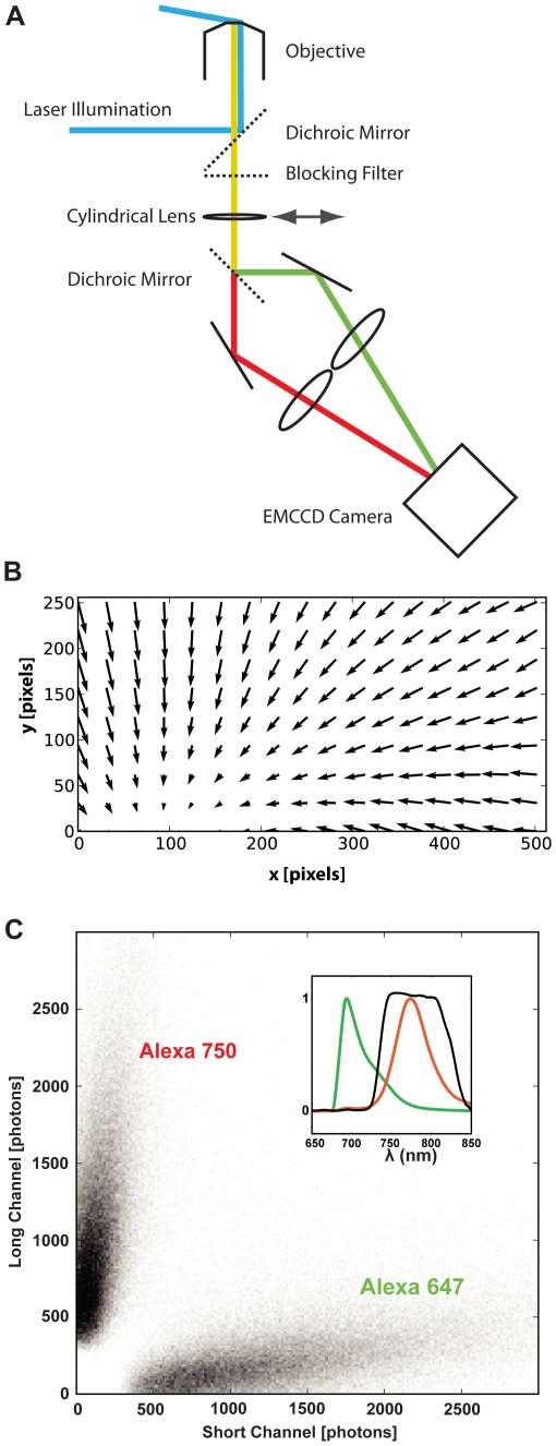
Figure 1
Schematic depicting optical setup for ratiometric localisation imaging.
A. A single laser at 671 nm is used to provide excitation and the collected light is split into two bands using a dichroic mirror. The bands are imaged side by side on an electron multiplying CCD (EMC...
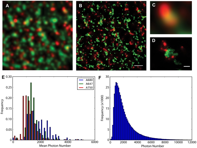
Figure 2
Two-dimensional super-resolution imaging of the distribution of Ryanodine receptors (red) and Caveolin (green), using Alexa 680 and Alexa 750 secondaries, in the periphery of isolated rat cardiac myocytes and overview of dye properties.
Panel A shows the sample at conventional resolution, panel B the super-resolved image. Comparison of enlarged detail ( C & D ) shows that apparent overlap in the diffraction-limited images is not seen...
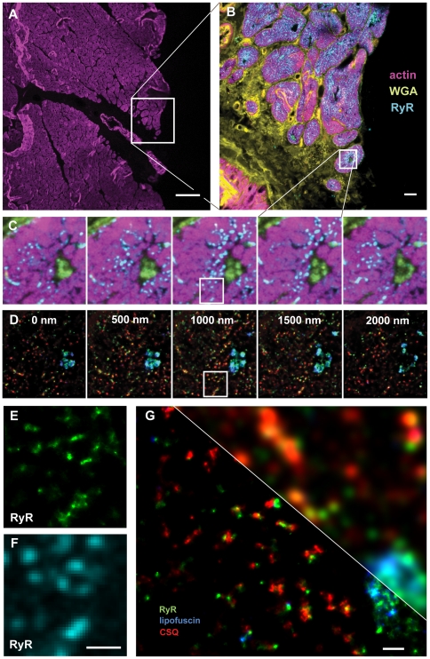
Figure 3
Correlative confocal and super-resolution imaging of a human cardiac tissue section.
The section was u223c10 u00b5m thick and was labeled with phalloidin for f-Actin (Alexa 488), WGA for the cell membrane and extracellular matrix (Alexa 594), along with antibodies for the ryanodine re...
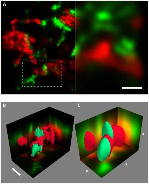
Figure 4
3D super-resolution imaging of GFP-alpha-SAP97 (antibody labelled with an Alexa 647 secondary, green) and Synapsin (with an Alexa 750 secondary, red) in a primary hippocampal culture.
4D imaging was performed using dual-colour 3D localisation based on astigmatism in conjunction with the ratiometric multi-colour approach. A . Comparison between detail in a super-resolution image (le...
Figure images are served from the NIH/NLM PubMed Central Open Access Subset or Europe PMC; copyright remains with the publishers and authors.
💬 Discussion
0 commentsNo comments yet. Be the first to start a discussion!
Leave a Comment