Abstract
Abstract Optical microscopy has emerged as a key driver of fundamental research since it provides the ability to probe into imperceptible structures in the biomedical world. For the detailed investigation of samples, a high-resolution image with enhanced contrast and minimal damage is preferred. To achieve this, an automated image analysis method is preferable over manual analysis in terms of both speed of acquisition and reduced error accumulation. In this regard, deep learning (DL)-based image processing can be highly beneficial. The review summarises and critiques the use of DL in image processing for the data collected using various optical microscopic techniques. In tandem with optical microscopy, DL has already found applications in various problems related to image classification and segmentation. It has also performed well in enhancing image resolution in smartphone-based microscopy, which in turn enablse crucial medical assistance in remote places. Graphical abstract
🔬 Techniques
🧬 Organisms
✨ Fluorophores
🧪 Sample Preparation
🔬 Cell Lines
🏭 Microscope Brands
🧪 Reagent Suppliers
🔎 Objectives
💻 Software Details
🏛️ Research Organizations (ROR)
Affiliated research institutions:
📊 Figures
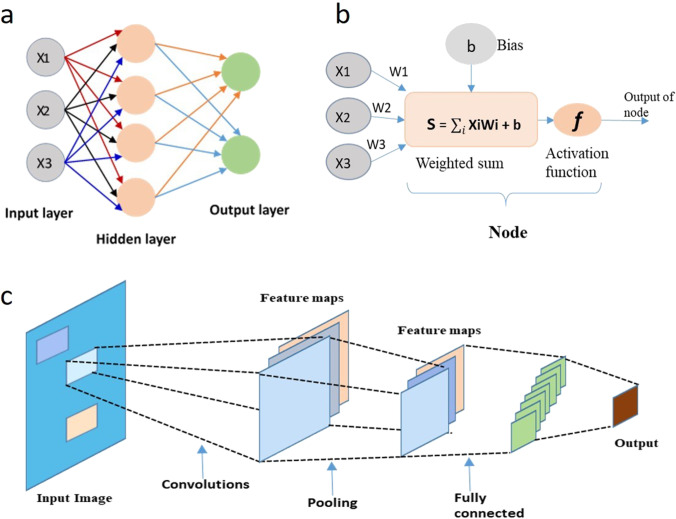
Fig. 1
a Block diagram of a simple ANN architecture, b function of a node, c architecture of CNN in image classification of hepatocellular carcinoma graded into well, moderately, and poorly differentiated ty...
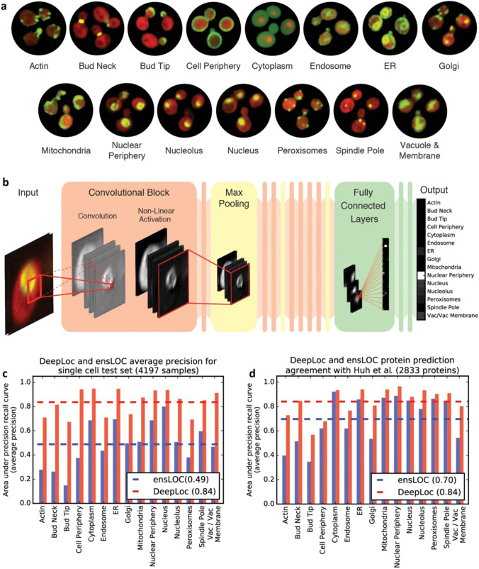
Fig. 2
DeepLoc input data, architecture, and performance. a Example micrographs of yeast cells expressing GFP-tagged proteins that localize to the 15 subcellular compartments sed to train DeepLoc. b Architec...
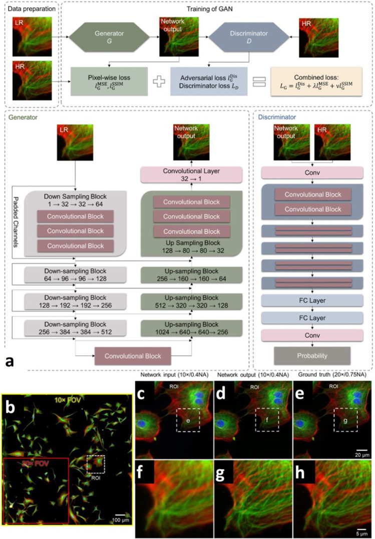
Fig. 3
a The training process and the architecture of the generative adversarial network that we used for super-resolution of images of bovine pulmonary artery endothelial cells (BPAEC). b Network input imag...
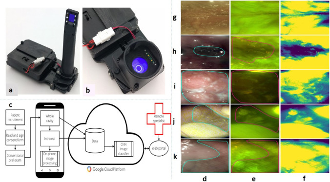
Fig. 4
Smartphone-based oral cancer screening device using both WLI and AFI. a Intraoral imaging device. b Whole cavity imaging device. c Field testing workflow for smartphone-based oral screening. d White l...
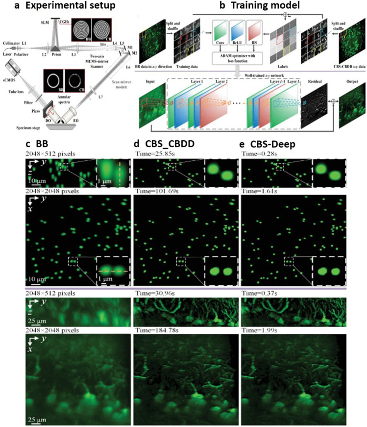
Fig. 5
a Schematics of the CBS-based LSFM imaging system. b Training of the network using a BB image and a corresponding CBS-CBDD reference image in the x u2013 y direction and then reconstructing the high-q...
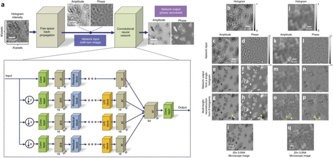
Fig. 6
a Architecture of deep neural network is composed of convolutional layers, residual blocks, and upsampling blocks which blindly outputs artefact-free phase and amplitude images of the object using onl...
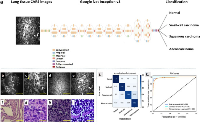
Fig. 7
Coronal mouse brain SRS images acquired at 2990u00a0cm u22121 . a Low-power image acquired at 1 mW Stokes and 20 mW pump. b The low-power image denoised with VST. c The low-power image is denoised wit...
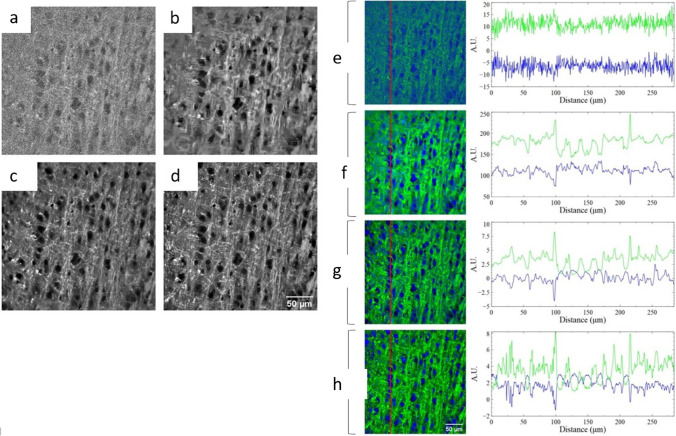
Fig. 8
a Transfer learning layout. A GoogleNet Inception v3 CNN, pre-trained on the ImageNet data is fine-tuned with CARS images comprising four classes: normal, small-cell carcinoma, squamous carcinoma, and...
Figure images are served from the NIH/NLM PubMed Central Open Access Subset or Europe PMC; copyright remains with the publishers and authors.
💬 Discussion
0 commentsNo comments yet. Be the first to start a discussion!
Leave a Comment