Abstract
Abstract Expansion microscopy (ExM) is a highly effective technique for super-resolution fluorescence microscopy that enables imaging of biological samples beyond the diffraction limit with conventional fluorescence microscopes. Despite the development of several enhanced protocols, ExM has not yet demonstrated the ability to achieve the precision of nanoscopy techniques such as Single Molecule Localization Microscopy (SMLM). Here, to address this limitation, we have developed an iterative ultrastructure expansion microscopy (iU-ExM) approach that achieves SMLM-level resolution. With iU-ExM, it is now possible to visualize the molecular architecture of gold-standard samples, such as the eight-fold symmetry of nuclear pores or the molecular organization of the conoid in Apicomplexa. With its wide-ranging applications, from isolated organelles to cells and tissue, iU-ExM opens new super-resolution avenues for scientists studying biological structures and functions.
🔬 Techniques
🔭 Microscopes
🧬 Organisms
💻 Software
✨ Fluorophores
🧪 Sample Preparation
🔬 Cell Lines
🏭 Microscope Brands
🧪 Reagent Suppliers
📷 Detectors
🔎 Objectives
💻 Software Details
💾 Data Repositories
🏛️ Research Organizations (ROR)
Affiliated research institutions:
📋 Methods
Enhancing iU-ExM by combining with TREx protocol
The iU-ExM technique enables an approximate 15-fold expansion, derived from the initial expansion of the first DHEBA gel. This gel expands by a factor of around 5.8 when denatured at 85 °C. When reintroduced into the neutral gel, it undergoes slight shrinkage, resulting in an expansion factor of approximately 3.5/4 fold. Subsequently, the second gel expands by a factor of 4–4.5. Multiplying these two expansion factors gives an average final expansion factor of 14–18 times. Currently, it is not feasible to perform a third iteration due to the last gel’s inability to cleave. To explore the potential for even greater expansion factors, we decided to combine iU-ExM with the polymers used in the TREx method, which allows a maximum expansion of tenfold 8 . Since TREx employs a gel based on BIS-acrylamide cross-linker, we replaced the third gel in our protocol with the TREx recipe. To evaluate the expansion factor, we used again Toxoplasma conoid, Chlamydomonas basal body (with defined dimensions, 225 nm wide and 500 nm long 48 ), and nuclear pores as molecular rulers (Fig. 6 ). In all three cases, the combination of iU-ExM with the TREx-based gel resulted in average expansions of approximately 22.4× for the conoid (Fig. 6a, b ), 26× for the Chlamydomonas basal bodies (Fig. 6 c, d), and 22.6× for the nuclear pores (Fig. 6e, f ). The average values presented here are based on three replicates, and it should be noted that variations in the expansion factor were observed in each experiment as exemplified in Supplementary Fig. 12 . These variations can be primarily attributed to the challenge of accurately controlling the shrinkage of the first gel within the neutral gel. The final expansion factor can be explained by the first gel shrinking at 3.5–4×, which, when multiplied with the 7× TREx-based gel, theoretically gives an expansion factor between 22 and 28×. Furthermore, we did not observe any deformation in the molecular structure, as demonstrated by the organization of the microtubule triplets in the Chlamydomonas basal body (Fig. 6c , insets). Overall, this combination of protocols demonstrates that the iU-ExM procedure is compatible with other types of gels. Additionally, the iterative nature of this approach allows for a multiplier effect on the expansion factors, where even a small difference can result in a significant change in the final outcome. Fig. 6 Coupling TREx to iU-ExM. a Widefield full field of view of T. gondii tachyzoites stained for tubulin (magenta) expanded with either iU-ExM (left) or iU-ExM-T (right). Scales bar: 30 μm non-corrected. b Quantification of the diameter of the conoid, highlighting the 1.6-fold improvement of the expansion factor. iU-ExM: N = 15 conoids (average ± standard error = 5.3 ± 0.4 μm), iU-ExM-T: N = 9 conoids (M = 8.5 ± 0.7 μm).
Show full methods section
Enhancing iU-ExM by combining with TREx protocol
The iU-ExM technique enables an approximate 15-fold expansion, derived from the initial expansion of the first DHEBA gel. This gel expands by a factor of around 5.8 when denatured at 85 °C. When reintroduced into the neutral gel, it undergoes slight shrinkage, resulting in an expansion factor of approximately 3.5/4 fold. Subsequently, the second gel expands by a factor of 4–4.5. Multiplying these two expansion factors gives an average final expansion factor of 14–18 times. Currently, it is not feasible to perform a third iteration due to the last gel’s inability to cleave. To explore the potential for even greater expansion factors, we decided to combine iU-ExM with the polymers used in the TREx method, which allows a maximum expansion of tenfold 8 . Since TREx employs a gel based on BIS-acrylamide cross-linker, we replaced the third gel in our protocol with the TREx recipe. To evaluate the expansion factor, we used again Toxoplasma conoid, Chlamydomonas basal body (with defined dimensions, 225 nm wide and 500 nm long 48 ), and nuclear pores as molecular rulers (Fig. 6 ). In all three cases, the combination of iU-ExM with the TREx-based gel resulted in average expansions of approximately 22.4× for the conoid (Fig. 6a, b ), 26× for the Chlamydomonas basal bodies (Fig. 6 c, d), and 22.6× for the nuclear pores (Fig. 6e, f ). The average values presented here are based on three replicates, and it should be noted that variations in the expansion factor were observed in each experiment as exemplified in Supplementary Fig. 12 . These variations can be primarily attributed to the challenge of accurately controlling the shrinkage of the first gel within the neutral gel. The final expansion factor can be explained by the first gel shrinking at 3.5–4×, which, when multiplied with the 7× TREx-based gel, theoretically gives an expansion factor between 22 and 28×. Furthermore, we did not observe any deformation in the molecular structure, as demonstrated by the organization of the microtubule triplets in the Chlamydomonas basal body (Fig. 6c , insets). Overall, this combination of protocols demonstrates that the iU-ExM procedure is compatible with other types of gels. Additionally, the iterative nature of this approach allows for a multiplier effect on the expansion factors, where even a small difference can result in a significant change in the final outcome. Fig. 6 Coupling TREx to iU-ExM. a Widefield full field of view of T. gondii tachyzoites stained for tubulin (magenta) expanded with either iU-ExM (left) or iU-ExM-T (right). Scales bar: 30 μm non-corrected. b Quantification of the diameter of the conoid, highlighting the 1.6-fold improvement of the expansion factor. iU-ExM: N = 15 conoids (average ± standard error = 5.3 ± 0.4 μm), iU-ExM-T: N = 9 conoids (M = 8.5 ± 0.7 μm).
Data from one experiment. c
Widefield image of C. reinhardtii isolated basal bodies stained for tubulin (magenta) expanded using iU-ExM (right) or iU-ExM-T (left). Scale bars: 5 μm non-corrected. d Quantifications of the centriole diameter in the proximal region, further illustrate the 1.6-fold improvement of the expansion factor. iU-ExM: N = 43 centrioles (average ± standard error = 3.5 ± 0.2 μm), iU-ExM-T: N = 34 centrioles (average ± standard error = 5.7 ± 0.4 μm) centrioles from one experiment. e iU-ExM widefield full field of view image of NPCs from isolated U2OS NUP96-GFP nuclei (left) and expanded with iU-ExM-T (right). Scale bars: 10 μm non-corrected. f Quantification of the expanded NUP96 diameter between iU-ExM and iU-ExM-T. iU-ExM: N = 55 NPCs (average ± standard error = 1.7 ± 0.2 μm), iU-ExM-T: N = 63 NPCs (average ± standard error= 2.4 ± 0.3 μm) from 3 independent experiments. Expansion factors are calculated by dividing the expanded average diameter by 107 nm.
Methods Ethical statement
The research performed in this study complies with ethical regulations (authorization VD1367 to the Kostic and Arsenijevic laboratories (Hospital Jules Gonin, Lausanne, Switzerland) regarding mouse retinas). Organelles, cell lines, strains and tissue samples Chlamydomonas reinhardtii To ensure an efficient expansion, we used a cell wall-free C. reinhardtii CW15 − strain. The liquid culture (TAP buffer 50 ) is inoculated from the solid agarose culture of the algae and grown for 3 days at RT under light exposure and slow shaking. The cells were sedimented on 12 mm Poly-D-Lysine coated coverslips for 10 min, then the excess was removed, and the cells were fixed with either cold methanol (see cell fixations) or no fixation. The purified Chlamydomonas basal bodies were prepared and spun on coverslips as previously described 50 . Briefly, deflagellated CW15- Chlamydomonas cells were lysed 1 h at 4 °C in presence of 1 mM HEPES (pH 7), 0.5 mM MgCl 2 , 1% NP-40, and 5000 units of DNase. After centrifugation at 600 × g for 10 min at 4 °C to remove the cell debris, basal bodies were further purified and concentrated, first using a centrifugation at 10,000 × g with a 60% sucrose cushion and second a centrifugation at 68,320 × g on a 40–70% sucrose gradient. Isolated basal bodies were collected at the 70% sucrose interface. Toxoplasma gondii T. gondii tachyzoites were amplified in human foreskin fibroblasts (HFFs, ATCC-CRL-2429, CCD-1112Sk, Lot/Batch No: 70014723) in Dulbecco’s Modified Eagle’s Medium (DMEM, Gibco) supplemented with 5% of Fetal Bovine Serum (FCS, Gibco), 2 mM glutamine and 25 µg/ml gentamicin (Gibco). Tachyzoites were transfected by electroporation 51 . To target any gene of interest, 40 µg of specific plasmid driving the expression of the gRNA and Cas9 protein was transfected alongside a PCR product flanked by homology regions (gRNA sequences: DCX: GTGGGGAGCGTGTCACTCAT, SAS6-L: ACTTATGTACGAGTGCACGG). Transfected parasites then carrying an HXGPRT cassette 52 were selected with 25 mg/ml of mycophenolic acid and 50 mg/ml of xanthine. In brief, SAS6L-mAiD-3HA (HXGPRT) and DCX-mAiD-3HA have been generated by transfecting a Cas9-gRNA encoding vector alongside a PCR fragment encoding for mAiD-HA and HXGPRT cassette. For immuno-staining as well as for U-ExM and iU-ExM, freshly egressed tachyzoites were sedimented for 10 min on pre-coated Poly-L-Lysine coverslips before starting the protocols described in this study. Note that the T. gondii tachyzoites used in the paper have the following genetic background: RHΔhxgprtΔKu80 - Tir1 (Parental line for all -mAiD-HA strains generated in this study).
Human cell lines Homo sapiens bone osteosarcoma U2OS
ATCC-HTB-96 and NUP96-GFP U2OS cell line (from Jonas Ries lab 13 ) were grown in Dulbecco’s modified Eagle’s medium and GlutaMAX, supplemented with 10% fetal calf serum and penicillin and streptomycin (100 μg/ml) at 37 °C in a humidified 5% CO 2 incubator. U2OS expressing GFP-sec61β (133–291) were transiently transfected with JetPRIME following the manufacturer’s instructions. After 24 h of expression, cells were cryo-fixed as described in ref. 33 . All cell cultures were regularly tested for mycoplasma contaminations. Mouse retina Retinas from Adult mice (2 months) C57BL/6J were dissected as previously described 40 . Two eyes were used from two different mice. Briefly, after enucleation, eyes were directly incubated in 4% PFA (paraformaldehyde, P6148, Sigma-Aldrich) in PBS for 15 min at RT. Then, the eyes were put in a Matek dish (P35G-1.5-10-C, MatTek) for their dissection where the cornea and lens were cut and removed, and the sclera was carefully detached from the retina and discarded. From here, retinas were then directly processed for expansion microscopy. Isolated nuclei To prepare isolated nuclei 53 , cells were harvested from a T75 flask with trypsin and centrifuged at 200 × g for 5 min. The pellet was resuspended in 5 mL of complete DMEM and aliquoted with 1 mL in 1.5 mL Eppendorf tubes. Cells were pelleted with 200 × g for 5 min and resuspended in hypotonic buffer (20 mM Tris-HCl pH 7.4, 10 mM KCl, 2 mM MgCl 2 , 1 mM EGTA, 0.5 mM DTT, 0.5 mM PMSF) for 3 min on ice. Then, NP-40 was added to a final concentration of 0.3% for 3 min on ice to lyse the cells with vortexing. The suspension was next centrifuged at 200 g for 5 min and the supernatant (cytoplasmic material) removed. The pellet (nuclei) was resuspended in isotonic buffer (20 mM Tris-HCl pH 7.4, 150 mM KCl, 2 mM MgCl 2 , 1 mM EGTA, 0.5 mM DTT, 0.5 mM PMSF) supplemented with NP-40 to reach 0.3% final concentration for 3 min on ice with vortexing to further purify the nuclei. Nuclei were then pelleted at 200 g for 5 min and resuspended in 1× PBS. Finally, purified nuclei were pelleted on 12 mm Poly-D-lysine coverslips at 250 × g for 15 min, by adding one 12 mm coverslip on a well with 500 μL of nuclei preparation. The nuclei were then fixed with 2.4% FA in 1× PBS for 20 min at RT and the coverslips were either expanded or mounted for regular immunofluorescence. Cell fixations According to the type of structure that we investigated, different fixations were used: Glutaraldehyde-formaldehyde fixation Immediately after removing the plate from the incubator, the medium was removed from the well and 4 mL of fixing solution (0.1% Glutaraldehyde (GA); 3% formaldehyde (FA) in PBS 1×) was poured for 15 min at RT or at 37 °C (for the mitochondria) in the well without any rinsing to ensure proper fixation. Next, cells were rinsed 3 times with PBS and expanded shortly after.
Cold methanol fixation
The coverslips with cells were plunged into cold methanol (−20 °C) and incubated in the freezer (−20 °C) for 7 min. Cryo-fixation U2OS cells were cryo-fixed, and freeze substituted as previously described 33 , with some minor modifications. Briefly, the coverslips containing the sample were held halfway with a thin tweezer (Dumont 5, Sigma F6521-1EA). After blotting the remaining medium, the coverslips were plunged with a homemade plunge freezer in an ethane/propane mix cooled with liquid nitrogen. Coverslips were then transferred into a 5-ml Eppendorf tube containing 1.5 ml of liquid nitrogen-chilled acetone supplemented with paraformaldehyde glutaraldehyde (PFA-GA) at 0.5–0.02% respectively. Tubes were placed on dry ice with a 45° angle and agitated overnight to allow the temperature to rise to −80 °C and further incubated without dry ice for 1 h until the temperature reached ~0 °C. Samples were then rehydrated in successive ethanol: water solutions supplemented with PFA-GA (0.5–0.02%), as follows: ethanol 100%, ethanol 100%, ethanol 95%, ethanol 95%, ethanol 70%, ethanol 50%, ethanol 25% and PBS. Note that coverslips were incubated in the ethanol 100% solutions for 5 min and 3 min for the following. Cells were directly processed for expansion. Fixation to visualize nuclear pores Cells were pre-extracted as follows 13 . First cells were pre-fixed with 2.4% FA in PBS for 30 s and permeabilized for 3 min with 0.4% Triton X-100 in 1× PBS and washed 2 times with PBS for 5 min. Then, cells were fixed with 2.4% FA in PBS for 20 min at RT and washed 2 times for 5 min with 1× PBS. Finally, a second permeabilization with 0.2% Triton X100 in 1× PBS was performed for 10 min and followed by 2 washes of 1× PBS for 5 min. The coverslips were then stored in 1× PBS before expansion.
Iterative expansion iU-ExM protocol
Before starting, a gelation chamber was prepared: on a glass microscope slide, two stacks of two 22 × 22 mm coverslips (no. 1.5) were glued to the slide with enough space for a 12 mm round coverslip in between. Next, one 22 × 22 mm coverslip was added as a lid and secured with 2 coverslips on the side and two on top to create a rack where the lid coverslip could slide in. In this configuration, the lid coverslip cannot move, and the gel thickness would be approximately 170 μm (Supplementary Fig. 13 ). Then proceed to the first expansion following the described steps: Fixation : Dependent on the sample or imaged organelle. See cell fixations. Anchoring: The sample on coverslip was incubated in the anchoring solution (2% AA; 1.4% FA in 1× PBS) for 3 h at 37 °C. Gelation : After anchoring, the excess of the anchoring solution was removed using Kimwipes and the coverslip was sealed in the gelation chamber (Supplementary Fig. 13 ). The gelation chamber is next put on humid chamber on ice. Using a gelation chamber is important to ensure a controlled and homogeneous height of the gel (around 170 μm). Next, a monomer solution (MS) (10% AA, 19% Sodium Acrylate (SA), 0.1% DHEBA, 0.25% tetramethylethylenediamine (TEMED)/Ammonium Persulfate (APS)) was added to fill the space between the coverslip and the lid of the gelation chamber so that it covers entirely the 12 mm coverslip. After 15 min on ice, the humid chamber was placed at 37 °C for 45 min to complete the gelation. Denaturation : After gelation, the coverslip with the gel was carefully removed from the imaging chamber and dipped in 2 mL of denaturation buffer (200 mM Sodium Dodecyl Sulfate (SDS); 200 mM NaCl; 50 mM Tris-BASE; pH = 6.8) in a 6-well plate under shaking until the gel detaches from the coverslip. Next, the gel was transferred in a 1.5 mL Eppendorf tube with 1 mL of fresh denaturation buffer and incubated for 1 h30 at 85 °C. The temperature was carefully watched with an external thermometer. A few degrees below can cause expansion anisotropy while a few degrees higher can cause gel disintegration. 1st Expansion step : after denaturation at 85 °C, the gel was dipped into ddH 2 O in a 12 cm petri dish. The water was changed every 20–30 min until the expansion of the gel plateaus. We observed that the expansion factor is around 5–6×, depending on the crosslinker purity, pH of the denaturation buffer, temperature, and/or time of denaturation. Note that the DHEBA crosslinker is sensible to pH and temperature, and at 85 °C some crosslinkers are cleaved explaining the gain in expansion factor compared to a 73 °C denaturation. Intermediate antibody staining : the immunolabelling is performed after the 1 st expansion step (see staining procedures). Then process to the 2nd expansion following the described steps: Neutral gel embedding : The first expanded gel was cut into approximately 1 cm 2 pieces and placed in a 6-well plate on ice. Then, a piece of gel was incubated 3 times 10 min under shaking and on ice, with activated neutral gel (10% AA; 0.05% DHEBA; 0.1% APS/TEMED in ddH2O). Due to the APS salt content, the gel is expected to shrink about 1.5x. The gel was then put on a microscope slide, and the excess of monomer solution was gently removed using Kimwipes and next, the gel was covered by a 22 × 22 mm coverslip and incubated in a humid chamber for 1 h at 37 °C. 2nd anchoring : Note that while this step can be avoided in the pan-ExM original protocol 12 , it is required for iU-ExM to retain the antibody staining in the 2nd gel. The gel embedded in the neutral gel was incubated in the anchoring solution (1.4% FA/2% AA) for 3–5 h under shaking at 37 °C. The gel was then washed in PBS 1× for 30 min. 2nd monomer solution embedding : In a 6-well plate, the gel was washed 3 times for 10 min under shaking and on ice with the 2nd expansion monomer solution (10% AA, 19% SA, 0.1% BIS, 0.1% TEMED/APS) for a ±16× expansion factor. Next, the excess of monomer solution was gently removed using Kimwipes and the gel was covered by a 22 × 22 mm coverslip and incubated in a humid chamber for 1 h at 37 °C. Dissolution of the first and neutral gels: After final polymerization, the entire gel was incubated in 200 mM NaOH solution for 1 h under shaking at RT followed by washes of ±20 min with PBS 1× until the pH drops to 7. Final expansion : The gel was next dipped in ddH 2 O and the water was changed until the expansion of the gel plateaus. iU-ExM coupled to TREx For iU-ExM-T, which gives rise to 22-26X of expansion factor, the final monomer solution is replaced with the TREx 8 monomer solution (14.5% AA; 10.5% SA; 0.01%BIS; 0.1% APS/TEMED in ddH 2 O). This monomer solution gave in our hands 7–8× of linear expansion factor in single expansion and not 10×. We attribute this 3–2× discrepancy to the amount of crosslinker in the monomer solution: a lower concentration would have given a more expanded but too fragile gel to be compatible with iU-ExM. iU-ExM for mouse retina tissue expansion The 1st expansion of the sample was processed as described in ref. 40 . Briefly, in a 35 mm Mattek dish with 10 mm microwell (P35G-1.5-10-C), the retina was first embedded in anchoring solution (1.4% FA/2% AA) overnight at 37°C. After that, the tissue was incubated for 45 min in a monomer solution (19% SA; 10% AA; 0,2% DHEBA in 1x PBS) without APS/TEMED to ensure a proper diffusion of the monomer solution in the tissue. Note that the DHEBA concentration is doubled to strengthen the gel to ensure proper expansion. Then fresh activated MS was added, and a 22 * 22 mm coverslip was placed on top of the Matek well for 45 min on ice followed by 1 h at 37 °C in a humid chamber. Denaturation buffer was next added to the polymerized gel until the gel popped out of the well. Next, the gel was placed on a 1.5 mL Eppendorf with fresh 1 mL of denaturation buffer and incubated at 85 °C for 2 h. The gel was then expanded in ddH 2 O and sliced as previously described 40 . The 2nd expansion was performed on the slices as described for the regular iU-ExM protocol. pan-ExM Protocol The pan-ExM protocol was done as described 12 . Briefly, to analyze centrioles, cells were incubated without fixation in 0.7% formaldehyde + 1% acrylamide (w/v) in 1× PBS for 6 h at 37 °C. After washing the cells in PBS, in a gelation chamber (Supplementary Fig. 13 ) cells were incubated in the monomer solution containing (19% (w/v) sodium acrylate (SA) + 10% acrylamide (AA) (w/v) + 0.1% (w/v) DHEBA (N,N′-(1,2-dihydroxyethylene) bisacrylamide) + 0.25% (v/v) TEMED (N,N,N′,N′-tetramethylethylenediamine) + 0.25% (w/v) Ammonium persulfate (APS) in PBS and incubated for 1 h at 37 °C in a humid chamber to reach complete polymerization. Then, the gel was dipped in 2 mL denaturation buffer under shaking until the gel detached from the coverslip. The gel was next transferred in a 1.5 mL Eppendorf tube with 1 mL of fresh denaturation buffer and incubated for 1 h at 73 °C. Then, the gel was expanded in ddH 2 O with at least 3 washes, until the expansion of the gel plateaus. The gel should expand 4–4.5× according to the DHEBA purity. Then, the gel was cut into a 1 cm 2 piece and embedded in a neutral gel (10% AA; 0.05% DHEBA; 0.05% APS/TEMED in ddH 2 O). Embedded gels were incubated in a third monomer solution containing (19% (w/v) SA + 10% AA (w/v) + 0.1% (w/v) BIS + 0.05% (v/v) TEMED + 0.05% (w/v) APS in PBS). After polymerization, the first gel containing DHEBA crosslinkers was dissolved by incubating it in 0.2 M NaOH for 1 h. After several washes, the gels were subjected to immunostaining (see immunostaining below), and placed in distilled water for the final expansion procedure. Ultrastructure expansion microscopy (U-ExM) Expansion of unfixed and cryo-fixed cells was performed as previously described 6 , 33 . Briefly, cells were incubated for 3 h in anchoring solution (2% AA, 1.4% FA in 1× PBS) at 37 °C before gelation in U-ExM monomer solution (10% AA, 19% SA, 0.1% BIS in 1× PBS) containing 0.5% TEMED and APS. Next, cells were incubated for 5 min on ice followed by 1 h at 37 °C and incubated for 1 h30 at 95 °C in denaturation buffer at pH = 9. Gels were washed from the denaturation buffer twice in ddH 2 O. Labelling and immunostainings iU-ExM intermediate staining/U-ExM gels After the first expansion, the gels were shrunk in 1× PBS and stained for 3 h at 37 °C in 1× PBS-BSA 2% for both primary and secondary antibodies, both steps followed by 3 washes for 15 min with 1× PBS-Tween 0.1%. For NPCs, the primary antibody staining was done overnight at 4 °C and secondary for 6 h at 37 °C, both steps followed by 3 washes for 15 min with 1× PBS-Tween 0.1% (see Tables 1 and 2 for concentrations and antibody reference). The gel was next re-expanded in ddH 2 O. Table 1 Reagents used in this study Product Supplier Reference N,N′-(1,2-Dihydroxyethylene)-bisacrylamide (DHEBA) Merck–Sigma Aldrich 294381 Bis-acrylamide (BIS) Merck–Sigma Aldrich M1533 Acrylamide 40% w/w Merck–Sigma Aldrich A4058 Formaldehyde 35-38% Merck–Sigma Aldrich F8775 Sodium acrylate AK Scientific R624 Sodium Hydroxide Merck–Sigma Aldrich 206060010 Ammonium persulfate (APS) Thermo Fisher 17874 Tetramethylethylenediamine (TEMED) Thermo Fisher 17919 DMEM supplemented with glutamax Thermo Fisher 61965-026 Trypsin Thermo Fisher 25300-054 Fetal Bovine Serum Thermo Fisher 10270 Penicillin-Streptomycin Thermo Fisher 15140122 Poly-D-Lysine Merck–Sigma Aldrich A38904-01 Sodium Dodecyl Sulfate Pan Reac Applichem A7219 Tris-Base Roth 2449.3 NP-40 Merck–Sigma Aldrich I8896 Tween 20 Roth 9127-2 Nuclease Free water Invitrogen AM9937 Bovine Serum Albumin (BSA) Merck–Sigma Aldrich 10735086001 Twinsil Picodent Picodent Twinsil Glutaraldehyde Merck–Sigma Aldrich G5882 Paraformaldehyde 16% Electron Microscopy Science 15710 Table 2 Antibodies and dyes used in this study Antibody/Dye Supplier-Reference Dilution/Concentration α Tubulin monobody ABCD Antibodies - AA344 1:100 (iU-ExM), 1:250 (U-ExM) β Tubulin monobody ABCD Antibodies - AA345 1:100 (iU-ExM), 1:250 (U-ExM) HsPOC5 Bethyl – A303-341A 1:200 (iU-ExM & U-ExM) GFP Torrey Pine-TP401 1:200 (iU-ExM), 1:250 (U-ExM) NUP98-96 ProteinTech-12329-1-AP 1:200 (iU-ExM) NUP205 ProteinTech-24439-1-AP 1:200 (iU-ExM) Rat Anti-HA Roche– 11 867 423 001 1:250 (iU-ExM) Rhodopsin Thermoscientific-MA5-11741 1:250 (iU-ExM) WGA CF568 Biotium-29077-1 20 μg/mL (iU-ExM) NHS-Ester Atto-Tec-AD594-31 20 μg/mL(iU-ExM/U-ExM) Rat 488 Invitrogen – A21208 1:250 (iU-ExM), 1:400 (U-ExM) Rabbit 488 Invitrogen-A11008 1:250 (iU-ExM), 1:400 (U-ExM) Mouse 488 Invitrogen-A11029 1:250 (iU-ExM), 1:400 (U-ExM) Mouse 568 Invitrogen-A11004 1:250 (iU-ExM), 1:400 (U-ExM) Guinea Pig 568 Invitrogen-A11075 1:250 (iU-ExM), 1:400 (U-ExM) Centrin Millipore – 04-1624 1:250 (iU-ExM) LCA5 Proteintech – 19333-1-AP 1:250 (iU-ExM) CEP290 Proteintech – 22490-1-AP 1:250 (iU-ExM and U-ExM) iU-ExM/pan-ExM post staining After the last expansion in the iterative protocols, the gel was first shrunk in 1× PBS. The primary antibodies were diluted in 1× PBS – BSA 2% (see Tables 1 and 2 for the dilutions). The gel was next incubated with the antibodies for at least 12 h at 37 °C under shaking. The secondary antibodies were diluted in 1× PBS-BSA 2% and incubated with the gel for 6–12 h minimum at 37 °C. Lastly, the gel was washed 3 times for 30 min minimum with 1× PBS-Tween 0.1% and expanded in ddH 2 O before imaging.
WGA staining
For iU-ExM, the WGA staining is done after the antibody labelling on the 1st gel. The gels are shrunk in 1× PBS and incubated under shaking at 37 °C for 1 h30 min with 10 μg/mL of WGA CF-568 in 1× PBS followed by 3 washes for 15 min with 1× PBS, Tween 0.1%.
NHS-Ester staining
The final gels (from either U-ExM, iU-ExM or pan-ExM protocols) were incubated 1 h30 at RT under shaking with NHS-Ester ATTO 594 diluted at 20 μg/mL in 1× PBS. The gels were next washed 3 times with PBS-Tween 0.1% for at least 30 min under shaking. Note that the NHS-Ester staining post-expansion can lead to some unspecific signal by additionally labelling the antibodies used for the intermediate staining procedure. For U-ExM/first expanded iU-ExM/pan-ExM gels, the NHS-Ester staining is done after immuno-labelling as NHS-Ester binding might cover the epitopes.
DAPI staining
The gel was shrunk in 1× PBS and then stained with DAPI at 1 μg/mL in 1× PBS for 15 min under shaking at RT followed by 3 washes with PBS-Tween 0.1%. Note that for iU-ExM gels, the DAPI staining shows better intensity when labelled with an intermediate staining procedure.
Gel mounting
For all stages of expansion (1st or 2nd), gels were cut with a razor blade into squares to fit in a 36 mm metallic imaging chamber. The excess of water was carefully removed using kimwipes, being careful not to dry the gel to avoid shrinking. Next, the gel was placed on a Poly-D-Lysine coated 24 mm coverslips to prevent drifting. Note that the 2 nd expanded gels are prone to drift even on Poly-D-Lysine coated coverslips. In case of excessive drift, the gel was stabilized by embedding it in a Twinsil Picodent in the imaging chamber.
Image acquisition & analysis
Confocal and widefield images were acquired using either Leica SP8, Leica Stellaris 8, or Leica Thunder, using 63×/1.4 NA oil, 20X/0.4 NA air, 100x/1.47 NA oil objectives. Microscope parameters are controlled using the Suite X software (LAS X; Leica Microsystems). In the figures, if not specifically specified, the images were treated with either LVCC for large images or SVCC for small ROI with widefield images and with Lightning noise reduction for confocal images (Leica Microsystems). Images were processed and quantifications were done with ImageJ (FIJI) 54 . Gel conservation Either stained or non-stained expanded gels can be stored for at least a year in 50% glycerol at −20 °C. For storage, the expanded gel should be washed at minimum 3 times for 1 h with 50% glycerol, until the glycerol completely diffuses in the gel. To thaw the gels, gels are placed in 1× PBS causing their shrinkage, expelling the glycerol. Next, the gels are washed 2 times for 30 min minimum with PBS1× and then expanded in ddH2O at least once before staining. Insufficient wash would cause weak and noisy antibody labelling for unstained gel and optic aberrations for stained gel due to the altered refractive index due to the presence of glycerol.
Quantifications Nuclei area measurement
When the intensity of the DAPI allows it (mostly for 1st expanded gels and non-expanded cells, S/N ratio is often too low with iterative gels), the images were automatically binarized and the particles were selected with the particle tool of ImageJ with a home-made ImageJ macro. For iterative gels, when the DAPI staining did not allow automatic segmentation of the nuclei, areas were manually measured on ImageJ 55 . As the nucleus can be distinguishable with NHS-Ester ATTO 594, Sec61β, or NUP96 staining, the nuclei area could also be manually measured with those staining (at least 10 nuclei).
Expansion factor measurement
For iteratively expanded gels, it is difficult to determine the expansion factor by measuring the gel as all the intermediary steps add too many variations. Thus, we relied on biological rulers. Therefore, we have used ground truth values to measure the expansion factor. For membranous structures (mitochondria, endoplasmic reticulum), the expanded cross section of the nuclei was divided by the non-expanded average cross-section. To obtain the NPC expansion factor, the diameter of the NUP96 signal was measured and divided by 107 nm as previously published 13 to obtain the expansion factor that will be further used for all the other measurements. Between a NUP96 and NUP205 staining of a same experiment, we controlled that the expanded WGA diameter was identical before extending the calculated NUP96 expansion factor to the NUP205 measurements. For T. gondii , the expanded diameter of the conoid at the apical part was divided by 380 nm 28 . For C. reinhardtii centrioles, the proximal diameter was divided by 225 nm 56 . For retina expansion, the expanded diameter of the basal body at the proximal region was divided by 230 nm, for 50% maximal intensity 40 . Note that we assumed that the linkage error, due to antibody labelling, was neglectable. Automatic detection of nuclear pores corners To detect the number of corners labelled per nuclear pores, we implemented an adaptation of the algorithm proposed in 13 , originally designed only for SMLM data. The input 3D stacks that contain several NPCs are first transformed in 2D max Z-projections. Then individual nuclear pores with ring shape are manually detected and cropped. For each 2D NPC cropped, the number of corners dection was divided in three main steps. Firstly, the 2D image is transformed into a point cloud, defined as the set of pixel coordinates higher than a given threshold. To prevent the influence of outlier points, we kept only a proportion of the initial number of points (set to 0.9 by default) by discarding the furthest points from the center of the NPC. The set of kept points is denoted documentclass[12pt]{minimal} usepackage{amsmath} usepackage{wasysym} usepackage{amsfonts} usepackage{amssymb} usepackage{amsbsy} usepackage{mathrsfs} usepackage{upgreek} setlength{oddsidemargin}{-69pt} begin{document}$$P$$end{document} P . Secondly, we estimated the orientation of the NPC, in order to align it with the other cropped NPCs. To this end, we applied a rotation to each point of P, to position all the points inside the same sector of the 8-fold symmetry, and we found the average rotation of the points in polar coordinates. This can be formulated as (1) documentclass[12pt]{minimal} usepackage{amsmath} usepackage{wasysym} usepackage{amsfonts} usepackage{amssymb} usepackage{amsbsy} usepackage{mathrsfs} usepackage{upgreek} setlength{oddsidemargin}{-69pt} begin{document}$${theta }^{ast}=mathop{{{{{rm{argmin}}}}}}limits_{theta in [0,2pi ]} mathop{sum}limits_{pin P}nu left(vartheta (p)left[frac{2pi }{s}right],theta right)$$end{document} θ * = argmin θ ∈ [ 0 , 2 π ] ∑ p ∈ P ν ϑ ( p ) 2 π s , θ where documentclass[12pt]{minimal} usepackage{amsmath} usepackage{wasysym} usepackage{amsfonts} usepackage{amssymb} usepackage{amsbsy} usepackage{mathrsfs} usepackage{upgreek} setlength{oddsidemargin}{-69pt} begin{document}$$nu ({theta }_{1},{theta }_{2})$$end{document} ν ( θ 1 , θ 2 ) is the angular difference between documentclass[12pt]{minimal} usepackage{amsmath} usepackage{wasysym} usepackage{amsfonts} usepackage{amssymb} usepackage{amsbsy} usepackage{mathrsfs} usepackage{upgreek} setlength{oddsidemargin}{-69pt} begin{document}$${theta }_{1}$$end{document} θ 1 and documentclass[12pt]{minimal} usepackage{amsmath} usepackage{wasysym} usepackage{amsfonts} usepackage{amssymb} usepackage{amsbsy} usepackage{mathrsfs} usepackage{upgreek} setlength{oddsidemargin}{-69pt} begin{document}$${theta }_{2}$$end{document} θ 2 , defined by (2) documentclass[12pt]{minimal} usepackage{amsmath} usepackage{wasysym} usepackage{amsfonts} usepackage{amssymb} usepackage{amsbsy} usepackage{mathrsfs} usepackage{upgreek} setlength{oddsidemargin}{-69pt} begin{document}$$nu ({theta }_{1},{theta }_{2})=({theta }_{1}-{theta }_{2}+pi )[2pi ]-pi,$$end{document} ν ( θ 1 , θ 2 ) = ( θ 1 − θ 2 + π ) [ 2 π ] − π , documentclass[12pt]{minimal} usepackage{amsmath} usepackage{wasysym} usepackage{amsfonts} usepackage{amssymb} usepackage{amsbsy} usepackage{mathrsfs} usepackage{upgreek} setlength{oddsidemargin}{-69pt} begin{document}$$vartheta (p)$$end{document} ϑ ( p ) is the rotation of point documentclass[12pt]{minimal} usepackage{amsmath} usepackage{wasysym} usepackage{amsfonts} usepackage{amssymb} usepackage{amsbsy} usepackage{mathrsfs} usepackage{upgreek} setlength{oddsidemargin}{-69pt} begin{document}$$p$$end{document} p in polar coordinates, and documentclass[12pt]{minimal} usepackage{amsmath} usepackage{wasysym} usepackage{amsfonts} usepackage{amssymb} usepackage{amsbsy} usepackage{mathrsfs} usepackage{upgreek} setlength{oddsidemargin}{-69pt} begin{document}$$s$$end{document} s is the symmetry of the object ( documentclass[12pt]{minimal} usepackage{amsmath} usepackage{wasysym} usepackage{amsfonts} usepackage{amssymb} usepackage{amsbsy} usepackage{mathrsfs} usepackage{upgreek} setlength{oddsidemargin}{-69pt} begin{document}$$s=8$$end{document} s = 8 in the particular case of NPC). Finally, we divided the image in documentclass[12pt]{minimal} usepackage{amsmath} usepackage{wasysym} usepackage{amsfonts} usepackage{amssymb} usepackage{amsbsy} usepackage{mathrsfs} usepackage{upgreek} setlength{oddsidemargin}{-69pt} begin{document}$$s=8$$end{document} s = 8 sectors that separated the corners of the NPC (see Fig. 2e ). The sector borders are defined in polar coordinates by (3) documentclass[12pt]{minimal} usepackage{amsmath} usepackage{wasysym} usepackage{amsfonts} usepackage{amssymb} usepackage{amsbsy} usepackage{mathrsfs} usepackage{upgreek} setlength{oddsidemargin}{-69pt} begin{document}$$B=left{left(R,{theta }^{*}-frac{pi }{s}+frac{2pi k}{s}right)kin [0,s-1]right},$$end{document} B = R , θ * − π s + 2 π k s k ∈ [ 0 , s − 1 ] , where documentclass[12pt]{minimal} usepackage{amsmath} usepackage{wasysym} usepackage{amsfonts} usepackage{amssymb} usepackage{amsbsy} usepackage{mathrsfs} usepackage{upgreek} setlength{oddsidemargin}{-69pt} begin{document}$$R$$end{document} R is the distance between the center and the furthest point of documentclass[12pt]{minimal} usepackage{amsmath} usepackage{wasysym} usepackage{amsfonts} usepackage{amssymb} usepackage{amsbsy} usepackage{mathrsfs} usepackage{upgreek} setlength{oddsidemargin}{-69pt} begin{document}$$P$$end{document} P . We counted the number of points that belong to each sector, and a sector was said to be activated if it contained a number of points higher than a given threshold, defined as a proportion of the total number of points. This procedure allowed us to produce the histograms of activated corners Fig. 2 and Supplementary Figs. 2 and 3 .
Statistics and reproducibility
The normality distribution of every data set was assessed with the Shapiro–Wilk test. If normality passed, ANOVA or student test was runed. If not, non-parametric statistical analysis was done on GraphPad Prism assuming equal SD. All tests were run two-sided. N indicates independent biological replicates from distinct samples. Data are all represented as scatter dot plots with the center line as the mean. The graphs with error bars indicate 1 SD (±) and the significance level is denoted as usual (* p < 0.05, ** p < 0.01, *** p < 0.001). All the statistical analyses were performed using Prism7 (GraphPad version 7.0a, April 2, 2016). All experiments were performed independently at least 3 times, with some exceptions: iU-ExM on mouse retina with CEP290: N = 2.
Quantification of the NUP96 dots per
NPC for NUP96 antibody only: N = 1 for quantification (N = 3 in total but 2 N where not used for quantification as the fluorescence signal was too weak giving a wrong automatic corner counting). For T. gondii micrographs showing the spiral organization of the conoid, micrographs are performed regularly with similar results. Reporting summary Further information on research design is available in the Nature Portfolio Reporting Summary linked to this article.
iU-ExM protocol
Before starting, a gelation chamber was prepared: on a glass microscope slide, two stacks of two 22 × 22 mm coverslips (no. 1.5) were glued to the slide with enough space for a 12 mm round coverslip in between. Next, one 22 × 22 mm coverslip was added as a lid and secured with 2 coverslips on the side and two on top to create a rack where the lid coverslip could slide in. In this configuration, the lid coverslip cannot move, and the gel thickness would be approximately 170 μm (Supplementary Fig. 13 ). Then proceed to the first expansion following the described steps: Fixation : Dependent on the sample or imaged organelle. See cell fixations. Anchoring: The sample on coverslip was incubated in the anchoring solution (2% AA; 1.4% FA in 1× PBS) for 3 h at 37 °C. Gelation : After anchoring, the excess of the anchoring solution was removed using Kimwipes and the coverslip was sealed in the gelation chamber (Supplementary Fig. 13 ). The gelation chamber is next put on humid chamber on ice. Using a gelation chamber is important to ensure a controlled and homogeneous height of the gel (around 170 μm). Next, a monomer solution (MS) (10% AA, 19% Sodium Acrylate (SA), 0.1% DHEBA, 0.25% tetramethylethylenediamine (TEMED)/Ammonium Persulfate (APS)) was added to fill the space between the coverslip and the lid of the gelation chamber so that it covers entirely the 12 mm coverslip. After 15 min on ice, the humid chamber was placed at 37 °C for 45 min to complete the gelation. Denaturation : After gelation, the coverslip with the gel was carefully removed from the imaging chamber and dipped in 2 mL of denaturation buffer (200 mM Sodium Dodecyl Sulfate (SDS); 200 mM NaCl; 50 mM Tris-BASE; pH = 6.8) in a 6-well plate under shaking until the gel detaches from the coverslip. Next, the gel was transferred in a 1.5 mL Eppendorf tube with 1 mL of fresh denaturation buffer and incubated for 1 h30 at 85 °C. The temperature was carefully watched with an external thermometer. A few degrees below can cause expansion anisotropy while a few degrees higher can cause gel disintegration. 1st Expansion step : after denaturation at 85 °C, the gel was dipped into ddH 2 O in a 12 cm petri dish. The water was changed every 20–30 min until the expansion of the gel plateaus. We observed that the expansion factor is around 5–6×, depending on the crosslinker purity, pH of the denaturation buffer, temperature, and/or time of denaturation. Note that the DHEBA crosslinker is sensible to pH and temperature, and at 85 °C some crosslinkers are cleaved explaining the gain in expansion factor compared to a 73 °C denaturation. Intermediate antibody staining : the immunolabelling is performed after the 1 st expansion step (see staining procedures). Then process to the 2nd expansion following the described steps: Neutral gel embedding : The first expanded gel was cut into approximately 1 cm 2 pieces and placed in a 6-well plate on ice. Then, a piece of gel was incubated 3 times 10 min under shaking and on ice, with activated neutral gel (10% AA; 0.05% DHEBA; 0.1% APS/TEMED in ddH2O). Due to the APS salt content, the gel is expected to shrink about 1.5x. The gel was then put on a microscope slide, and the excess of monomer solution was gently removed using Kimwipes and next, the gel was covered by a 22 × 22 mm coverslip and incubated in a humid chamber for 1 h at 37 °C. 2nd anchoring : Note that while this step can be avoided in the pan-ExM original protocol 12 , it is required for iU-ExM to retain the antibody staining in the 2nd gel. The gel embedded in the neutral gel was incubated in the anchoring solution (1.4% FA/2% AA) for 3–5 h under shaking at 37 °C. The gel was then washed in PBS 1× for 30 min. 2nd monomer solution embedding : In a 6-well plate, the gel was washed 3 times for 10 min under shaking and on ice with the 2nd expansion monomer solution (10% AA, 19% SA, 0.1% BIS, 0.1% TEMED/APS) for a ±16× expansion factor. Next, the excess of monomer solution was gently removed using Kimwipes and the gel was covered by a 22 × 22 mm coverslip and incubated in a humid chamber for 1 h at 37 °C. Dissolution of the first and neutral gels: After final polymerization, the entire gel was incubated in 200 mM NaOH solution for 1 h under shaking at RT followed by washes of ±20 min with PBS 1× until the pH drops to 7. Final expansion : The gel was next dipped in ddH 2 O and the water was changed until the expansion of the gel plateaus.
pan-ExM Protocol
The pan-ExM protocol was done as described 12 . Briefly, to analyze centrioles, cells were incubated without fixation in 0.7% formaldehyde + 1% acrylamide (w/v) in 1× PBS for 6 h at 37 °C. After washing the cells in PBS, in a gelation chamber (Supplementary Fig. 13 ) cells were incubated in the monomer solution containing (19% (w/v) sodium acrylate (SA) + 10% acrylamide (AA) (w/v) + 0.1% (w/v) DHEBA (N,N′-(1,2-dihydroxyethylene) bisacrylamide) + 0.25% (v/v) TEMED (N,N,N′,N′-tetramethylethylenediamine) + 0.25% (w/v) Ammonium persulfate (APS) in PBS and incubated for 1 h at 37 °C in a humid chamber to reach complete polymerization. Then, the gel was dipped in 2 mL denaturation buffer under shaking until the gel detached from the coverslip. The gel was next transferred in a 1.5 mL Eppendorf tube with 1 mL of fresh denaturation buffer and incubated for 1 h at 73 °C. Then, the gel was expanded in ddH 2 O with at least 3 washes, until the expansion of the gel plateaus. The gel should expand 4–4.5× according to the DHEBA purity. Then, the gel was cut into a 1 cm 2 piece and embedded in a neutral gel (10% AA; 0.05% DHEBA; 0.05% APS/TEMED in ddH 2 O). Embedded gels were incubated in a third monomer solution containing (19% (w/v) SA + 10% AA (w/v) + 0.1% (w/v) BIS + 0.05% (v/v) TEMED + 0.05% (w/v) APS in PBS). After polymerization, the first gel containing DHEBA crosslinkers was dissolved by incubating it in 0.2 M NaOH for 1 h. After several washes, the gels were subjected to immunostaining (see immunostaining below), and placed in distilled water for the final expansion procedure. Ultrastructure expansion microscopy (U-ExM) Expansion of unfixed and cryo-fixed cells was performed as previously described 6 , 33 . Briefly, cells were incubated for 3 h in anchoring solution (2% AA, 1.4% FA in 1× PBS) at 37 °C before gelation in U-ExM monomer solution (10% AA, 19% SA, 0.1% BIS in 1× PBS) containing 0.5% TEMED and APS. Next, cells were incubated for 5 min on ice followed by 1 h at 37 °C and incubated for 1 h30 at 95 °C in denaturation buffer at pH = 9. Gels were washed from the denaturation buffer twice in ddH 2 O.
Supplementary information Supplementary_information Peer Review File Description of Additional Supplementary Files Supplementary Movie 1 Supplementary Movie 2 Reporting Summary Source data Source Data
📊 Figures
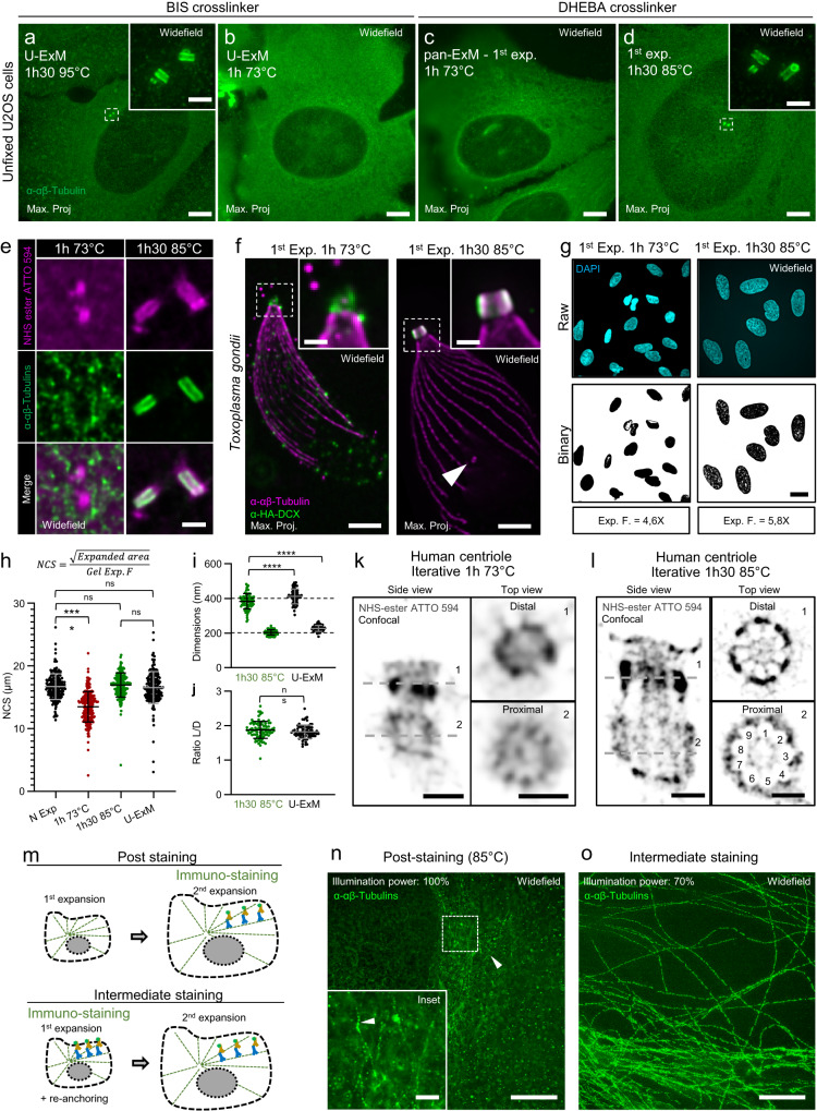
Fig. 1
iU-ExM development.
a u2013 d Expanded U2OS unfixed cells stained for u03b1 and u03b2 tubulins. Scale bars: 10 u03bcm non-corrected. e Centrioles stained for tubulin and NHS-ester ATTO 594 after the first expansion (unfi...
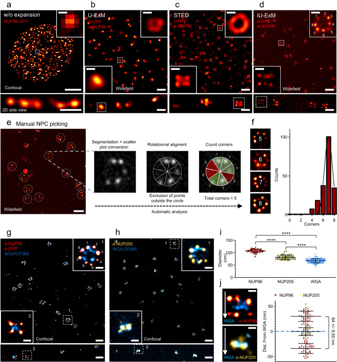
Fig. 2
iU-ExM reveals the 8-fold organization of the human Nuclear Pore Complexes.
a u2013 d Upper panel: 2D top view, lower: 2D side view. a Confocal image of NUP96-eGFP positive nucleus (red hot) without expansion. Scale bar: 4u2009u00b5m (upper image), 500u2009nm (lower), 100u200...
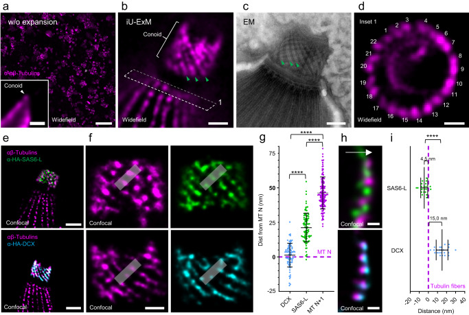
Fig. 3
Molecular organization of the conoid in Toxoplasma gondii tachyzoites.
a Widefield image of non-expanded T. gondii tachyzoites labelled with tubulin antibodies (magenta). Inset: one parasite with the conoid region (white arrowhead). Scale bars: 30u2009u00b5m (full pictur...
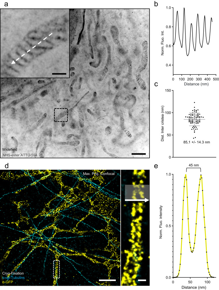
Fig. 4
iU-ExM on membranous organelles.
a iU-ExM widefield image of a U2OS cell fixed with 3% PFA, 0.1% GA, and stained with NHS-ester ATTO 594 to label the mitochondria. Note that iU-ExM allows the visualization of individual cristae (inse...
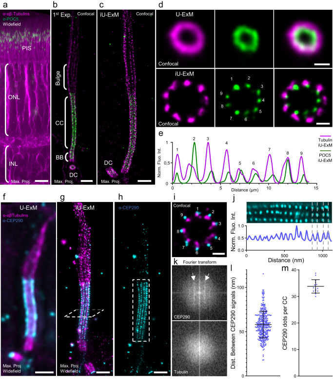
Fig. 5
iU-ExM is compatible with tissue expansion and reveals ciliary periodicity.
a widefield image with a 20u00d7 objective of an iU-ExM 1st gel retina tissue stained for tubulin (magenta) and POC5 (green), highlighting the preservation of the different retinal layers. PIS Photore...
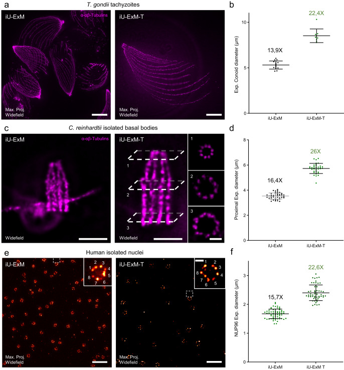
Fig. 6
Coupling TREx to iU-ExM.
a Widefield full field of view of T. gondii tachyzoites stained for tubulin (magenta) expanded with either iU-ExM (left) or iU-ExM-T (right). Scales bar: 30 u03bcm non-corrected. b Quantification of t...
Figure images are served from the NIH/NLM PubMed Central Open Access Subset or Europe PMC; copyright remains with the publishers and authors.
💬 Discussion
0 commentsNo comments yet. Be the first to start a discussion!
Leave a Comment