Abstract
Live imaging is a powerful tool, enabling scientists to observe living organisms in real time. In particular, when combined with fluorescence microscopy, live imaging allows the monitoring of cellular components with high sensitivity and specificity. Yet, due to critical challenges (i.e., drift, phototoxicity, dataset size), implementing live imaging and analyzing the resulting datasets is rarely straightforward. Over the past years, the development of bioimage analysis tools, including deep learning, is changing how we perform live imaging. Here we briefly cover important computational methods aiding live imaging and carrying out key tasks such as drift correction, denoising, super-resolution imaging, artificial labeling, tracking, and time series analysis. We also cover recent advances in self-driving microscopy.
🔬 Techniques
💻 Software
✨ Fluorophores
🧪 Sample Preparation
📷 Detectors
💻 Software Details
🏛️ Research Organizations (ROR)
Affiliated research institutions:
📊 Figures
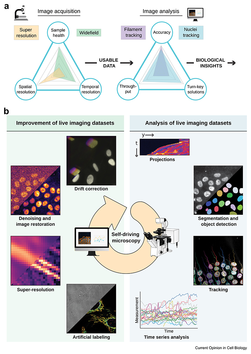
Figure 1
Live-cell imaging main challenges and computational solutions.
( a ) Live fluorescence imaging presents unique challenges that require a careful balance between managing light sensitivity and ensuring optimal spatial, temporal, and spectral resolution to observe ...
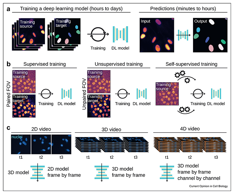
Figure 2
Deep learning and video analysis.
( a ) The DL pipeline. A DL model must first be trained using a training dataset. This step is generally time-consuming and takes hours to weeks, depending on the size of the training dataset. Once tr...
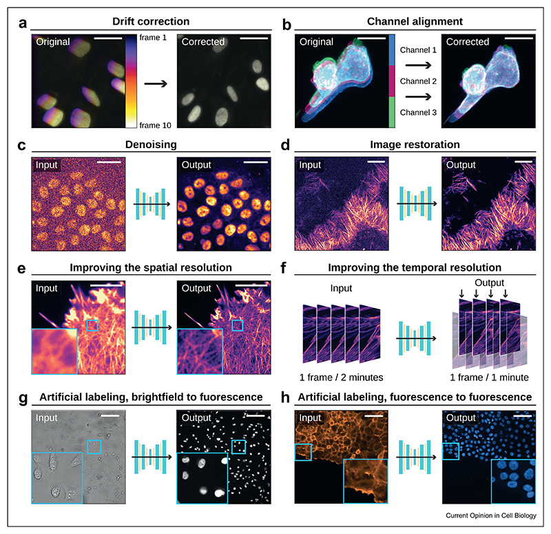
Figure 3
Example of computational tools that can improve live cell imaging movies.
This figure illustrates the power and versatility of computational tools in enhancing the quality, resolution, and content of various types of microscopy images. ( a ) Time projection of drifting live...
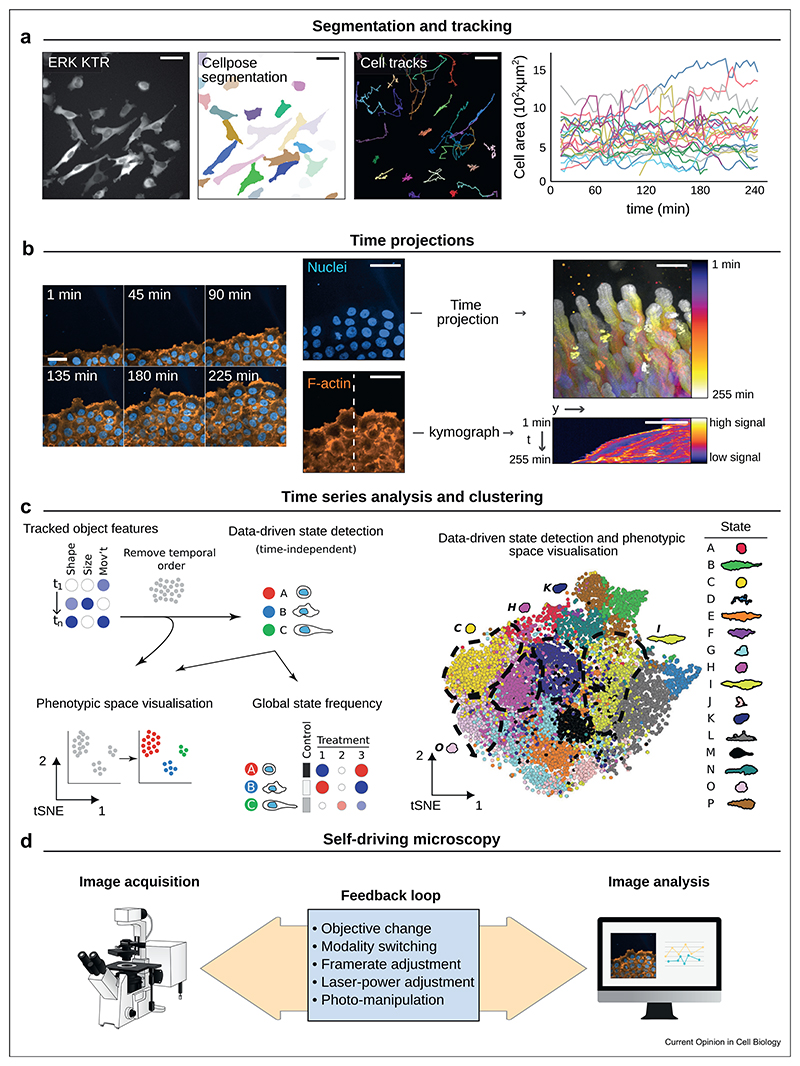
Figure 4
Extracting temporal information from live imaging data.
( a ) Widefield fluorescence microscopy was used to image breast cancer cells expressing a GFP-tagged ERK-reporter (dataset described in Ref. [ 48 ]). The cytoplasm was segmented using a custom CellPo...
Figure images are served from the NIH/NLM PubMed Central Open Access Subset or Europe PMC; copyright remains with the publishers and authors.
💬 Discussion
0 commentsNo comments yet. Be the first to start a discussion!
Leave a Comment