Abstract
Abstract Histological analysis of biological tissues by mechanical sectioning is significantly time‐consuming and error‐prone due to loss of important information during sample slicing. In the recent years, the development of tissue clearing methods overcame several of these limitations and allowed exploring intact biological specimens by rendering tissues transparent and subsequently imaging them by laser scanning fluorescence microscopy. In this review, we provide a guide for scientists who would like to perform a clearing protocol from scratch without any prior knowledge, with an emphasis on DISCO clearing protocols, which have been widely used not only due to their robustness, but also owing to their relatively straightforward application. We discuss diverse tissue‐clearing options and propose solutions for several possible pitfalls. Moreover, after surveying more than 30 researchers that employ tissue clearing techniques in their laboratories, we compiled the most frequently encountered issues and propose solutions. Overall, this review offers an informative and detailed guide through the growing literature of tissue clearing and can help with finding the easiest way for hands‐on implementation.
🔬 Techniques
🔭 Microscopes
💻 Software
✨ Fluorophores
🧪 Sample Preparation
🔬 Cell Lines
🏭 Microscope Brands
🧪 Reagent Suppliers
📷 Detectors
💻 Software Details
🏛️ Research Organizations (ROR)
Affiliated research institutions:
📊 Figures
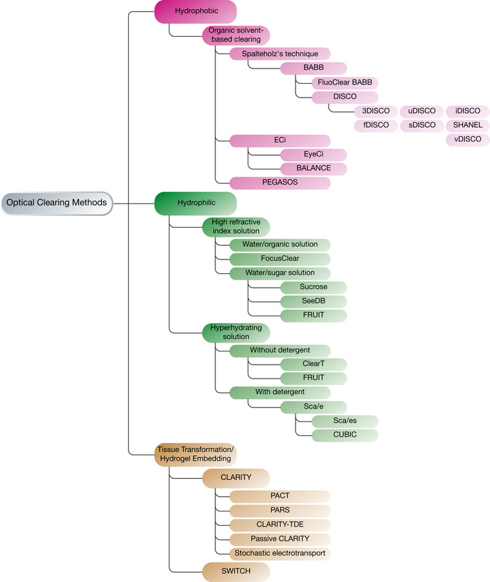
Figure 1
The most common tissue clearing methods
Tissue clearing methods can be separated into three main categories based on their chemistry: hydrogelu2010based, hydrophilic, and hydrophobic. The subtypes mainly differ according to reagents/approac...
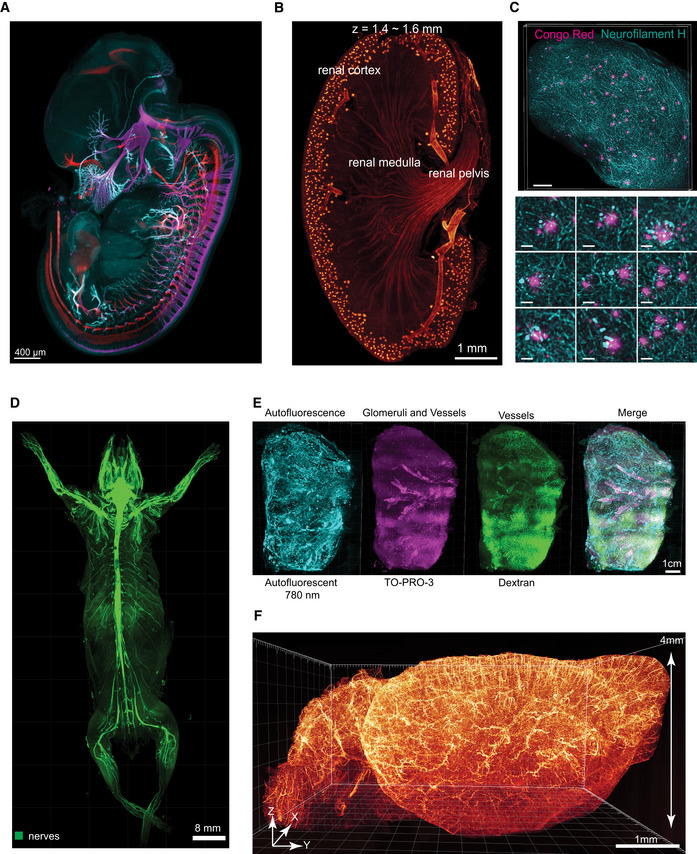
Figure 2
Tissue clearing can be applied to samples ranging from organs, to mouse embryos to adult rodent bodies and reveals macroscopic and microscopic details
(A) Nerve and nerve endings are revealed in an E12.5 mouse embryo cleared with iDISCO protocol and immunostained for motor (red) and sensory (magenta) nerves and nerve endings (cyan). (B) Kidney vascu...
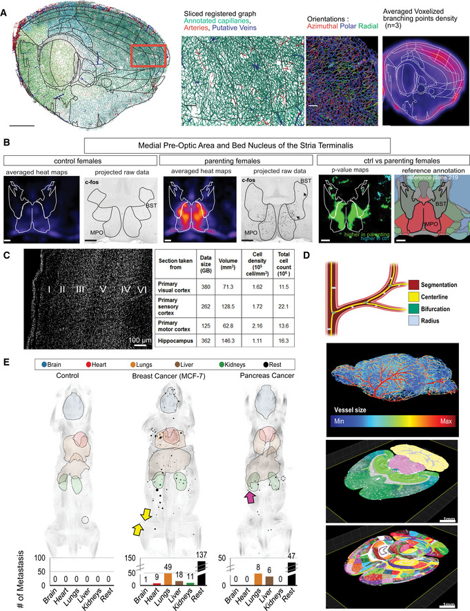
Figure 3
Analysis tools for tissue clearing applications
Analysis tools include: (A) TubeMap pipeline which demonstrates the fineu2010scale organization on the brain vasculature. In this study, Kirst et al ( 2020 ) demonstrated how stroke affects the brain,...
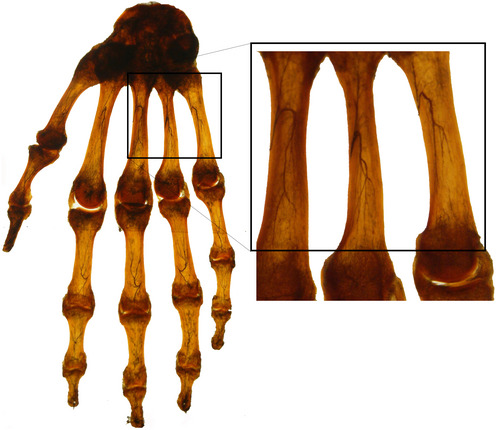
Figure 4
Hand sample cleared by Spalteholz himself in 1911
Spalteholz injected Indian ink to the hand's arteries via the A. nutriciae. Decalcification allowed observing the arterial branching in the bone. The magnified image depicts the inner arterial supply ...
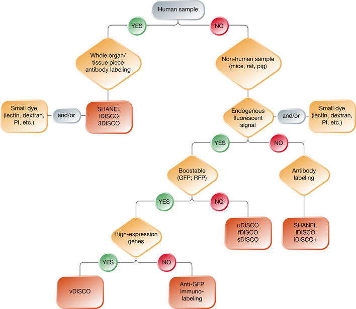
Figure 5
Decision tree for choosing the optimal tissue clearing protocol
Several parameters should be weighed to achieve optimal results when choosing the clearing protocol. This flowchart demonstrates an easy way to decide which clearing method would suit a potential rese...
Figure images are served from the NIH/NLM PubMed Central Open Access Subset or Europe PMC; copyright remains with the publishers and authors.
💬 Discussion
0 commentsNo comments yet. Be the first to start a discussion!
Leave a Comment