Abstract
Abstract Super-resolution microscopy (SRM) enables non-invasive, molecule-specific imaging of the internal structure and dynamics of cells with sub-diffraction limit spatial resolution. One of its major limitations is the requirement for high-intensity illumination, generating considerable cellular phototoxicity. This factor considerably limits the capacity for live-cell observations, particularly for extended periods of time. Here, we give an overview of new developments in hardware, software and probe chemistry aiming to reduce phototoxicity. Additionally, we discuss how the choice of biological model and sample environment impacts the capacity for live-cell observations.
🔬 Techniques
✨ Fluorophores
🧪 Sample Preparation
🔬 Cell Lines
📷 Detectors
🏛️ Research Organizations (ROR)
Affiliated research institutions:
📊 Figures
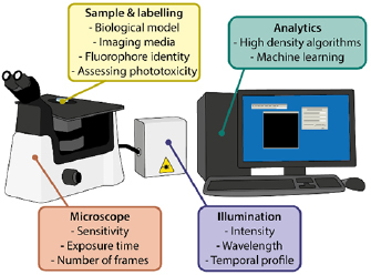
Figure 1.
Summary of the factors that can be optimised to reduce phototoxicity innSRM.
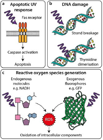
Figure 2.
Interactions of light with cellular components leading to phototoxicity. (a)nUV light can trigger apoptosis by inducing Fas receptor-mediated signallingnpathways. (b) UV light can directly damage DNA ...
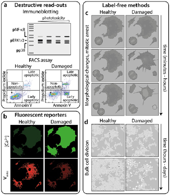
Figure 3.
Methods for measuring phototoxicity. (a) u2018Destructive read-outsu2019 arentechniques prohibiting further imaging of the sample. These include blottingnfor phosphorylated forms of proteins present i...
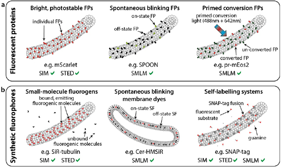
Figure 4.
Low phototoxicity fluorescent probes and labelling for live-cell SRM. Variousnrecently-developed fluorescent protein (a) and synthetic fluorophore (b)nbased methods for labelling in live-cell super-re...
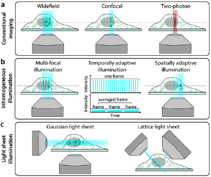
Figure 5.
Hardware modalities for conventional and low-phototoxicity SRM. (a)nMicroscopy illumination regimes for conventional fluorescence imaging. (b)nExamples of regimes that reduce light dose to the sample ...
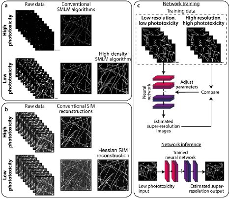
Figure 6.
Analytics to complement low-phototoxicity imaging regimes. (a) Top: typicalnSMLM images are successfully reconstructed from sparse blinking raw datanacquired under high phototoxic illumination. Bottom...
Figure images are served from the NIH/NLM PubMed Central Open Access Subset or Europe PMC; copyright remains with the publishers and authors.
💬 Discussion
0 commentsNo comments yet. Be the first to start a discussion!
Leave a Comment