Abstract
Neurons have a membrane periodic skeleton (MPS) composed of actin rings interconnected by spectrin. Here, combining chemical and genetic gain- and loss-of-function assays, we show that in rat hippocampal neurons the MPS is an actomyosin network that controls axonal expansion and contraction. Using super-resolution microscopy, we analyzed the localization of axonal non-muscle myosin II (NMII). We show that active NMII light chains are colocalized with actin rings and organized in a circular periodic manner throughout the axon shaft. In contrast, NMII heavy chains are mostly positioned along the longitudinal axonal axis, being able to crosslink adjacent rings. NMII filaments can play contractile or scaffolding roles determined by their position relative to actin rings and activation state. We also show that MPS destabilization through NMII inactivation affects axonal electrophysiology, increasing action potential conduction velocity. In summary, our findings open new perspectives on axon diameter regulation, with important implications in neuronal biology.
🔬 Techniques
🔭 Microscopes
💻 Software
✨ Fluorophores
🧪 Sample Preparation
🔬 Cell Lines
🏭 Microscope Brands
🧪 Reagent Suppliers
📷 Detectors
🔎 Objectives
💻 Software Details
💻 Code & Software
MATLAB script for calculating action potentials propagation velocities using spikes timestamps in specified electrodes in a MEA device (coupled with...
MATLAB script for calculating action potentials propagation velocities using spikes timestamps in specified electrodes in a MEA device (coupled with...
💾 Data Repositories
🏷️ Research Resource Identifiers (RRIDs)
Verified research resources used in this paper:
🏛️ Research Organizations (ROR)
Affiliated research institutions:
📋 Methods
Key resources table
Reagent type (species) or resource Designation Source or reference Identifiers Additional information Antibody Rabbit polyclonal anti-NMIIA Sigma-Aldrich Cat# M8064, RRID: AB_260673 WB_1:1000; IF_1:200 Antibody Rabbit polyclonal anti-NMIIB Sigma-Aldrich Cat# M7939, RRID: AB_260669 WB_1:1000; IF_1:200 Antibody Rabbit polyclonal canti-α-adducin Abcam Cat# ab51130, RRID: AB_867519 WB_1:1000 Antibody Rabbit monoclonal anti-vinculin Abcam Cat# ab129002, RRID: AB_11144129 WB_1:1000 Antibody Mouse monoclonal anti-βactin Sigma-Aldrich Cat# A5441, RRID: AB_476744 WB_1:5000 Antibody Mouse monoclonal anti-αtubulin Sigma-Aldrich Cat# T6199, RRID: AB_477583 WB_1:1000 Antibody Peroxidase-AffiniPure donkey polyclonal anti-rabbit IgG (H+L) Jackson Immuno Research Labs Cat# 711-035-152, RRID: AB_10015282 WB_1:5000 Antibody Peroxidase-AffiniPure donkey polyclonal anti-mouse IgG (H+L) Jackson Immuno Research Labs Cat# 715-035-151, RRID: AB_2340771 WB_1:5000 Antibody Mouse monoclonal anti-βII-spectrin BD Transduction Cat# 612563,RRID: AB_399854 IF_1:200 Antibody Rabbit polyclonal anti-MAP2 Synaptic Systems Cat# 188002, RRID: AB_2138183 IF_1:20000 Antibody Rabbit polyclonal anti-NMIIC Robert Adelstein NHLBI, Bethesda, USA N/A IF_1:40 Antibody Rabbit polyclonal anti-pMLC2 Thr18/Ser19 Cell Signaling Cat# 3674, RRID: AB_2147464 IF_1:50 Antibody Rabbit polyclonal anti-MYPT1 Cell Signaling Cat# 2634, RRID: AB_915965 IF_1:50 Antibody Rabbit monoclonal anti-MLC2 Cell Signaling Cat# 1678505S IF_1:40 Antibody Goat polyclonal anti-mouse STAR 635P Abberior GmbH Cat# 2-0002-007-5 IF_1:200 Antibody Goat polyclonal anti-rabbit STAR 635P Abberior GmbH Cat# 2-0012-007-2 IF_1:200 Antibody Goat polyclonal anti-mouse STAR 580 Abberior GmbH Cat# 2-0002-005-1, RRID: AB_2620153 IF_1:200 Antibody Goat polyclonal anti-mouse STAR 580 Abberior GmbH Cat# 2-0012-005-8 IF_1:200 Antibody Goat polyclonal anti- mouse Alexa Fluor 532 Thermo Fischer Scientific Cat# A-11002, RRID: AB_2534070 IF_1:200 Antibody Donkey polyclonal anti- rabbit Alexa Fluor 647 Jackson ImmunoResearch Cat# 711-605-152 IF_1:1000 STORM_ 1:200 Chemical compound, drug Blebbistatin Sigma-Aldrich Cat# B0560 3 µM Chemical compound, drug ML-7 Sigma-Aldrich Cat# I2764 4 µM Chemical compound, drug Myovin1 Calbiochem Cat# 475984 4 µM Chemical compound, drug Calyculin A Sigma-Aldrich Cat# C5552 5 nM Chemical compound, drug Dimethyl sulfoxide (DMSO) VWR International Cat# A3672.0050 N/A Chemical compound, drug Phalloidin 635P Abberior GmbH Cat# 2-0205-002-5 0.33 µM Cell line CAD (mouse) ECACC through Sigma-Aldrich Cat# 08100805, RRID: CVCL_0199 N/A Cell line PC-12 (rat) ATCC Cat# CTL-1721, RRID: CVCL_F659 N/A Cell line SH-SY5Y (human) ATCC Cat# CRL-2266, RRID: CVCL_0019 N/A Recombinant DNA reagent pEGFP-C1 Addgene N/A N/A Recombinant DNA reagent pLKO.1 Addgene N/A N/A Recombinant DNA reagent shRNA NMIIA Rai et al., 2017 N/A 5’ GCGATACTACTCAGGGCTTAT 3’ Recombinant DNA reagent shRNA NMIIB This paper N/A 5‘ GCCAACATTGAAACATACCT 3’ Recombinant DNA reagent shRNA NMIIC This paper N/A 5’ CCGGGCTCATTTATACCTACT 3’ Recombinant DNA reagent shRNA RLC Wang et al., 2008 N/A 5′ GCACGGAGCGAAAGACAAA 3′ Recombinant DNA reagent shRNA MYPT1 This paper N/A 5′ GAGCCTTGATCAGAGTTATAAC 3′ Recombinant DNA reagent shRNA α-adducin Sigma-Aldrich Cat# TRCN0000108809 5’ GCAGAAGAAGAGGGTGTCTAT 3’ Recombinant DNA reagent Human mutated ShRNA-resistant NMIIA This paper N/A Vectorbuilder Page 20_line 374–376 Recombinant DNA reagent Human mutated ShRNA-resistant NMIIB This paper N/A Vectorbuilder Page 20_line 374–376 Recombinant DNA reagent Human ShRNA-resistant RLC Addgene Cat #35680 N/A Recombinant DNA reagent Human mutated ShRNA-resistant MYPT1 This paper N/A Vectorbuilder Page 20_line 374–376 Recombinant DNA reagent CMV-eGFP-NMIIA Addgene Cat #11347 N/A Recombinant DNA reagent CMV-NMIIA-mApple John Hammer, NHLBI, Bethesda, USA N/A N/A Commercial assay or kit NZY Total RNA Isolation Kit NZY Tech Cat# MB13402 N/A Commercial assay or kit SuperScript First-Strand Synthesis System for RT-PCR Thermo Fisher Scientific Cat# 11904018 N/A Sequence-based reagent NMIIC sense primer This paper N/A 5’ CCTGGCTGAGTTCTCCTCAC 3’ Sequence-based reagent NMIIC antisense primer This paper N/A 5’ TGCTTCTGCTCCATCATCTG 3’ Sequence-based reagent RLC sense primer This paper N/A 5’ CCTTTGCCTGCTTTGATGAG 3’ Sequence-based reagent RLC antisense primer This paper N/A 5’ GTGACTGGGATGGGGTGTAG 3’ Sequence-based reagent MYPT1 sense primer This paper N/A 5’ AAGGGAACGAAGAGCTCTAGAAA 3' Sequence-based reagent MYPT1 antisense primer This paper N/A 5’ TGACAGTCTCCAGGGGTTCT 3’ Sequence-based reagent β-actin sense primer This paper N/A 5’ ACCACACCTTCTACAATGAG 3’ Sequence-based reagent β-actin antisense primer This paper N/A 5’ TAGCACAGCCTGGATAGC 3’ Sequence-based reagent GADPH sense primer This paper N/A 5’ AGGCACCAAGATACTTACAAAAAC 3’ Sequence-based reagent GADPH antisense primer This paper N/A 5’ TGTATTGTAACCAGTCATCAGCA 3' Software, algorithm MATLAB R2018a MATLAB RRID: SCR_001622 N/A Software, algorithm Fiji NIH SRRID: SCR_002285 N/A Software, algorithm Leica LAS X software Leica RRID: SCR_013673 N/A Software, algorithm µSpikeHunter software Heiney et al., 2019 N/A N/A Software, algorithm GraphPad Prism GraphPad RRID: SCR_002798 N/A Software, algorithm Huygens Software Scientific Volume Imaging RRID: SCR_014237 N/A Hippocampal neuron cultures Mice and rat hippocampal neuron cultures were performed as described previously ( Kaech and Banker, 2006 ). Briefly, the hippocampus of each individual E18 embryo was digested 15 min in 0.06% trypsin from porcine pancreas solution (Sigma-Aldrich, cat# T4799) and triturated. Either 12,500 cells/coverslip (for STED imaging) or 50,000 cells/coverslip (for SMLM imaging) were plated onto 50 µg/mL poly-L-lysine hydrobromide (Sigma-Aldrich, cat# P2636-100MG) pre-coated 1.5H glass 13 mm rounded coverslips (Marienfeld, for STED imaging) or 22 × 22 mm coverslips (Corning, for SMLM imaging) in 24- or 6-well plates (Nunc). Neurons were cultured in Neurobasal medium (Thermo Fisher Scientific, cat# 21103–049) supplemented with 2% B-27 (Thermo Fisher Scientific, cat# 0080085SA), 1% penicillin/streptomycin (Thermo Fisher Scientific, cat# 15140–122) and 2 mM L-glutamine (Thermo Fisher Scientific, cat# 25030024). In all conditions, cells were fixed with 4% (w/v) paraformaldehyde (PFA) in phosphate-buffered saline (PBS) at pH7.4 for 20 min at room temperature.
Show full methods section
Key resources table
Reagent type (species) or resource Designation Source or reference Identifiers Additional information Antibody Rabbit polyclonal anti-NMIIA Sigma-Aldrich Cat# M8064, RRID: AB_260673 WB_1:1000; IF_1:200 Antibody Rabbit polyclonal anti-NMIIB Sigma-Aldrich Cat# M7939, RRID: AB_260669 WB_1:1000; IF_1:200 Antibody Rabbit polyclonal canti-α-adducin Abcam Cat# ab51130, RRID: AB_867519 WB_1:1000 Antibody Rabbit monoclonal anti-vinculin Abcam Cat# ab129002, RRID: AB_11144129 WB_1:1000 Antibody Mouse monoclonal anti-βactin Sigma-Aldrich Cat# A5441, RRID: AB_476744 WB_1:5000 Antibody Mouse monoclonal anti-αtubulin Sigma-Aldrich Cat# T6199, RRID: AB_477583 WB_1:1000 Antibody Peroxidase-AffiniPure donkey polyclonal anti-rabbit IgG (H+L) Jackson Immuno Research Labs Cat# 711-035-152, RRID: AB_10015282 WB_1:5000 Antibody Peroxidase-AffiniPure donkey polyclonal anti-mouse IgG (H+L) Jackson Immuno Research Labs Cat# 715-035-151, RRID: AB_2340771 WB_1:5000 Antibody Mouse monoclonal anti-βII-spectrin BD Transduction Cat# 612563,RRID: AB_399854 IF_1:200 Antibody Rabbit polyclonal anti-MAP2 Synaptic Systems Cat# 188002, RRID: AB_2138183 IF_1:20000 Antibody Rabbit polyclonal anti-NMIIC Robert Adelstein NHLBI, Bethesda, USA N/A IF_1:40 Antibody Rabbit polyclonal anti-pMLC2 Thr18/Ser19 Cell Signaling Cat# 3674, RRID: AB_2147464 IF_1:50 Antibody Rabbit polyclonal anti-MYPT1 Cell Signaling Cat# 2634, RRID: AB_915965 IF_1:50 Antibody Rabbit monoclonal anti-MLC2 Cell Signaling Cat# 1678505S IF_1:40 Antibody Goat polyclonal anti-mouse STAR 635P Abberior GmbH Cat# 2-0002-007-5 IF_1:200 Antibody Goat polyclonal anti-rabbit STAR 635P Abberior GmbH Cat# 2-0012-007-2 IF_1:200 Antibody Goat polyclonal anti-mouse STAR 580 Abberior GmbH Cat# 2-0002-005-1, RRID: AB_2620153 IF_1:200 Antibody Goat polyclonal anti-mouse STAR 580 Abberior GmbH Cat# 2-0012-005-8 IF_1:200 Antibody Goat polyclonal anti- mouse Alexa Fluor 532 Thermo Fischer Scientific Cat# A-11002, RRID: AB_2534070 IF_1:200 Antibody Donkey polyclonal anti- rabbit Alexa Fluor 647 Jackson ImmunoResearch Cat# 711-605-152 IF_1:1000 STORM_ 1:200 Chemical compound, drug Blebbistatin Sigma-Aldrich Cat# B0560 3 µM Chemical compound, drug ML-7 Sigma-Aldrich Cat# I2764 4 µM Chemical compound, drug Myovin1 Calbiochem Cat# 475984 4 µM Chemical compound, drug Calyculin A Sigma-Aldrich Cat# C5552 5 nM Chemical compound, drug Dimethyl sulfoxide (DMSO) VWR International Cat# A3672.0050 N/A Chemical compound, drug Phalloidin 635P Abberior GmbH Cat# 2-0205-002-5 0.33 µM Cell line CAD (mouse) ECACC through Sigma-Aldrich Cat# 08100805, RRID: CVCL_0199 N/A Cell line PC-12 (rat) ATCC Cat# CTL-1721, RRID: CVCL_F659 N/A Cell line SH-SY5Y (human) ATCC Cat# CRL-2266, RRID: CVCL_0019 N/A Recombinant DNA reagent pEGFP-C1 Addgene N/A N/A Recombinant DNA reagent pLKO.1 Addgene N/A N/A Recombinant DNA reagent shRNA NMIIA Rai et al., 2017 N/A 5’ GCGATACTACTCAGGGCTTAT 3’ Recombinant DNA reagent shRNA NMIIB This paper N/A 5‘ GCCAACATTGAAACATACCT 3’ Recombinant DNA reagent shRNA NMIIC This paper N/A 5’ CCGGGCTCATTTATACCTACT 3’ Recombinant DNA reagent shRNA RLC Wang et al., 2008 N/A 5′ GCACGGAGCGAAAGACAAA 3′ Recombinant DNA reagent shRNA MYPT1 This paper N/A 5′ GAGCCTTGATCAGAGTTATAAC 3′ Recombinant DNA reagent shRNA α-adducin Sigma-Aldrich Cat# TRCN0000108809 5’ GCAGAAGAAGAGGGTGTCTAT 3’ Recombinant DNA reagent Human mutated ShRNA-resistant NMIIA This paper N/A Vectorbuilder Page 20_line 374–376 Recombinant DNA reagent Human mutated ShRNA-resistant NMIIB This paper N/A Vectorbuilder Page 20_line 374–376 Recombinant DNA reagent Human ShRNA-resistant RLC Addgene Cat #35680 N/A Recombinant DNA reagent Human mutated ShRNA-resistant MYPT1 This paper N/A Vectorbuilder Page 20_line 374–376 Recombinant DNA reagent CMV-eGFP-NMIIA Addgene Cat #11347 N/A Recombinant DNA reagent CMV-NMIIA-mApple John Hammer, NHLBI, Bethesda, USA N/A N/A Commercial assay or kit NZY Total RNA Isolation Kit NZY Tech Cat# MB13402 N/A Commercial assay or kit SuperScript First-Strand Synthesis System for RT-PCR Thermo Fisher Scientific Cat# 11904018 N/A Sequence-based reagent NMIIC sense primer This paper N/A 5’ CCTGGCTGAGTTCTCCTCAC 3’ Sequence-based reagent NMIIC antisense primer This paper N/A 5’ TGCTTCTGCTCCATCATCTG 3’ Sequence-based reagent RLC sense primer This paper N/A 5’ CCTTTGCCTGCTTTGATGAG 3’ Sequence-based reagent RLC antisense primer This paper N/A 5’ GTGACTGGGATGGGGTGTAG 3’ Sequence-based reagent MYPT1 sense primer This paper N/A 5’ AAGGGAACGAAGAGCTCTAGAAA 3' Sequence-based reagent MYPT1 antisense primer This paper N/A 5’ TGACAGTCTCCAGGGGTTCT 3’ Sequence-based reagent β-actin sense primer This paper N/A 5’ ACCACACCTTCTACAATGAG 3’ Sequence-based reagent β-actin antisense primer This paper N/A 5’ TAGCACAGCCTGGATAGC 3’ Sequence-based reagent GADPH sense primer This paper N/A 5’ AGGCACCAAGATACTTACAAAAAC 3’ Sequence-based reagent GADPH antisense primer This paper N/A 5’ TGTATTGTAACCAGTCATCAGCA 3' Software, algorithm MATLAB R2018a MATLAB RRID: SCR_001622 N/A Software, algorithm Fiji NIH SRRID: SCR_002285 N/A Software, algorithm Leica LAS X software Leica RRID: SCR_013673 N/A Software, algorithm µSpikeHunter software Heiney et al., 2019 N/A N/A Software, algorithm GraphPad Prism GraphPad RRID: SCR_002798 N/A Software, algorithm Huygens Software Scientific Volume Imaging RRID: SCR_014237 N/A Hippocampal neuron cultures Mice and rat hippocampal neuron cultures were performed as described previously ( Kaech and Banker, 2006 ). Briefly, the hippocampus of each individual E18 embryo was digested 15 min in 0.06% trypsin from porcine pancreas solution (Sigma-Aldrich, cat# T4799) and triturated. Either 12,500 cells/coverslip (for STED imaging) or 50,000 cells/coverslip (for SMLM imaging) were plated onto 50 µg/mL poly-L-lysine hydrobromide (Sigma-Aldrich, cat# P2636-100MG) pre-coated 1.5H glass 13 mm rounded coverslips (Marienfeld, for STED imaging) or 22 × 22 mm coverslips (Corning, for SMLM imaging) in 24- or 6-well plates (Nunc). Neurons were cultured in Neurobasal medium (Thermo Fisher Scientific, cat# 21103–049) supplemented with 2% B-27 (Thermo Fisher Scientific, cat# 0080085SA), 1% penicillin/streptomycin (Thermo Fisher Scientific, cat# 15140–122) and 2 mM L-glutamine (Thermo Fisher Scientific, cat# 25030024). In all conditions, cells were fixed with 4% (w/v) paraformaldehyde (PFA) in phosphate-buffered saline (PBS) at pH7.4 for 20 min at room temperature.
Cell lines
CAD cells (mouse catecholaminergic neuronal cell line¸ ECACC through Sigma-Aldrich cat# 08100805; authenticated by DNA barcoding; mycoplasma contamination testing status: negative), PC-12 cells (rat adrenal gland pheochromocytoma, ATCC cat# CTL-1721; mycoplasma contamination testing status: negative) and SH-SY5Y cells (human neuroblastoma, ATCC cat# CRL-2266; mycoplasma contamination testing status: negative) were used in specific experiments as detailed below. Both CAD and PC-12 cells were maintained in Dulbecco’s modified Eagle’s medium (DMEM, Sigma-Aldrich, cat# D6429-500ML) supplemented with 10% fetal bovine serum (FBS, Sigma-Aldrich, cat#F9665-500ML), and 1% penicillin/streptomycin, while SH-SY5Y cells were cultured in DMEM:F12 (Sigma-Aldrich, cat# D8437−6 × 500 ML) 1:1, with 10% FBS, and 1% penicillin/streptomycin. SH-SY5Y were differentiated as detailed ( Encinas et al., 2002 ). Briefly, 6500 cells were plated on 10 µg/mL poly-D-lysine hydrobromide (Sigma-Aldrich, cat# P0899) and 5 µg/mL laminin (Sigma-Aldrich, cat# L2020) pre-coated 1.5H glass 13 mm rounded coverslips (Marienfeld, for STED imaging) in a 24-well plate (Nunc). In the following day, media was supplemented with 10 µM retinoic acid (Sigma-Aldrich, cat# R2625) and renewed every other day for 5 days. At day 6, the media was changed for DMEM:F12 with 2% B-27, 1% penicillin/streptomycin and brain derived neurotrophic (BDNF, Sigma-Aldrich, cat# B3795, 50 ng/ml); at day 9 differentiated cells were used for imaging. Modulation of MNII activity using pharmacological agents To modulate myosin activity, blebbistatin (Sigma-Aldrich, cat# B0560, 3 µM), ML-7 (Sigma-Aldrich, cat# I2764, 4 µM), or myovin1 (Calbiochem, cat# 475984, 4 µM), were added to hippocampal neurons at DIV3 and at DIV8 1 hr prior to fixation. Calyculin A (Sigma-Aldrich, cat# C5552, 5 nM) was added to hippocampal neurons at DIV8, 25 min prior to fixation. For all drugs, dimethyl sulfoxide (DMSO, VWR International, cat# A3672.0050) was used as a vehicle. In control conditions, vehicle alone was used at the same concentration as in the respective experimental condition. For the analysis of the effect of blebbistatin in in vitro thinning, the drug or its vehicle were additionally added at DIV16 and at DIV22 1 hr prior to fixation. To evaluate the reversibility of the effect of blebbistatin, at DIV8 the drug was either replaced by vehicle, or fresh drug was added to the culture. To confirm the inhibitory effect of ML-7 on RLC phosphorylation, hippocampal neurons were incubated with either vehicle or ML-7 (4 µM) at DIV8 1 hr prior fixation and immunofluorescence against pMLC was conducted as detailed below. To evaluate reversibility of the effect of calyculin A on axonal diameter, hippocampal neurons were treated with blebbistatin (3 µM) and calyculin A (5 nM) at DIV8; 25 min later the cells were fixed. To evaluate a possible cumulative effect of adducin and NMII in axonal diameter, sh-mediated knockdown of α-adducin was performed at DIV3 (detailed below) and either blebbistatin or vehicle were added at DIV4 and DIV8 1 hr before fixation. shRNA-mediated downregulation shRNA constructs against NMIIA, NMIIB, NMIIC, RLC, MYPT1 and α-adducin were used. Sequences were the following: for NMIIA 5’- GCGATACTACTCAGGGCTTAT -3’ ( Rai et al., 2017 ); for NMIIB 5’- GCCAACATTGAAACATACCT -3’ (VectorBuilder); for NMIIC 5’- CCGGGCTCATTTATACCTACT -3’ (VectorBuilder); for RLC 5′- GCACGGAGCGAAAGACAAA -3′ ( Wang et al., 2008 ); for MYPT1 5′- GAGCCTTGATCAGAGTTATAAC -3′ (Vector Builder) and for α-adducin 5’- GCAGAAGAAGAGGGTGTCTAT -3’ (TRCN0000108809, Sigma). In each case, rescue experiments were performed using shRNA-resistant constructs containing 2–3 mismatches in relation to the respective shRNA. For NMIIA, NMIIB and MYPT1 these were designed and ordered from Vector Builder; for RLC, the shRNA-resistant construct was obtained from Addgene (#35680). In control conditions, the pLKO.1 empty vector (Addgene) or a shRNA scramble plasmid (VectorBuilder) were used. At DIV3 hippocampal neurons were co-transfected with specific shRNAs and pEGFP-C1 (0.5 µg:0.25 µg/wells) or in the case of rescue experiments, additionally with the respective shRNA-resistant construct (0.5 µg/well) using Lipofectamine 3000 (Thermo Fisher Scientific, cat# L3000001) according to the manufacturer’s instructions. Cells were fixed at DIV8. In all conditions, transfected (EGFP-positive) and non-transfected (EGFP-negative) cells in the same well were used for analysis. To validate shRNAs, CAD cells, PC-12 cells and hippocampal neurons were used. CAD and PC-12 cells were seeded at a density of 250000 cells/well while hippocampal neurons at a density of 12500 cells/well in 24-well plates and co-transfected with specific shRNAs and pEGFP-C1 (1.5 µg:0.5 µg) using Lipofectamine 2000 (Thermo Fisher Scientific, cat# 116678030) for cell lines, and Lipofectamine 3000 for hippocampal neurons. To evaluate shRNA efficiency, either Western blot analysis of CAD or PC-12 cells (for adducin, NMIIA and NMIIB) or RT-PCR (for NMIIC and MYPT-1) were performed. Additionally, immunofluorescence of primary hippocampal neurons was conducted for NMIIA, NMIIB, NMIIC, RLC and MYPT1, as detailed below. For immunoblotting, cell extracts were prepared in lysis buffer (0.3% Triton X-100 (Sigma-Aldrich, cat# T9284-100ML), protease inhibitors (cOmplete, Mini, Roche, Merck, cat# 04693124001) and 2 mM orthovanadate (Sigma-Aldrich, cat# S6508-10G) in PBS), run in 10% SDS-PAGE gels and transferred to Amersham Protran Premium 0.45 μm nitrocellulose membranes (GE Healthcare Life Sciences, VWR International, cat# GEHE10600003). The following primary antibodies were used: rabbit anti-NMIIA (Sigma-Aldrich, cat# M8064, 1:1000), rabbit anti-NMIIB (Sigma-Aldrich, cat# M7939, 1:1000), rabbit anti-α-adducin (Abcam, cat# ab51130, 1:1000); rabbit anti-vinculin (Abcam cat# ab129002, 1:1000), mouse anti-β-actin (Sigma-Aldrich, cat# A5441, 1:5000), and mouse anti-α-tubulin (Sigma-Aldrich, cat# T6199, 1:1000). The secondary antibodies donkey anti-rabbit IgG conjugated with horseradish peroxidase (HRP) (Jackson Immuno Research, cat# 711-035-152, 1:5000) and donkey anti-mouse IgG conjugated with HRP (Jackson Immuno Research, cat# 715-035-151, 1:5000) were employed. Quantifications were performed using Quantity One 1-D Analysis Software version 4.6 (Bio-Rad). In the case of RT-PCR, RNA from CAD and PC12 cells was extracted using NZY Total RNA Isolation Kit (NZY Tech, cat# MB13402). RNA concentration and purity were determined by NanoDrop spectrophotometry, and integrity was assessed using BioRad's Experion RNA chip. cDNA synthesis was performed with SuperScript First-Strand Synthesis System for RT-PCR (Thermo Fisher Scientific, cat# 11904018). RT-PCR was done using the following specific primers: for NMIIC (forward 5’- CCTGGCTGAGTTCTCCTCAC -3’ and reverse 5’- TGCTTCTGCTCCATCATCTG -3’, for amplification of a 207 bp fragment); for MYPT1 (forward 5’- AAGGGAACGAAGAGCTCTAGAAA -3’ and reverse 5’- TGACAGTCTCCAGGGGTTCT -3’, for amplification of a 242 bp fragment; β-actin (forward 5’- ACCACACCTTCTACAATGAG -3’ and reverse 5’- TAGCACAGCCTGGATAGC -3’, for amplification of a 161 bp fragment) and GADPH (forward 5’- AGGCACCAAGATACTTACAAAAAC -3’ and reverse 5’- TGTATTGTAACCAGTCATCAGCA -3’, for amplification of a 193 bp fragment). Immunolabeling Primary hippocampal neurons were fixed with 4% PFA, in PBS at pH 7.4 for 20 min at room temperature. Fixed cells were permeabilized with 0.1% (v/v) triton X-100 (in PBS) for 5 min and autofluorescence was quenched with 0.2M ammonium chloride (Merck, cat# 1.01145.0500). Non-specific labeling was blocked by incubation with blocking buffer (5% FBS in PBS) for 1 hr. Primary antibodies diluted in blocking buffer were incubated overnight at 4°C: mouse anti-βII-spectrin (BD Transduction, cat# 612563, 1:200), rabbit anti-MAP2 (Synaptic Systems, cat# 188002, 1:20000), rabbit anti-NMIIA (Sigma-Aldrich, cat# M8064, 1:200), rabbit anti-NMIIB (Sigma-Aldrich, cat# M7939, 1:200), rabbit anti-NMIIC (a kind gift from Dr Robert Adelstein, 1:40), rabbit anti-pMLC2 Thr18/Ser19 (Cell Signaling, cat# 3674, 1:50), rabbit anti-MLC2 (Cell Signaling, cat# 1678505S, 1:40), and rabbit anti-MYPT1 (Cell Signaling, cat# 2634, 1:50). After three 5 min washes in PBS, incubation with secondary antibody was performed for 1 hr at room temperature. For STED microscopy, the following secondary antibodies were used: goat anti-mouse STAR 635P (Abberior GmbH, cat# 2-0002-007-5, 1:200); goat anti-rabbit STAR 635P (Abberior GmbH, cat# 2-0012-007-2, 1:200); goat anti-mouse STAR 580 (Abberior GmbH, cat# 2-0002-005-1, 1:200) and goat anti-rabbit STAR 580 (Abberior GmbH, cat# 2-0012-005-8, 1:200). For actin staining, 0.3 µM phalloidin 635P (Abberior GmbH, cat# 2-0205-002-5, in PBS), was used. Phalloidin was incubated in cells for 1 hr at 37°C following the secondary antibody incubation. For SMLM dSTORM the following secondaries were used: goat-anti mouse Alexa Fluor 532 (Thermo Fischer Scientific, cat# A-11002, 1:200) and donkey-anti rabbit Alexa Fluor 647 (Jackson ImmunoResearch, cat# 711-605-152, 1:200). After three 5 min PBS washes, coverslips were mounted in 80% glycerol for STED microscopy; for SMLM imaging, coverslips were mounted in GLOX/MEA buffer (detailed below) in depression slides and sealed with twinsil (Picodent, cat# 13001000) and for immunofluorescence, the coverslips were mounted in fluoroshield with DAPI (Vector Laboratories, cat# H-1200) and sealed with nail varnish. In the case of shRNA validations, images were acquired using a Zeiss Axio ImagerZ1 widefield microscope (Carl Zeiss) equipped with oil-immersion 63x/1.4 (Plan-Apochromat) and with Differential Interference Contrast (DIC) and a TCS Leica SP8 confocal microscope (Leica Microsystems). To validate the effectiveness of shRNA-mediated downregulation of NMIIA, NMIIB, NMIIC, RLC and MYPT1, the cell body from transfected and non-transfected cells, was delineated with the segmented line tool from Fiji, and the mean fluorescence intensity was measured. To confirm that ML-7 inhibits the phosphorylation of NMII, the AIS was delineated with the segmented line from Fiji and the mean fluorescence intensity was compared in treated and non-treated cells.
STED imaging
STED imaging was performed on an inverted Leica TCS SP8 STED 3X (Leica Microsystems), using DIV8 hippocampal neurons, unless otherwise indicated. Hippocampal neurons were imaged, at a fixed distance of 80–100 µm from the cell body, with a HC PLAPO CS2 100x NA 1.4 STED WHITE oil immersion objective (Leica Microsystems) using confocal and STED modes. The 2D vortex STED images with lateral resolution enhancement were recorded with 20 nm pixel size in xy and dwell times of typically 600 ns. First, the STED far-red channel (Abberior STAR 635P) was recorded with 633 nm excitation using the pulsed white light laser with 80 MHz repetition rate and STED depletion was performed with a synchronized pulsed 775 nm depletion laser. The detection bandpass was set to 650 to 750 nm and the pinhole was set to 0.93AU. The following acquisition settings were applied: 16 x line averaging and detector gating on a Hybrid Detector (HyD, Leica Microsystems) of 0.3 ns to 6 ns. The second STED channel (Abberior STAR580) was recorded in line sequential mode with 561 nm excitation and 775 nm depletion using a detection window from 580 to 620 nm. All other settings remained constant. We alternatively used an Abberior Instruments ‘Expert Line’ gated-STED coupled to a Nikon Ti microscope with an oil-immersion 60x 1.4 NA Plan-Apo objective (Nikon, Lambda Series) and a pinhole size set at 0.8 Airy units. The system features 40 MHz modulated excitation (405, 488, 560 and 640 nm) and depletion (775 nm) lasers. The microscope’s detectors are avalanche photodiode detectors (APDs) which were used to gate the detection between ~700 ps and 8ns. To analyze ring periodicity, the maximum intensity of peaks was determined and the interpeak distance was measured. To determine axon diameter, the distance between the outer points (brighter, in the focus plane) that formed the MPS was determined. Only axons unequivocally focused in the maximum wide plan were considered. Under distinct control conditions axon diameter varied, which is probably inherent to the different cultures used throughout the study. Given the use of specific controls in each experimental setting, this variation did not interfere with the interpretation of results. The tilting of actin rings was determined by measuring the angle of each actin ring regarding the axonal axis using the angle tool from Fiji. The tilting angle α was measured relative to the longitudinal axon axis. Angles larger than 90° were mirrored to the first quadrant to yield an effective angle α eff =90-|90-α|. SMLM imaging with dSTORM/GSDIM For super-resolution SMLM-imaging with the Leica SR GSD using the dSTORM/GSDIM protocol, 18 mm coverslips (50000 cells/slide) were stored in PBS after fixation and immunolabelling at 4°C. The coverslips were mounted onto a single depression slide (76 mm ×26 mm) and the cavity filled with 90–100 µl GLOX-MEA buffer (0.5 mg/ml glucose oxidase (Sigma-Aldrich, cat# G7141 , 40 μg/ml catalase (Sigma-Aldrich, cat# 02071 ) 10% w/v glucose (Sigma-Aldrich, cat# 49163 ), 50 mM Tris-HCl pH 8.0, 10 mM NaCl and 10 mM β-mercaptoethylamine (Sigma-Aldrich, cat# M9768-5G)). The buffer was freshly prepared before imaging. Imaging was performed with a Leica SR GSD system using a HC PL APO 160×/NA 1.43 oil objective. The images were recorded with an Andor iXon 897 EMCCD camera at 40 Hz using a central 180 pixel x 180 pixel subregion. For excitation, a 532 nm laser (500 mW maximum power output) and a 642 nm laser (500 mW maximum power output) were used and attenuated using an AOTF when appropriate. The two fluorophores were recorded sequentially and image acquisition, single molecule analysis and image reconstruction was performed with Leica LAS X 1.9.0.13747. Spinning disk imaging Differentiated SH-SY5Y cells (6500 cells/well) and DIV6-hippocampal neurons (50000 cells/well) were co-transfected with CMV-eGFP-NMIIA (Addgene, cat# 11347) and NMIIA-mApple (a kind gift from Dr John Hammer) using 1 µg:1 µg of each construct/well and Lipofectamine 3000 following the manufacturer’s instructions. Two days later, in the case of SH-SY5Y cells, and four days later (at DIV10) in the case of primary hippocampal neurons, the cells were fixed. Transfected cells were then imaged using an Olympus SpinSR10 spinning disk confocal super‐resolution microscope (Olympus, Tokyo, Japan) equipped with an PlanAPON 60 ×/1.42 NA oil objective (Olympus), a CSU-W1 SoRa-Unit (Yokogawa, Tokyo, Japan) with 3.2x magnification and ORCA‐Flash 4.0 V3 Digital CMOS Camera (Hamamatsu, Hamamatsu City, Japan).
Preparation of microelectrode–microfluidic devices and electrophysiology recordings
Custom designed µEF devices were prepared following Lopes et al. (2018) . Briefly, coated MEA chips (MultiChannel Systems MCS GmbH, Germany), with 252 recording electrodes of 30 µm in diameter and a center-to-center inter-electrode spacing of 100 µm, were combined with polydimethylsiloxane (PDMS) microfluidic chambers with an appropriate microgroove spacing for compartmentalization and monitoring of axonal activity. MEAs were coated with 0.01 mg/ml of poly-D-lysine (PDL, Corning) overnight at 37°C, and then washed with sterile water. Microfluidic devices were sterilized with 70% ethanol and were gently attached to PDL-coated MEAs, creating a µEF chamber composed of two separate compartments connected by 700 μm length ×9.6 μm height ×10 μm width microchannels. The medium reservoirs were loaded with 150 μl of 5 μg/ml laminin isolated from mouse Engelbreth-Holm-Swarm sarcoma (Sigma-Aldrich Co.) and incubated overnight at 37°C. The unbound laminin-1 was removed, and chambers were refilled with Neurobasal medium and left to equilibrate for at least 2 hr at 37°C before cell seeding. Hippocampal neurons at DIVs 11, 12 and 14 were used in the electrophysiology experiments, where either blebbistatin (3 μM), or vehicle were added. Recordings were performed using a MEA2100 recording system (MultiChannel Systems MCS GmbH, Germany). The µEF devices prepared with these MEAs had 16 microchannels with 7 electrodes positioned along each microchannel, as well as 126 electrodes dedicated to the somal compartment. For each time point, recordings were obtained at a sampling rate of 20 kHz for the characterization of the overall network activity. Then, high-temporal resolution recordings were obtained at a sampling rate of 50 kHz, for a duration of 60 or 120 s, for the calculation of the conduction velocity. Throughout the experiments, the temperature was maintained at 37°C and all recorded activity was spontaneous activity. Data analysis was carried out in MATLAB R2018a (The MathWorks Inc) using custom scripts (available in GitHub at: https://github.com/paulodecastroaguiar/Calculate_APs_velocities_in_MEAs ; copy archived at https://github.com/elifesciences-publications/Calculate_APs_velocities_in_MEAs ; Aguiar, 2020 ) and the µSpikeHunter tool ( Heiney et al., 2019 ). Raw signals were band-pass filtered (200–3000 Hz) and spikes were detected by a threshold set to 6 × STD of the electrode noise. Electrodes with a mean firing rate (MFR) of at least 0.1 Hz were considered active. For the propagation velocity calculations, the extracted spike times were further corrected based on the voltage waveforms. To be considered a propagating event the following requirements had to be fulfilled: detection over the entire microchannel (7 electrodes); time delay between electrode pairs lower than or equal to 1 ms (minimum propagation velocity of 0.1 m/s); isolated spike in a 3 ms time window (as to ensure spike identity in all electrodes). Propagation velocity was then calculated by dividing the first-to-last electrode distance (600 μm span) by the delay between spike times. This stringent detection method eliminated any ambiguity during bursts and excluded sequences with missing spike times on at least one electrode, which drastically reduced the size but strengthened the quality of the action potentials dataset.
Statistical analysis
All measurements were performed with the researcher blinded to the experimental condition. Data are shown as mean ± s.e.m, which the exception of propagation velocity values which are shown as median ± s.d. Statistical significance was determined by Student’s t-test using Prism (GraphPad Software), with exception of actin ring measurements in hippocampal neuron cultures, where one-way ANOVA was used (GraphPad Software). Sample sizes are indicated in Figure legends and significance was defined as p*
📊 Figures
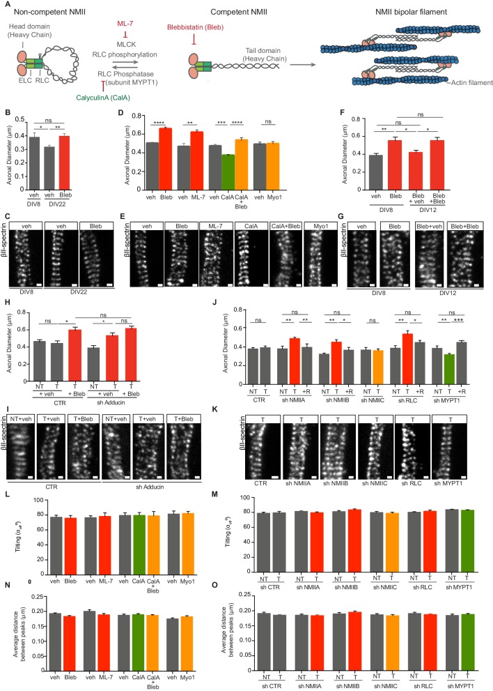
Figure 1.
Modulation of NMII activity regulates axon diameter.
( A ) Representation of NMII structure and activity regulation. NMII contains two heavy chains (HC), two RLCs and two ELCs. The head domain includes an actin-binding site and an ATPase motor domain. E...
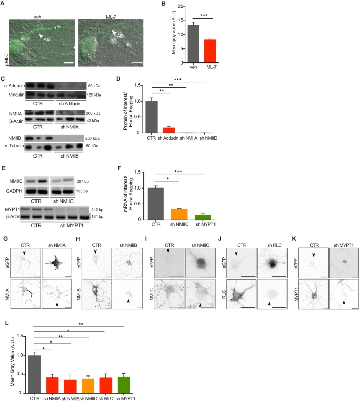
Figure 1u2014figure supplement 1.
Analysis of ML-7 activity and ShRNA-mediated downregulation.
( A ) Representative images of DIC and pMLC immunolabeling of hippocampal neurons treated with either vehicle (veh) or 4 u03bcM ML-7 at DIV3 and DIV8, (1 hr before fixation). Scale bar: 10 u03bcm. ( B...
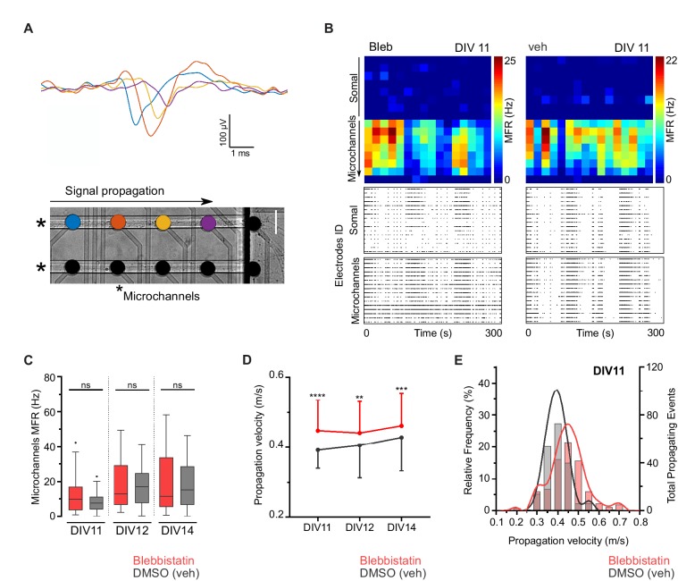
Figure 2.
Blebbistatin treatment increases axonal signal propagation velocity.
( A ) Single propagating event recorded along four electrodes inside a microchannel. Each colored signal trace corresponds to an electrode from a single microchannel. Two microchannels are indicated b...
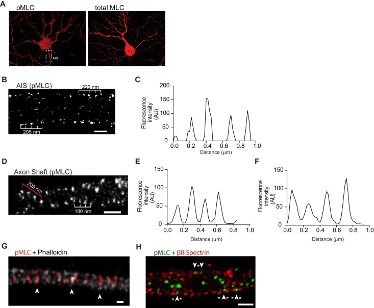
Figure 3.
Phosphorylated NMII light chains are organized as circular periodic structures persisting throughout the axon shaft.
( A ) Immunolabeling of DIV8 hippocampal neurons with rabbit anti-pMLC (left) and rabbit anti-total MLC (right) using a secondary anti- rabbit Alexa Fluor 647 antibody. Scale bar: 5 u03bcm. ( B ) Sing...
Video 1.
3D single colour SMLM of pMLC distribution in the AIS of a DIV8 hippocampal neuron.
Scale bar: 500 nm.
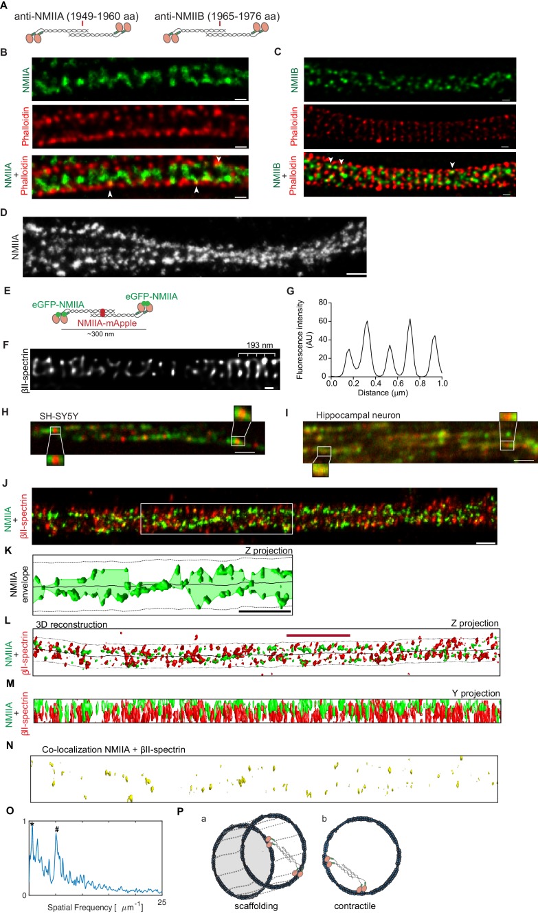
Figure 4.
NMII heavy chains organize into filaments distributed in multiple orientations along the axon shaft.
( A ) Representation of recognition sites of antibodies against NMIIA and NMIIB (NMIIA: 1949u20131960 aa; NMIIB: 1965u20131976 aa). ( B ) Representative STED analysis of a DIV8 hippocampal neuron co-s...
Video 2.
3D two-color SMLM of DIV8 hippocampal neurons immunostained against u03b2II-spectrin (red) and NMIIA (green).
Scale bar: 500 nm.
Figure images are served from the NIH/NLM PubMed Central Open Access Subset or Europe PMC; copyright remains with the publishers and authors.
💬 Discussion
0 commentsNo comments yet. Be the first to start a discussion!
Leave a Comment