Abstract
BACKGROUND: Excess storage of lipids in ectopic tissues, such as skeletal muscle, liver, and heart, seems to associate closely with metabolic abnormalities and cardiac disease. Intracellular lipid storage occurs in lipid droplets, which have gained attention as active organelles in cellular metabolism. Recent developments in high-resolution microscopy and microscopic spectroscopy have opened up new avenues to examine the physiology and biochemistry of intracellular lipids. SCOPE OF REVIEW: The aim of this review is to give an overview of recent technical advances in microscopy, and its application for the visualization, identification, and quantification of intracellular lipids, with special focus to lipid droplets. In addition, we attempt to summarize the probes currently available for the visualization of lipids. MAJOR CONCLUSIONS: The continuous development of lipid probes in combination with the rapid development of microscopic techniques can provide new insights in the role and dynamics of intracellular lipids. Moreover, in situ identification of intracellular lipids is now possible and promises to add a new dimensionality to analysis of lipid biochemistry, and its relation to (patho)physiology.
🔬 Techniques
🧬 Organisms
✨ Fluorophores
🧪 Sample Preparation
🔬 Cell Lines
🏭 Microscope Brands
🏛️ Research Organizations (ROR)
Affiliated research institutions:
📊 Figures
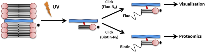
Figureu00a01
Example of a bifunctional lipid . Upon activation of a photoactivatable (clickable) fatty acid, the fatty acid can be turned into a variety of bifunctional phospholipids. These functional phospholipid...
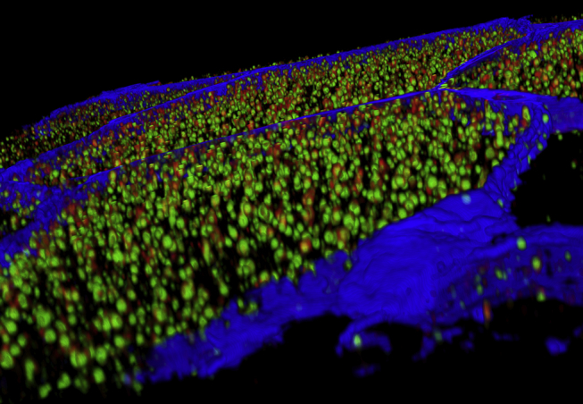
Figureu00a02
3D reconstruction of the cross-section of human skeletal muscle fibers . 3D reconstruction of the cross-section of human skeletal muscle fibers (cell membrane in blue, stained for laminin), illustrati...
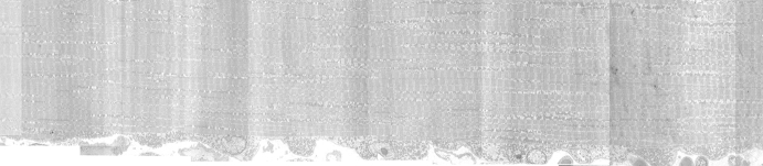
Figureu00a03
Merging of multiple transmission electron microscopy images . Merging of multiple transmission electron microscopy images from a longitudinal section of a muscle fiber located in m. vastus lateralis f...
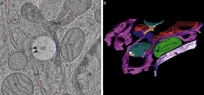
Figureu00a04
Direct membrane contact between ER and LD . (A) Direct membrane contact between ER and LD could be observed in reconstructed tilt-series from high-pressure frozen, freeze-substituted (acetone containi...
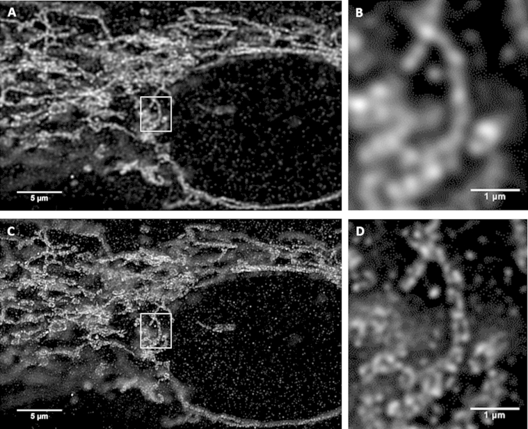
Figureu00a05
Comparative imaging of the mitochondrial outer membrane protein TOMM20 . Comparative imaging of the mitochondrial outer membrane protein TOMM20 in cultured human muscle cells with CLSM (A, B) and STED...
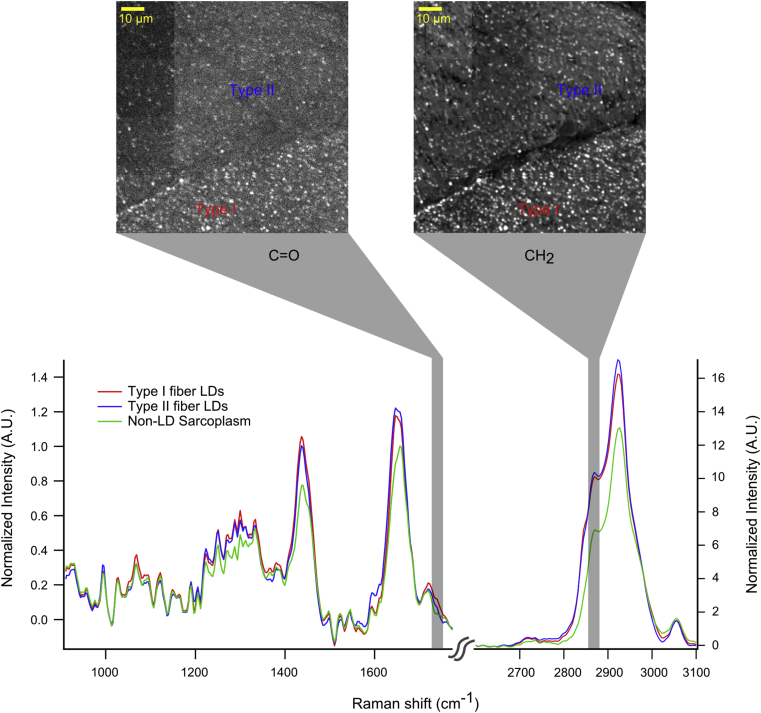
Figureu00a06
Quantitative imaging of lipid droplet chemistry in muscle using hyperspectral CARS microscopy . One complete hyperspectral dataset contained spectral data from Type I and Type II muscle fibers in a hu...
Figure images are served from the NIH/NLM PubMed Central Open Access Subset or Europe PMC; copyright remains with the publishers and authors.
💬 Discussion
0 commentsNo comments yet. Be the first to start a discussion!
Leave a Comment