Abstract
Since its initial demonstration in 2000, far-field super-resolution light microscopy has undergone tremendous technological developments. In parallel, these developments have opened a new window into visualizing the inner life of cells at unprecedented levels of detail. Here, we review the technical details behind the most common implementations of super-resolution microscopy and highlight some of the recent, promising advances in this field.
🔬 Techniques
🧬 Organisms
✨ Fluorophores
🧪 Sample Preparation
🔬 Cell Lines
📷 Detectors
💻 Software Details
🏛️ Research Organizations (ROR)
Affiliated research institutions:
📊 Figures
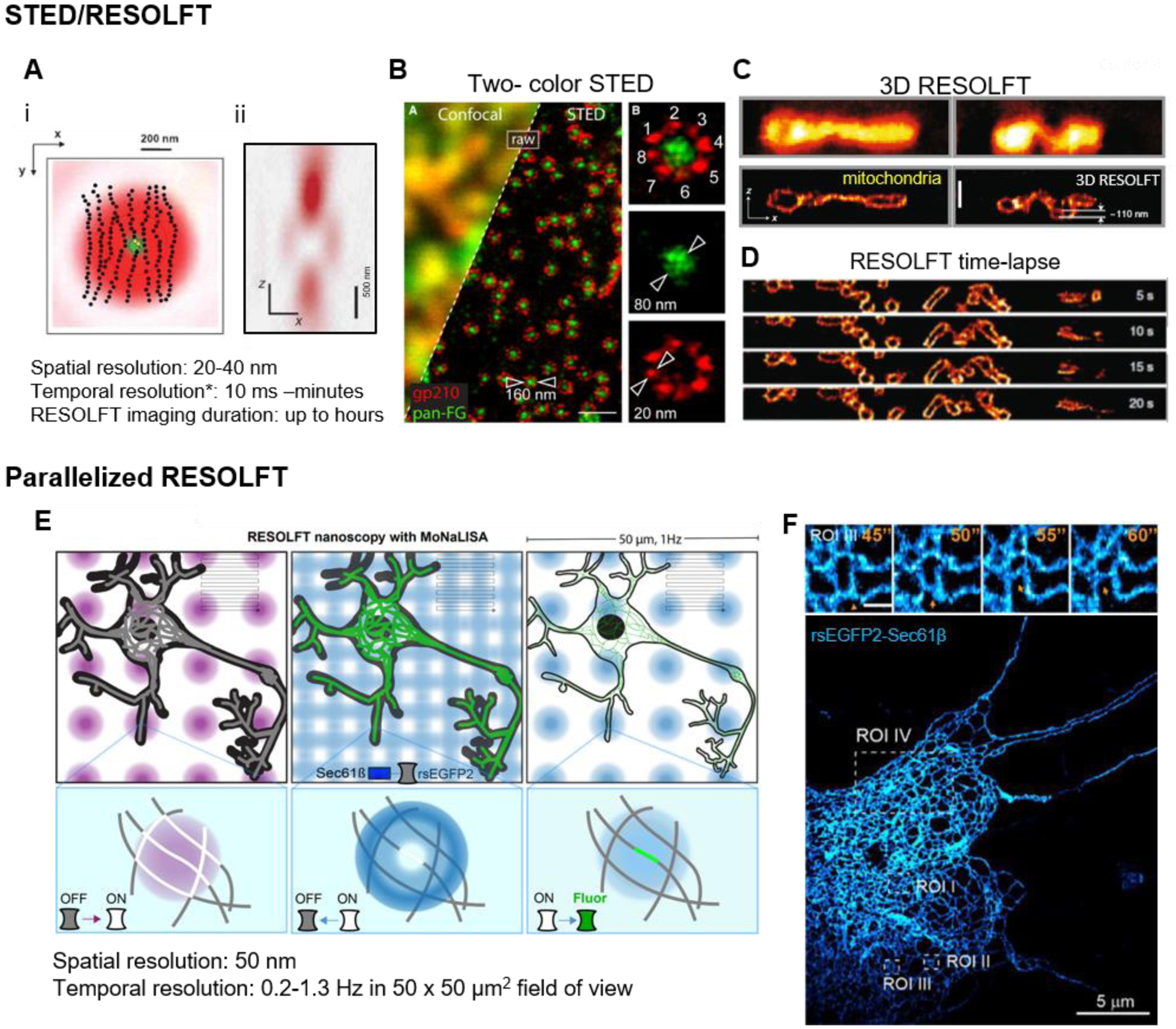
Figure 1:
STED and RESOLFT.
(A) (i) STED depletion beam (red) geometry in lateral (x,y) dimension overlayed with the excitation beam (green), and (ii) cross-section of the z depletion beam to improve axial resolution ( Hell, 201...
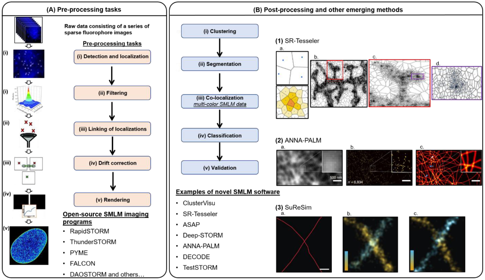
Figure 2:
Single Molecule Localization Microscopy and analysis.
(A) The pre-processing tasks of SMLM data consist of: (i) peak detection and localization, (ii) linking of localizations, (iii) filtering, (iv) drift correction, and (v) rendering. Currently, there ar...
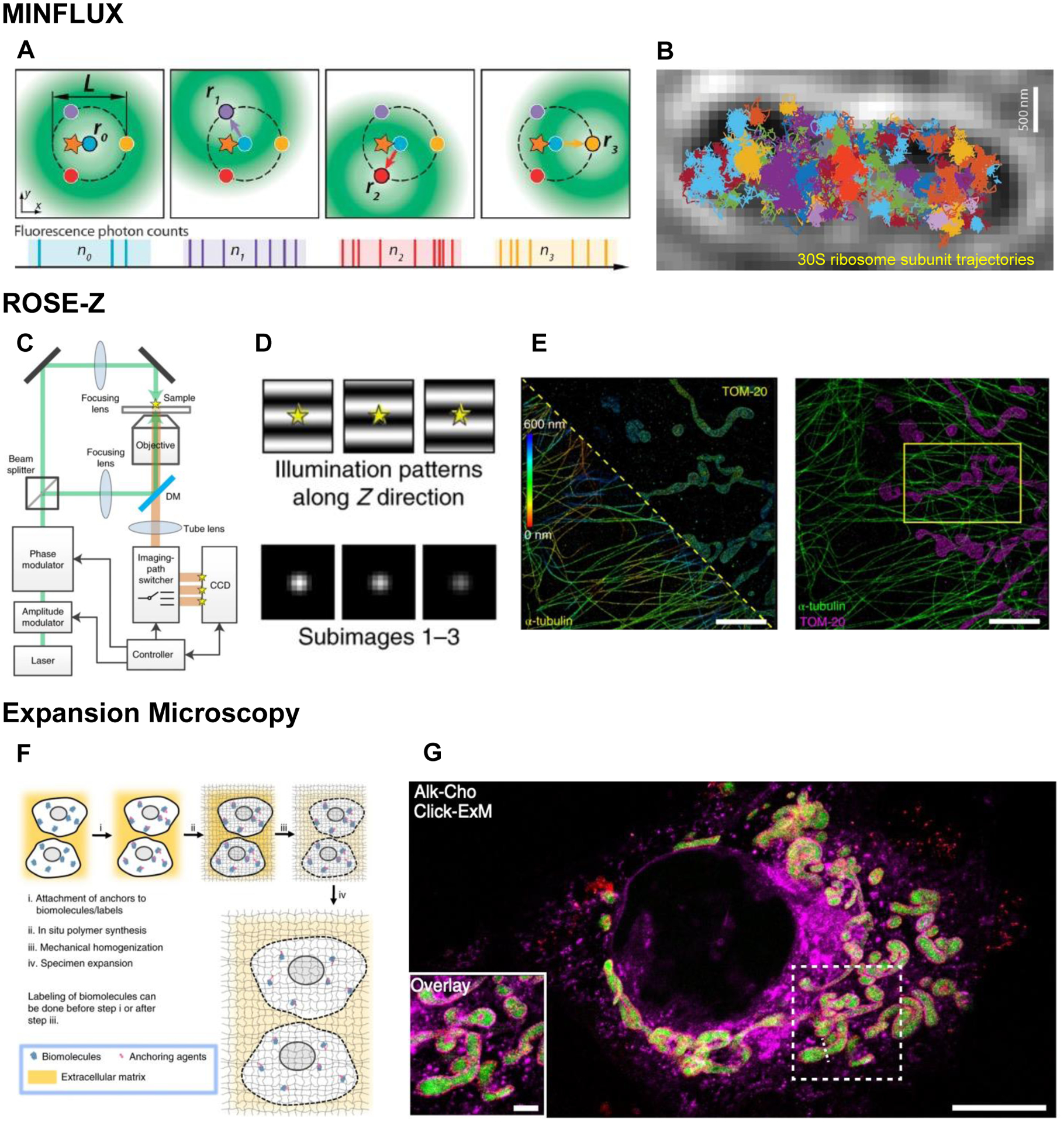
Figure 3:
Emerging super-resolution modalities.
( A ) Graphic representation of a fluorescent molecule (star) excited by a moving, donut-shaped excitation beam (green) at 4 different positions (r 0 , r 1 , r 2 , r 3 ) based on the donut minimum in ...
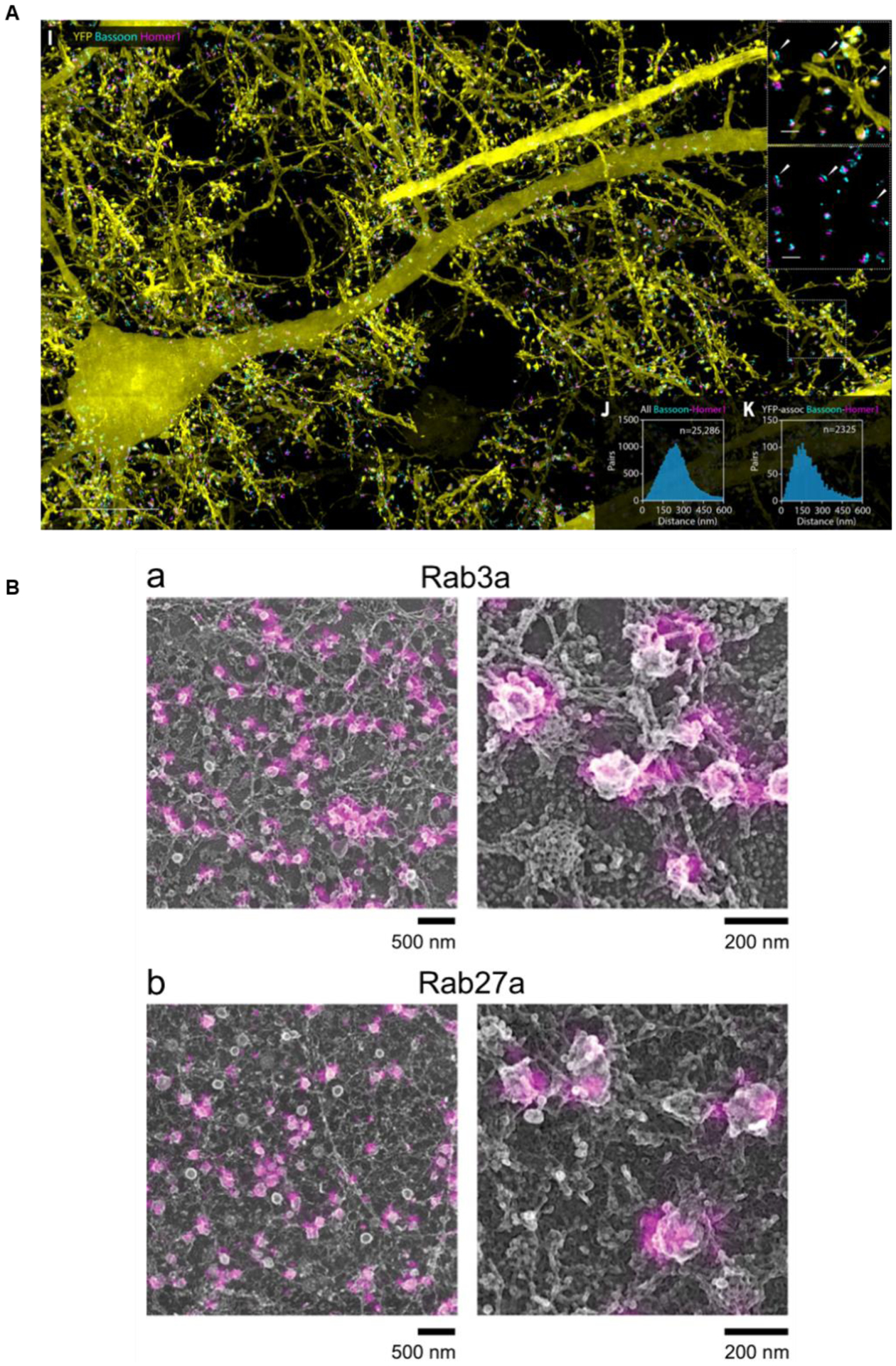
Figure 4:
Combined and correlative approaches.
(A) Combined expansion and lattice light sheet microscopy showing individual neurons marked with YFP, as well as two individual synaptic proteins, Bassoon and Homer1, in a mouse cortical brain slice. ...
Figure images are served from the NIH/NLM PubMed Central Open Access Subset or Europe PMC; copyright remains with the publishers and authors.
💬 Discussion
0 commentsNo comments yet. Be the first to start a discussion!
Leave a Comment